ClpX interactions with ClpP, SspB, protein
substrate and nucleotide
by
Greg Louis Hersch B.S. Biochemistry
University of California at Davis, 2001
Submitted to the Department of Biology in partial of the requirements for the degree of
OF ECHNOLOGY
FEB 0 1RIE2006 LIBRAR IES
fulfillment
Doctor of Philosophy in Biochemistry at the
Massachusetts Institute of Technology
McM0.l
February 2006
© 2005 Greg L. Hersch. All rights reserved
The authors hereby grants to MITpermission to reproduce and to distribute publicly Paper and electronic copies of the thesis document in whole or in part.
Signature of Author: Certified by: Accepted by: .1 - · - -- Department of Biology I/ Robert T. Sauer Salvador E. Luria Professor of Biology
Thesis Supervisor / - t R11 (_2~ ,tenhen P RPellI Co-Chair, Biology Professor of Biology Graduate Committee
CIpX interactions with CIpP, SspB, protein substrate and nucleotide
By
Greg Louis Hersch
Submitted to the Department of Biology on February 6th, 2006 in partial fulfillment of the
requirements for the degree of doctor of philosophy in biochemistry ABSTRACT
ClpXP and related ATP-dependent proteases are implements of cytosolic protein destruction. They couple chemical energy, derived from ATP hydrolysis, to the selection, unfolding, and degradation of protein substrates with the appropriate degradation signals. The ClpX component of ClpXP is a hexameric enzyme that recognizes protein substrates and unfolds them in an ATP-dependent reaction. Following unfolding, ClpX translocates the unfolded substrate into the ClpP peptidase for degradation.
The best characterized degradation signal is the ssrA-degradation tag, which contains a binding site for ClpX and an adjacent binding site for the SspB adaptor protein. I show that the close proximity of these
binding elements causes SspB binding to mask signals needed for ssrA-tag recognition by ClpX. The
SspB dimer overcomes this signal masking by tethering itself and bound substrate to ClpX, via docking sites located in the dimeric N-terminal domain of ClpX. Because this N-domain dimer binds only a single SspB subunit, the ClpX hexamer can accommodate just one SspB dimer per hexamer. Other adaptor proteins that use these same tethering sites must compete with SspB for access to ClpXP. Substrates bearing ssrA tags with increased spacing between the SspB and ClpX binding elements are degraded more efficiently at low concentrations by ClpXP. This mechanism in which the adaptor first obstructs and then stimulates substrate recognition may have evolved to permit an additional level of regulation of substrate choice. SspB binding to ssrA-tagged substrate is a highly dynamic process, allowing rapid
transfer of substrates from SspB to ClpX.
Although the ClpX hexamer is composed of six identical polypeptides, individual subunits assume at least three distinct conformations. Using a hexamer that was engineered to prevent nucleotide hydrolysis, I show that some nucleotide-binding sites in ClpX release ATP rapidly, others release ATP slowly, and at
least two sites remain nucleotide free. Occupancy of both the slow sites by ATP and the fast sites by
either ATP or ADP is required to bind the degradation tags of protein substrates. The ability of ClpX to retain binding of substrate with ATP or ADP in the fast sites suggests that nucleotide hydrolysis in the
fast sites, but not in the slow sites, will allow repeated unfolding attempts without substrate release over multiple ATPase cycles. My results rule out ATPase models including ClpX6eATP6 or ADP6 and also
suggest that the enzyme hydrolyzes only a fraction of bound ATP in a single turnover event.
Short peptide motifs of ClpX, known as IGF loops, interact with ClpP and change conformation as a response to nucleotide binding by ClpX. As ClpX varies its nucleotide content during the ATP hydrolysis cycle, it also varies its affinity for ClpP. Processing of substrates is coupled to the ATP-hydrolysis cycle of ClpX and appears to modulate ClpX's affinity for ClpP by changing how long each ClpX subunit
spends in each nucleotide state.
Thesis Supervisor: Robert T. Sauer
TABLE OF CONTENTS Page 2 Abstract Chapter One Chapter Two Chapter Three Chapter Four Appendix One
An introduction to ClpX, ClpP, SspB, and the AAA+
superfamily of ATPases
Part I: Introduction
Part II: Interactions between ClpX, ClpP, adaptors, and substrate Part III: The AAA+ superfamily
Part IV: Nucleotide utilization by AAA+ and related proteins Part V: Interpretations and Conclusions
SspB Delivery of Substrates for ClpXP Proteolysis Probed
by the Design of Improved Degradation Tags Introduction and Methods
Design of extended-spacing ssrA tags Improved SspB delivery to ClpXP
Dynamic interactions between SspB and ssrA tags Discussion
Characterization of contacts with the adaptor protein, SspB
mediated by the N-Domain dimer of ClpX Introduction
Interactions of SspB with the ClpX N Domain Discussion
Where does the XB peptide bind on the N-Domain? Is SspB a ClpX specific adaptor protein?
7 8 14 19 24 39 56 58 63 65 69 72 84 86 87 89 92 94
Asymmetric interactions of ATP with the AAA+ ClpX6unfoldase:
allosteric control of a protein machine 99
An ATP-hydrolysis defective ClpX variant 102
Strength and stoichiometry of ATP binding 104 Cooperative interactions in wild-type ClpX 107
Multiple classes of ATP sites 108
Linkage between ClpX binding to the ssrA tag and to Mg++/ATP 109 ATP binding and structural changes in the ClpX pore 110 ADP substitutes for ATP in "fast" nucleotide binding sites 112
Discussion 114
Communication between ClpX and ClpP during substrate processing and degradation
ClpP interaction requires more than two IGF loops Substrate processing strengthens ClpX-ClpP affinity Active-site communication between ClpP and ClpX Discussion 133 138 141 143 149 3
LIST OF FIGURES AND TABLES Chapter One: Figure 1 Figure 2 Figure 3 Figure 4 Figure 5 Figure 6 Table I Chapter Two: Figure 1 Table I Figure 2 Figure 3 Figure 4 Figure 5
The ATP-dependent protease, ClpXP and nucleotide
utilization by AAA+ ATPases
Architecture of the compartmentalized protease, ClpXP and a mechanical model for protein unfolding
Asymmetric docking of ClpX's six "IGF" loops with ClpP* A general model for adaptor proteins
Architecture of a AAA+ active site
Three potential mechanisms of ATP hydrolysis by AAA+ hexamers
A postulated mechanism for ATP synthesis by F1F ATP synthase Referenced crystal structures of AAA+ and related proteins
SspB Delivery of Substrates for ClpXP Proteolysis Probed by the Design of Improved Degradation Tags
- SspB delivery of ssrA-tagged substrates to ClpXP
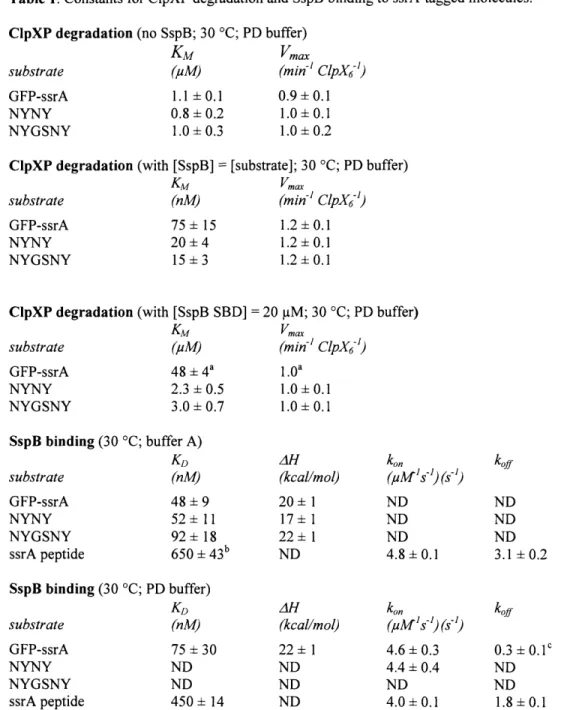
- Constants for ClpXP degradation and SspB binding to ssrA-tagged molecules
- ClpXP degradation of ssrA-tagged GFP variants in the presence of SspB
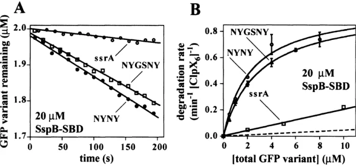
- ClpXP degrades the extended-tag substrates significantly faster than GFP-ssrA in the presence of the substrate-binding domain of SspB
- Equilibrium binding of SspB to GFP-ssrA
- Kinetics of dissociation and association of ssrA-tagged molecules with SspB 10 15 16 20 25 30 43 59 64 65 68 69 70
Chapter Three: Figure 1Figure 2 Figure 3 Figure 4 -Chapter Four: Figure 1 Figure 2 Figure 3 -Figure 4-Figure 5Figure 6
-Characterization of contacts with the adaptor protein, SspB
mediated by the N-Domain dimer of ClpX
Binding of the N Domain of ClpX and XB modules of SspB Binding stoichiometry of the N Domain of ClpX to XB modules of
SspB
Cartoon representation of SspB / ClpX interaction Perpendicular views of the N-domain dimer of ClpX
Asymmetric interactions of ATP with the AAA+ C1pX6
unfoldase: allosteric control of a protein machine
A ClpX variant defective in ATP-hydrolysis ATP binding by various methods
Dissociation kinetics reveals two classes of sites Nucleotide dependence of ClpX substrate binding and conformation
ADP and substrate binding
Models for ClpX binding to ATP and the ssrA tag of substrates
5 88 89 90 94 103 105 109 111 113 117
Appendix: Figure 1Figure 2 Figure 3 Figure 4 Figure 5 -Figure 6-Figure 7Figure 8 -* work of Shilpa
Communication between CIpX and CIpP during substrate processing and degradation
Symmetry mismatch between ClpX and ClpP* Properties of ClpX loopless
An assay for ClpX-ClpP interaction in solution*
ClpX-ClpP affinity changes in an ATPase-dependent fashion during substrate denaturation and translocation*
Modification of the ClpP active sites strengthens ClpX binding* ClpP rescues the unfolding defects of ClpX mutants*
The "ATP" state of ClpX is required for strong ClpP interactions* Model for the interaction of ClpX and ClpP
A. Joshi 137 139 141 143 144 146 147 151
CHAPTER ONE
An introduction to ClpX, ClpP, SspB, and the
AAA+ superfamily of ATPases
Part I: Introduction
The roles of energy dependent proteolysis
Many cytosolic proteins ultimately meet their fate at the hands of ATP-dependent proteases. This destruction is required to remove and recycle proteins that have become damaged by oxidation, heat unfolding and aggregation, or were simply translated from damaged mRNA (Gottesman, 1996). Energy-dependent proteases also destroy folded, native proteins as a signaling event. Examples of this occur as a response to DNA damage. environmental cues, and during the cell cycle (Jenal and Stephens, 2002; Gottesman, 2003; Jenal and Hengge-Aronis, 2003). Some of these same proteases function in quality assurance for the cell. Without quality control, translation of aberrant mRNA messages could result in partial protein products that aggregate or even actively interfere with vital cell processes. Instead, these proteins are recognized and removed by ATP-dependent proteases. Thus, energy dependent proteolysis allows the cell to dynamically control its proteome and respond rapidly to environmental cues. Energy-dependent proteases are all closely related, and belong to a much larger class of proteins called the AAA+ (ATPases associated with a variety of cellular activities) superfamily whose members all function in macromolecular disassembly (Neuwald et al., 1999; Vale, 2000; Glover and Tkach, 2001; Sauer et al., 2004).
E. coli ClpXP is an ATP-dependent protease, which degrades a wide range of substrates (Flynn et al., 2003). The best characterized substrates for ClpXP are those bearing the C-terminal ssrA peptide tag. The eleven-residue ssrA tag (AANDENYALAA), when appended to the C-terminus, can direct any protein to ClpXP for either in vivo or in vitro
degradation. In the cell, the ssrA tag is co-translationally attached to nascent polypeptides on ribosomes that stall at the end of mRNA lacking a stop codon or stall for other reasons (Keiler et al., 1996). Once tagged, these proteins are recognized by ClpXP and degraded. ClpXP also recognizes other substrates that do not bear the ssrA tag. It has been proposed that there are at least five distinct classes of ClpXP degradation tags, although other substrates which do not appear to fall into these classes have been identified (Flynn et al., 2003).
Below follows a brief introduction of how ClpX, ClpP, and the adaptor protein SspB interact with one another to select and degrade protein substrates. Although much is known about these proteins, many critical, unanswered questions remain. ClpX is a hexameric ATPase and understanding how ATP binding and hydrolysis support its function is an essential step towards understanding how it and related AAA+ proteins operate. How does nucleotide binding affect the conformation and properties of ClpX? Does ATP bind to all six nucleotide-binding sites on a hexamer? Are all bound ATPs equivalent? Do subunits hydrolyze all bound nucleotides simultaneously? Many models have been put forth in an attempt to explain the observed properties of AAA+ enzymes and various mutants. By summarizing what is known about ATP binding and utilization by members of the AAA+ superfamily, I will emphasize commonalities between the members as well as evidence both supporting and contradicting current models. Finally, by discussing the limitations of the experimental methods employed, I hope to highlight the inadequacy of narrow approaches to this problem
A
Strained substrateFigure 1
B
I
ClpP (protease)c
ClpP cutaway Sequestered serine active sites Compartmentalized proteolysis by ClpPProteolysis is not inherently energy dependent, as the hydrolysis of peptide bonds is an exergonic process. For instance, chymotrypsin and trypsin require no energy other than thermal energy to degrade substrates. Why do cytoplasmic proteases such as ClpXP require energy? ClpXP is capable of degrading stable substrates that possess significant secondary and tertiary structure and are resistant to proteolysis by non-energy requiring proteases. ClpXP achieves this by using energy from ATP hydrolysis to disrupt the native structure of protein substrates prior to proteolysis. Potential mechanisms for this disruption are discussed below. Importantly, because of ClpXP's unfolding activity, substrate selection is not subject to the same conformational constraints as
energy-independent proteases like chymotrypsin and trypsin. Because ClpXP is capable of destroying any cellular protein, substrate selection must be tightly controlled.
ClpXP is a complex of two proteins, ClpX and ClpP (Wojtkowiak et al., 1993). Substrate selection, unfolding, and ATP hydrolysis are carried out by ClpX. ClpP contains the proteolytic active sites and is made up of 14 subunits which form two seven-subunit rings that stack back-to-back (Fig. B). This arrangement results in a barrel-like structure with a large central cavity. Each of ClpP's 14 subunits contains a complete serine active site for peptide hydrolysis with a classical Ser, Asp, His catalytic triad. ClpP sequesters these active sites inside of its barrel-like structure (Fig. 1C). Access to the active sites is restricted by two axial portals on either end of the barrel. These portals have a minimum diameter of about 10 A, disallowing passage of native, folded proteins (Wang et al., 1997). Consistently, isolated ClpP displays very little proteolytic activity towards even unfolded substrates. Small peptides that are able to diffuse through the axial portals are hydrolyzed efficiently by ClpP, demonstrating that lack of proteolysis for larger substrates is due to limited access rather than active site orientation or activation state (Thompson et al., 1994). Thus, ClpP's robust proteolytic ability is controlled by its small axial portals which restrict proteolysis to substrates that have been translocated into ClpP by ClpX.
ClpX and potential mechanisms of unfolding
As discussed above, ClpX recognizes substrates, unfolds them if needed, and then translocates them into ClpP for proteolysis in an ATP-dependent process. ClpXP's
substrate selection is carried out entirely by ClpX. Six identical polypeptides make up the ClpX hexamer, forming a cylindrical, hex-nut structure with a central pore. In the ClpXP complex, the pores of ClpX and ClpP are aligned, and substrates are translocated through this central pore (Ortega et al., 2000). Like ClpP's axial portals, the central pore of ClpX is too small to allow passage of folded proteins.
ClpXP, like other energy-dependent proteases, disrupts the tertiary structure of protein substrates prior to proteolysis. ATP hydrolysis by ClpX is coupled to the unfolding reaction, although the details of this coupling are far from clear. The most prominent model involves coupling the chemical energy of ATP hydrolysis by ClpX to an exertion of mechanical force on the bound protein, driving disruption of structure. However, other plausible models exist. In one such model, ClpX binds its substrate, but then simply waits to capture or trap a spontaneous unfolding event without exerting any mechanical stress on the protein. In this model, ClpX quickly binds to exposed, unfolded portions of the protein as they become available in a ratchet-like mechanism.
The ratchet model seems insufficient to explain ClpXP's potent protease activity. For instance, green fluorescent protein (GFP) with a C-terminal ssrA tag is degraded by ClpXP at a rate 10 million times faster than its spontaneous unfolding rate in solution (Kim et al., 2000). Although this observation makes the ratchet model unlikely, it does not render it completely untenable. Proteins in solution may exhibit substantially different properties than those bound to another protein. A substrate bound to ClpX likely experiences a substantially altered local environment, but no evidence exists to suggest
that this local environment is especially conducive to disruption of a protein's structure. Furthermore, if ClpX were quickly capturing unfolded portions of the protein as they were spontaneously exposed, one would expect a significantly higher affinity for unfolded over folded proteins. Contrary to this expectation, ClpX's affinity for an unfolded protein was found to be essentially unchanged with respect to its folded counterpart (Kenniston et al., 2003). One caveat to these experiments is that unfolding of the substrate protein was achieved by chemical modification of cysteines with a negatively charged carboxymethyl group, and thus this modified molecule does not mimic all aspects of a true unfolded, natural substrate. Nonetheless, one would expect some substantial difference in substrate affinity, which was not observed.
Instead, ClpX probably employs an active unfolding mechanism in which ATP binding and hydrolysis is coupled to the unfolding of substrates via mechanical stress. In a simplified version of the most current model, ClpX binds substrate and repeated ATP-hydrolysis cycles drive conformational changes in ClpX that disrupt the bound substrate's native fold (Fig. 1A). Later in chapter 5, I show direct evidence for conformational changes in ClpX that depend on the identity of bound nucleotide. How these conformational changes in ClpX are translated into substrate unfolding is unclear. One possibility is that substrates unfold during ClpX's attempt to engulf them. The pores of ClpX and ClpP are both too small to admit native proteins. In the steric collision model, ClpXP recognizes and binds the exposed degradation tag of a folded substrate. Subsequent attempts to translocate this tag and attached peptide sequence through the narrow central channel of ClpXP results in steric collisions of the folded portion of the
substrate with the central pore. In this model, unfolding of the protein substrate occurs when a steric collision occurs with the right geometry to unravel the protein's tertiary structure.
Part II: Interactions between C1pX, ClpP, adaptors, and substrate ClpX's interactions with C1pP
ClpX must associate with ClpP to form a functional protease. At least one site of interaction between these two proteins has been located. ClpX contains an internal tri-peptide of sequence Ile-Gly-Phe (IGF) which extends from one face of ClpX as part of a surface loop (Fig. 2). Many experiments have suggested that these loops dock with ClpP, likely in seven hydrophobic pockets on either side of ClpP (red, Fig. 2). When the IGF loops of ClpX are mutated or removed, ClpX neither associates with ClpP nor delivers substrates for degradation. Wild-type ClpX associates with ClpP in an ATP-dependent fashion and isolated IGF peptides interact very weakly with ClpP (Joshi et al., 2004; Hersch et al., 2005), suggesting that ClpX hexamers specifically orient IGF loops for interaction with ClpP as a response to ATP binding.
Structural studies suggest that ClpP has seven potential sites of interaction on each face for ClpX's IGF loops (Wang et al., 1997). However, ClpX is a hexamer and thus possesses only six IGF loops. This asymmetry must result in at least one potential IGF
Figure 2
ClpX
ClpP
B
Why would ClpP want to have open docking sites? One possibility is to provide a way for ClpX to rotate with respect to ClpP. This feature was once an attractive model for explaining how ClpX drives translocation of substrates into ClpP. In this model, substrates were translocated by mechanism that resembles the way threaded screws enter a piece of wood when twisted as ClpX rotated with respect to ClpP. However, ClpX is capable of unfolding substrates in the absence of ClpP. Moreover, a full complement of IGF loops appears to be unnecessary for degradation of substrates (Joshi et aI., 2004). Consistent with an asymmetric interaction not being an essential feature of the true mechanism, another ATP-dependent protease, HsIUV, does not possess this asymmetry (both the ATPase and peptidase are hexamers) but catalyzes the same types of reactions as ClpXP (Sousa et aI., 2000). The functional relevance of the asymmetric interaction of ClpX with ClpP remains an unanswered question in the field.
ClpX's interactions with the adaptor molecule, SspB and substrate
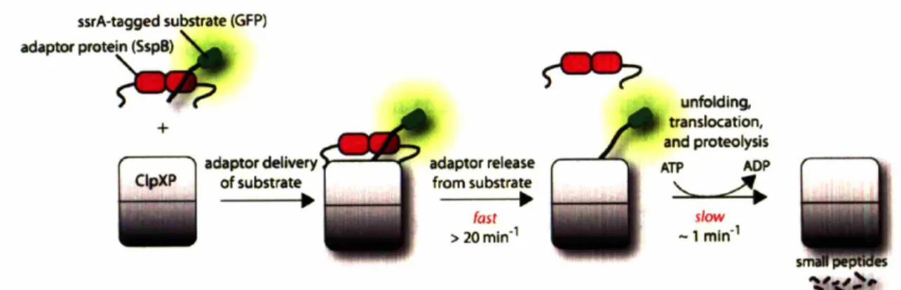
Proteins called adaptors can assist proteases in ATP-dependent degradation reactions. Adaptor proteins can bind substrate proteins in solution, and then dock with the protease. Substrate is then transferred to the protease for degradation and the adaptor protein is generally released, and not consumed by the reaction (Fig. 3). SspB is an 18 kDa protein that forms dimers in solution and can bind the ssrA tag of potential ClpXP substrates (Levchenko et aI., 2000; Wah et aI., 2002). It utilizes flexible C-terminal tails to tether itself to ClpXP and deliver substrates for degradation (Wah et aI., 2003). By bringing substrate and protease together, the efficiency of degradation is enhanced. This enhancement is achieved by significantly increasing the apparent affinity of substrate for ClpXP and modestly increasing the speed at which they are degraded.
Figure 3
ssrA-tagged substrate (GFP) adaptO< p"'te~ + ~ adaptor delivery C1pXP of substrate•
adaptor release from substrate•
fast > 20min-1 5 •• ~ unfolding, translocation, and proteolysis ATP ADP '----".
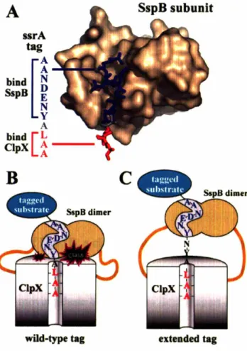
slow -1 mln-1SspB and ClpX bind distinct residues of the ssrA tag. Of the 11 residues composing the
recognized by ClpXP (bold) (Flynn et aI., 2001). The first four residues and the central Tyr7 are recognized by SspB (underlined). No single residue is crucial for both ClpX and SspB recognition. As I will describe later in this thesis, unfavorable interactions occur when ClpX and SspB simultaneously bind the ssrA tag. Consequently, when proteins
bearing the ssrA tag are bound by SspB, the ssrA tag is obstructed and no longer efficiently recognized by ClpX (Hersch et al., 2004). The inhibitory effect of this masking is overcome by the C-terminal tails of SspB, which tether it to ClpXP. This tethering drives tag engagement by ClpXP by means of a high local concentration. The net effect of this obstruct-then-stimulate interaction is a -20-fold increase in efficiency.
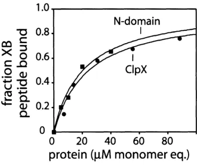
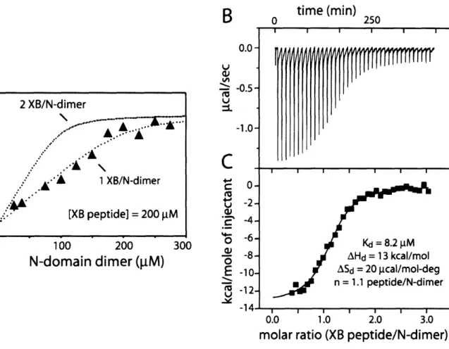
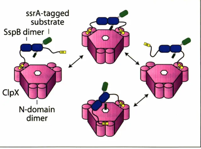
Central to SspB's activity as an adaptor is its ability to tether itself to ClpX. This interaction appears to be highly specific, because SspB does not deliver substrates to other closely related ATP-dependent proteases. ClpX and orthologs contain specialized regions which are not shared by other classes of AAA+ proteins (Neuwald et al., 1999). One such region, the N-terminal domain of ClpX, is essential for the enhancement of degradation by SspB. The isolated N-domain of ClpX forms dimers in solution (Wojtyra et al., 2003). Later in this thesis, I show that an isolated N-domain dimer of ClpX is sufficient for SspB recognition and that all of SspB's contacts with ClpX are made through this N-terminal domain (Bolon et al., 2004). The ClpX hexamer contains three N-domain dimers. Each dimer binds just one subunit of the SspB dimer, explaining why only a single SspB dimer binds efficiently to ClpX (Wah et al., 2002; Bolon et al., 2004).
SspB binds its ssrA-tagged substrates significantly more tightly than ClpX. How then are substrates transferred to ClpXP for degradation? As I discuss in chapter two, substrate dissociates from SspB much more rapidly than the processing (unfolding and degradation) of substrates by ClpXP (Figure 3). Since bound substrates are in a fast, dynamic equilibrium, ClpXP can simply wait for passive dissociation from SspB to
process substrates. In other words, waiting for substrate to dissociate will not perceptibly slow down proteolysis by ClpXP. Additionally, ClpXP could pull substrate from SspB in an active process, but based on the kinetic measurements there seems to be no need for it to do so.
In addition to enhancing substrate recognition, SspB causes a moderate increase in the rate at which substrates are degraded. This phenomenon remains essentially unexplained, although potential models do exist. On its own, ClpX is capable of binding only a single molecule of substrate, likely in the central pore of the hexamer (Piszczek et al., 2005). The turnover increase could be explained if ClpXP were able to degrade both substrate molecules bound to SspB simultaneously. A second way for SspB to increase the rate of substrate degradation would be to destabilize the substrates it delivers. For example, if binding of substrates to SspB resulted in minor disruption of structure near the ssrA tag, the rate at which those substrates are degraded could increase (Kenniston et al., 2003). Later, I show that GFP molecules fused to a C-terminal ssrA tag experience a reduction in fluorescence when bound to SspB in a manner consistent with a structural perturbation. If SspB truly does disrupt the structure of GFP-ssrA, then the perturbation must be minor since no major disruption of GFP's secondary structure is observed by circular dichroism measurements when bound by SspB (unpublished data). Also in conflict with this hypothesis, SspB enhances the rate at which carboxymethylated substrates, which are unstructured as determined by circular dichroism, are degraded (J. Kenniston, personal communication).
Part III: The AAA+ superfamily
The common purpose of AAA+ ATPases
ClpX contains a conserved AAA+ domain of roughly 200 amino acids. Many proteins that contain similar domains perform tasks that all seem to require the application of mechanical force (Vale, 2000; Glover and Tkach, 2001; Sauer et al., 2004). Enzymes of the AAA+ superfamily play important roles in life. They are involved in processive DNA replication, endoplasmic reticulum associated degradation (ERAD), vesicle fusion, cellular transport, viral genome replication, and a host of energy-dependent degradation pathways (Neuwald et al., 1999). The AAA+ domain encodes various elements that make up an ATPase active site. These include the well known Walker A and B sequences that mediate binding and hydrolysis of ATP, as well as two conserved arginine residues that have been implicated in sensing the identity of bound nucleotides. AAA+ proteins are typically hexameric and can contain either one or two copies of the AAA+ domain. Proteins that contain two AAA+ modules form two stacked hexameric rings, with each domain forming a subunit of each ring. In these cases, each ring often possesses distinct catalytic properties (Parsell et al., 1994; Singh and Maurizi, 1994; Nagiec et al., 1995; Seol et al., 1995; Watanabe et al., 2002; Song et al., 2003; Wang et al., 2003).
Useful AAA+ mutants
ClpX and other AAA+ enzymes use ATP hydrolysis to drive molecular disassembly processes that seem to require the exertion of mechanical force. ATP binding, hydrolysis, and product release at the ATPase active site of the enzyme must somehow result in allosteric conformational changes that perform these tasks. Mutations that disrupt the
ATPase cycle and communication network are critical for understanding how AAA+ proteins achieve their functions. Several classes of mutants have been used to probe this mechanism. Below, I outline the mutant classes and their characteristic enzymatic phenotypes which have provided the most information about the mechanism of AAA + proteins.
Figure 4
'"
'"
'"
'"'"
'"
'"
.... .... .... .... .... .... .... ....B
Walker A (P-Loop) KT Pore Walker B Glu185 Box VII Arg307 Arg370The ATPase sites of AAA+ proteins are located at the interfaces between subunits (Fig. 4A). Residues involved in nucleotide sensing and hydrolysis extend from both adjacent subunits. The Walker-A sequence motif is the canonical feature of P-loop ATPases and features residues which loop around the phosphate moiety of the bound nucleotide (Fig. 4A and B). This motif is critical for the binding of nucleotide, whereas residues of the
Walker-B motif are essential for ATP hydrolysis. The extended Walker-B motif contains a highly conserved sequence in ClpX Asp 84-Glul8 5-Ilel86-Asp187 and many related
proteins. Magnesium is an important cofactor of the ATP hydrolysis reaction, and is coordinated by Asp1 8 4 and Asp'8 7. Glu 8 5, shown in Figure 4A, serves as the catalytic base and activates a water molecule for hydrolysis of the gamma phosphate of ATP.
Mutation of the glutamate to a non-acidic residue is sufficient to disrupt ATP hydrolysis in most AAA+ and related proteins. By preventing hydrolysis, it is possible to study the static ATP-bound form that may normally not be substantially populated. In the wild-type ClpX, the ATP form of the enzyme is highly unstable due to a high basal ATP-hydrolysis rate (over 100 min-' enzyme-'). Hydrolysis of the ATP analog ATPyS occurs about 20-fold more slowly (Burton et al., 2003). Other ATP analogs fail to bind ClpX tightly or at all. Another concern is the possibility that analogs do not faithfully mimic ATP as reported for AMPPNP and GroEL (Rye et al., 1997). However, mutation of the Walker-B glutamate also has the potential to misreport characteristics of the wild-type enzyme. For instance, removal of the negatively charged glutamate from the active site could potentially change the rate at which nucleotide is bound and released or alter the conformational changes that typically accompany ATP binding and hydrolysis.
Arg3 70 and Arg307are important for ClpX activity, and arginines at homologous positions are common in other AAA+ proteins (Song et al., 2000; Hishida et al., 2004; Joshi et al., 2004; Schumacher et al., 2004). Arg3 70 is located in a region of ClpX known as the
sensor-II helix and is in close proximity to the gamma phosphate of ATP in some AAA+
structures, including that of Helicobacter pyrlori ClpX (Fig. 4A). In most, but not all AAA+ proteins, altering this residue results in a hydrolysis-defective mutant. In contrast to mutations in the Walker-B motif, enzymes lacking the sensor-II arginine do not usually display properties of the ATP state when ATP is bound. This observation suggests that the sensor-II arginine senses bound ATP and helps propagate resulting conformation changes.
Arg307 in ClpX is part of the box-VII sequence motif. This position has been described as an arginine finger in many AAA+ proteins, akin to those seen in GTPases (Ahmadian et al., 1997). The hydrolysis activity of GTPases is often activated by protein factors (GAPs) which supply a catalytic arginine to stabilize the transition state of nucleotide hydrolysis (Ahmadian et al., 1997). Similarly, the side chain of Arg3 0 7 in ClpX is
supplied in trans to the ATPase site of the adjacent subunit (Fig. 4A). The "arginine finger" of AAA+ proteins contacts the sensor-II arginine in some crystal structures and contacts the gamma phosphate of ATP in other structures (Bochtler et al., 2000). Rather than participating in catalysis, Arg307 in ClpX may serve a sensing function similar to
Arg3 7, except in trans; sensing the identity of the nucleotide bound to the adjacent subunit. As I discuss in chapter four, ATP hydrolysis by ClpX and many other AAA+ enzymes is cooperative. Therefore individual subunits of ClpX must have information about the nucleotide content of other subunits (Hattendorf and Lindquist, 2002; Burton et al., 2003; Hersch et al., 2005). The box-VII arginine seems a likely candidate for this role as mutants behave similarly to sensor-II mutants, although a role in transition state stabilization cannot be ruled out (Hishida et al., 2004).
Nucleotide utilization by AAA+ proteins
Nucleotide content determines the conformation of AAA+ proteins throughout the catalytic cycle. As I discuss in chapter four, ATP binding causes ClpX to adopt a conformation that can bind protein substrates and the ClpP peptidase tightly (Wah et al., 2002; Hersch et al., 2005). I also show that ATP/ATPyS binding to ClpX results in allosteric changes in the central pore, similar to those seen for other AAA+ enzymes (Schlieker et al., 2004; Hersch et al., 2005). Measuring the properties of an all ATP or all ADP form of the enzyme is useful, but does not report on the way energy is used in the enzymatic cycle. For instance, how many nucleotides are hydrolyzed in a single unfolding attempt? As a first step to answering this question, many laboratories have employed both crystallographic and biochemical methods to find the number of nucleotides bound. As a second step, it must be determined whether bound ATPs are hydrolyzed simultaneously or whether only a sub-population of bound ATP is hydrolyzed in a single enzymatic cycle. Data from different experimental systems can be compared to address the possibility that all hexameric AAA+ motor proteins use closely related mechanisms.
In the most straightforward ATP-hydrolysis scheme for ClpX, six ATP molecules are bound at the interface of the six subunits, simultaneously hydrolyzed to ADP + Pi, and then released (Fig. 5A). Some structural results for other AAA+ hexamers appear to support this concerted model of hydrolysis (Gai et al., 2004). However, many biochemical experiments and a few structures are inconsistent with this model and
suggest a significantly different mechanism. For example, several AAA+ and related hexameric proteins bind fewer than six molecules of ATP at saturation. Furthermore, functional as well as single turnover experiments suggest that bound ATPs are not all hydrolyzed at once, but instead are hydrolyzed individually or in other substoichiometric groups. What follows is an assemblage of the current evidence, highlighting unifying principles and the need for clarifying experiments in the hopes of illuminating the details of a shared catalytic mechanism.
Part IV: Nucleotide utilization by AAA+ and related proteins
ClpX and HslU - How AAA+ unfoldases utilize nucleotide
Later in this thesis, I examine ClpX's capacity for binding ATP by several methods including isothermal titration calorimetry (ITC). Only a subset of the six potential nucleotide-binding sites in ClpX bound nucleotide. Chromatography and filter-binding experiments confirmed that only 3-4 ATPs bound per ClpX hexamer. The remaining unbound sites could not be filled by ADP at the concentrations tested. Thus, biochemical experiments indicate that at least two of ClpX's subunits assume a conformation that prevents nucleotide binding at pM concentrations. This result is clearly in conflict with a model in which six ATPs are simultaneously hydrolyzed by ClpX. Figure 5B and 5C show the models of ATP hydrolysis consistent with the observed mode of ATP binding by ClpX.
Figure 5
A.Six fold symmetric, concerted ATPhydrolysis model
8e.
ADPIP,ReJease-~
'?
~~ smADP
8. Concerted hydolysis model with less than six ATPsbound
$
ATPHydro/ysis. ADPIP,R~fourADP
C.Partial hydrolysis model
$
ATPHydralys/s.~t
ADP/PIR~
~C6
(lJ
twoADP0)
I
II
III
Experiments show that ClpXeATP, but not ClpXeADP binds tightly to protein substrates and ClpP. This raises an interesting paradox. If a ClpX hexamer were to adopt the ADP state after hydrolysis of ATP (Fig. 5B, intermediate II), it would lose affinity for its
substrate and peptidase. This situation would present problems for a processive protease. Because ClpXP degrades ssrA-tagged proteins from the C-terminus, the degradation tag is degraded first (Lee et al., 2001; Kenniston et al., 2005). If the ClpXPosubstrate complex were to prematurely dissociate before completing degradation, the resulting
substrate fragment would be resistant to further proteolysis by ClpXP because it would lack a degradation signal. In this case, one would expect a build up of partially proteolyzed products. However, partially proteolyzed products are not normally observed, indicating that proteolysis by ClpXP is processive and rarely disrupted before completion. I discuss evidence for one potential solution to this paradox in chapter four. In short, I found that a hexamer with mixed nucleotide content (ATP/ADP) retained its tight association with substrate (Hersch et al., 2005). This mixed nucleotide intermediate is depicted in Figure 5C, II and suggests that the partial hydrolysis model depicted in Figure 5C is most consistent with the current data.
The crystal structure of Helicobacter pylori ClpX appears to contradict the partial hydrolysis model as every subunit in the crystal has an identical conformation and
contains bound ADP (Kim and Kim, 2003). However, the crystal did not contain a ring hexamer of ClpX. Instead, ClpX subunits are related to one another in the crystal by a screw axis (Table 1). This crystallographic arrangement may actually provide subtle evidence against a six-fold symmetric hexamer. With nucleotide bound to all subunits, the structures of the individual protomers may not be able to form a closed ring. Some empty subunits may be essential to provide the required kinks to form a closed ring.
Attempts to crystallize ClpX in the empty state or with bound ATPyS/magnesium produced an identical ADP-bound, helical arrangement (Kim and Kim, 2003).
HslU and ClpX are about 50% homologous and perform similar functions. Like ClpX, HslU is the unfoldase component of a bacterial ATP-dependent protease. Although biochemical testimony is lacking, the literature is rich with structural data for HslU. Unlike ClpX, HslU has been crystallized as a hexamer and in many different nucleotide states (Table 1). Some forms clearly have six nucleotides bound, which seems either to contradict the data for ClpX or possibly to suggest divergent mechanisms. The 1G4A structure of HslU was crystallized with six bound ADP molecules and the 1E94 structure was crystallized with six bound AMP-PNP molecules (Song et al., 2000; Wang et al., 2001a). However, the presence of AMP-PNP in the 1E94 structure has been called into question by showing that density for the gamma phosphate is mising from a recalculated electron-density map (Wang et al., 2001b).
Other HslU structures are not six-fold symmetric. For example, the 1DO0 structure consists of a dimer of trimers with two subunits containing ATP.Mg, two containing ATP, and the last two containing only sulfate ion (Bochtler et al., 2000). Each subunit pair exhibited a distinct conformation, with unique conformations of the pore residues. Another crystal form (1D02) contained a trimer of dimers with AMP-PNP bound in alternating subunits (Bochtler et al., 2000). This form was reminiscent of the Fl ATP synthase, in which catalytic and non-catalytic subunits alternate around the hexamer (Abrahams et al., 1994). In this case, differences in the structure and roles of different F1
subunits arise from differences in the primary sequence (discussed below). The two HslU hexamer structures which deviate from six-fold symmetry seem consistent with the biochemical data described for ClpX.
Unfortunately, some aspects of the HslU structures that deviate from six-fold symmetry are controversial. The identity of the bound nucleotide in the 1DO2 structure has been questioned because the temperature factor for the gamma phosphate is much higher than surrounding atoms (Wang et al., 2001b). This could simply reflect greater flexibility, or more ominously be indicating that the nucleotides are ADP and not ATP. An unusual syn conformation of the adenine base in this structure has also caused suspicion. An anti orientation of the base is typical, whereas the syn form is rarely observed in crystal structures including other HslU structures. If the ATP is incorrectly bound or is actually ADP, the resulting structure might not represent the true "ATP bound" state of the enzyme. A possible exception to the anti rule is the D2 AAA+ domain of NSF, which appears to bind ATP is the syn conformation (Lenzen et al., 1998; Yu et al., 1998). However, this domain has very low hydrolytic ability, so its relevance is unclear.
HslUV is the only ATP-dependent protease for which the crystal structure of the ATPaseepeptidase complex is known. HslUV structure 1G3I clearly shows how HslU aligns with HslV to form a shared central pore (Sousa et al., 2000). Although there are six ATPs bound in this structure, magnesium is not present. Biochemical experiments, some of which I will discuss in chapter four, have shown that magnesium is essential for ClpX, HslU, and likely all AAA+ ATPases to adopt the ATP-bound conformation (Burton et
al., 2005; Hersch et al., 2005). Furthermore, skepticism exists concerning the identity of the bound nucleotide as ATP rather than ADP. No structure of HslU exists with six bound molecules of Mg.ATP or Mg.ATP analogs. However, these structures do seem to show that HslU can form a closed ring with six bound molecules of ADP. Preliminary chromatography experiments with ClpX suggest that it cannot bind six ADP, but again only 3-4, similar to ATP. These data from ClpX seem far more congruent with crystal structures of HslU that deviate from six-fold symmetric nucleotide binding. Further experiments are needed to resolve this apparent contradiction.
F1Fo ATP Synthase - the best characterized molecular motor
The FFo ATP synthase is an important molecular machine found in the membranes of bacteria, chloroplasts, and mitochondria. This machinery is used to generate ATP from ADP and PI in the presence of an electrochemical gradient. It does this by coupling the downhill conductance of protons across the membrane to the endergonic synthesis of ATP. This complex is composed of two separable units termed F and F1. The Fo
complex is a membrane-spanning conduit for protons. The Fl component is composed of nine subunits of composition C3P37Y65. The a3 3 core of F is highly homologous to
AAA+ proteins. Indeed, the ATP-binding P-loop or Walker-A motif was first recognized in these proteins (Walker et al., 1982). Although neither the nor 3 subunits contain all of the sequence elements found in the AAA+ superfamily, there is significant structural and sequence homology (Saraste et al., 1981; Abrahams et al., 1994). For instance, (c313
forms a hexamer and subunits adopt a fold similar to AAA+ proteins including ATP-binding sites at the subunit interfaces. The oc and 3 subunits share 20% sequence identity,
but the a subunits are hydrolytically inactive due to the absence of a catalytic, basic residue in the active side. In the absence of the Fo channel, Fl can function as an ATPase. Together with sequence conservation, there is ample reason to believe that mechanistic similarities exist between the F1ATPase and AAA + proteins.
Crystal structures of F1 ATPase show that a hexamer is formed by alternating a and ~
subunits which surround a central y subunit (Bianchet et aI., 1991; Abrahams et al., 1994). In structure 1BMF, all three hydrolytically inactive asubunits are bound to AMP-PNP, but only one ~ subunit is bound to AMP-PNP. A second ~ subunit was bound to ADP and the final ~ subunit contained no bound nucleotide (Fig. 6A). Moreover, each ~ subunit assumed a slightly different conformation.
Figure 6
A
~ Loose (conformationfor ADP/Pi binding)
o
Open (conformation for ATP release)III
nght (conformation for ATP synthesis)B
ADPPi
'-
\~
The observation of asymmetry in nucleotide binding by the subunits was well suited to a postulated cyclical binding change mechanism (Cross, 1981; Boyer, 1993). In this mechanism, each 3 subunit can assume at least three conformations with different affinity for nucleotide called open (3o), loose (PL), and tight (T). Conversion of the loose site to a tight site requires conversion of the pre-existing tight site to an open site, and the open site to a loose site, all coupled to the orientation of the central subunit (Fig. 6B). In this way, subunits changed their affinity for nucleotide in a cyclical fashion during ATP synthesis and prevent hydrolysis of newly synthesized ATP. It is not entirely clear which of the three 3 subunits in the structure is tight, open, or loose but structural considerations seem to indicate the assignment designated in Figure 6A (Abrahams et al., 1994). The structure also suggested that these conformational changes were driven by rotation of the central y subunit, with the orientation of the gamma subunit preventing 0o from tight association with nucleotide. The rotation hypothesis was further bolstered by the direct observation of rotation by a fluorescent actin filament which had been cross-linked to the y subunit during ATP hydrolysis by Fl ATPase (Noji et al., 1997).
There is ample structural evidence to suggest that F operates by an asymmetric mechanism. In addition, biochemical evidence predating the structure also suggests the presence of only two nucleotide-binding sites per hexamer (Ackerman et al., 1987). Presumably, this stoichiometry reflects only the 3 subunits because the three c subunits do not readily exchange nucleotide. The field is not devoid of controversy, however. Figure 6B depicts a "bi-site" ATP synthesis mechanism that is likely an oversimplification, as many experiments suggest that F operates via a "tri-site"
mechanism where all sites must be filled with ATP (Weber and Senior, 2001). However, other experiments continue to challenge this notion and indicate that ATP synthesis is possible in a bi-site mechanism (Tomashek et al., 2004). Another interesting point is that Fl loses its asymmetry in the absence of nucleotide and becomes three-fold symmetric (Bianchet et al., 1991; Shirakihara et al., 1997). This result is important because it suggests that properly bound nucleotide is important for inducing asymmetry in the complex and warns us that the observed symmetry in other systems may not represent the functionally important states of the enzyme.
T7 and SV40 - DNA translocation machines
Many AAA+ and related motor proteins function as helicases to melt polynucleotide complexes. Helicases separate the strands of DNA and RNA in an energy-dependent process to facilitate DNA replication, RNA splicing, etc. Like ClpX and HslU, the T7 DNA helicase is a hexameric ATPase with a canonical P-loop active site. This helicase does not fit a strict definition of a AAA+ protein, but like F1 shares significant sequence
and structural homology with AAA+ family members. In addition to the motor domain, helicases typically have an N-terminal domain responsible for targeting the helicase to the site of strand separation (Hickman and Dyda, 2005). Occasionally this N-terminal domain also helps the helicase to oligomerize, but it is more commonly dispensable for this function.
Like the AAA+ proteins I have discussed, the T7 DNA helicase is a homohexamer with the nucleotide-binding sites located at subunit interfaces. Biochemical experiments have
indicated that the T7 helicase hexamer binds three nucleotides, but structural evidence suggests four are bound (Patel and Hingorani, 1995; Hingorani et al., 1997; Singleton et al., 2000). Distinguishing between three and four bound nucleotides may be critical for a detailed mechanistic understanding, but it seems clear that the hexamer does not bind six nucleotides. Asymmetric nucleotide binding is an essential feature of the binding change mechanism suggested for T7 DNA helicase. Whereas F ATP synthase binds the subunit in its central channel, T7 helicase binds its substrate DNA. In a reversal of the model for F. ATP synthase, different subunits vary their affinity for substrate based on the nucleotide bound. As nucleotide is bound, hydrolyzed, and released, each subunit of the helicase is postulated to vary its affinity for DNA causing the DNA substrate to be bound and translocated by a subset of T7 subunits, and then released. When one set of subunits releases the substrate DNA, another set of T7 subunits binds the DNA to prevent back sliding, as well as to execute the next translocation step. In this way, the helicase could propel itself along DNA. Strand separation likely occurs as a single strand is pulled through the central pore, thereby excluding or peeling off the complementary strand that was formerly bound.
For unknown reasons, T7 DNA helicase hydrolyzes dTTP more efficiently than any other nucleotide (Hingorani and Patel, 1996; Sawaya et al., 1999). Pre-steady state hydrolysis experiments have shown that the T7-helicase hexamer hydrolyzes 1 dTTP in a burst phase during the first -250 milliseconds of the reaction (Jeong et al., 2002). These data imply that only a single subunit of the T7 DNA helicase hydrolyzes nucleotide at a time. This hydrolysis is detected as a burst phase because ADP release is rate limiting. A
similar burst, but of two nucleotides per hexamer is seen in the RuvAB branch-migration motor protein (Marrione and Cox, 1995; Marrione and Cox, 1996).
An AAA+ helicase is encoded by the genome of simian virus 40 (SV40), a small DNA virus that encodes just two open reading frames (ORF). One ORF produces the capsid proteins and the other encodes an AAA+ helicase called the large tumor antigen (LTag) (Gai et al., 2004; Hickman and Dyda, 2005). Crystal structures are available for hexameric LTag in a variety of bound forms including ATP, ADP.BeF3'.Mg2+, ADP,
and no nucleotide (Gai et al., 2004 and Table 1). All structures were six-fold symmetric, seeming to suggest a six-fold symmetric, concerted hydrolysis mechanism (Fig. 5A). In the ATP-bound structure, magnesium was omitted to prevent hydrolysis. Although no biochemical data are available, we can assume by extension from ClpX and HslU that magnesium is essential for SV40 to sense and correctly respond to bound ATP. "ATP-bound" crystal forms in the absence of bound magnesium likely do not represent a biologically relevant conformation, or at least not a true Mg.ATP-bound state.
The LTag structure with ATP and no Mg2+ was very similar to a second structure with bound ADP.BeF3.Mg2+. Because of this similarity, the authors believe that the
ADPeBeF3eMg2 bound structure represents the ATP-bound state. However, it is unclear
what state this nucleotide analog truly induces in ATP-binding proteins. BeF3' (like
AlF3-) is a phosphoryl mimic sometimes assumed to mimic the transition state, occasionally the ATP-bound state, and at other times the post-hydrolysis state preceding phosphate release. A second point of contention is that the ADPeBeF3'.Mg2+-bound structure was
obtained by soaking BeF3/Mg2+ into a preformed ADP-bound crystal. Soaking techniques often allow crystallographers to identify ligand binding sites, but can also prohibit large structural changes that would result in perturbation of crystal contacts. Thus, both structures must be interpreted with extreme caution. However, like HslU, it seems clear that a ring hexamer with six bound nucleotides can be crystallized. The biological relevance of such structures, however, remains unclear.
The membrane motor - p97 / VCP
In mammals, the AAA protein p97 mediates membrane fusion, is essential for dislocation of proteins from the endoplasmic reticulum, and is required for reassembly of the ER and golgi (Ye et al., 2001; Meyer, 2005). It is found in all mammalian tissue types and has relatives in other species, including flies and yeast. p97 contains two AAA modules (D1 and D2) and assembles into a double hexameric ring. The D1 ring does not hydrolyze ATP at physiological temperatures, but nucleotide binding to this ring promotes hexamer stability (Song et al., 2003).
Small-angle x-ray scattering (SAXS) has provided low-resolution data on the solution geometry of p97. Large conformational changes were observed that depended on the identity of nucleotide bound to the D2 domain (Davies et al., 2005). The low-resolution reconstructions of p97 were largely symmetric, although some deviations from six-fold symmetry were detected. Larger deviations were observed in high-resolution crystal structures of p97, bound to different nucleotides (DeLaBarre and Brunger, 2005). The protein crystallized as a closed hexamer, with multiple subunits in the asymmetric unit of
some crystal forms. For instance, the ADP and ADP.A1F3- bound forms crystallized with three subunits in the asymmetric unit, similar to crystals of T7 DNA helicase. Each subunit assumed a different conformation, detected as a rotation of the D2 module with respect to the D1 module, clearly demonstrating a deviation from six-fold symmetry. Analysis of heterogeneity was not possible with the nucleotide-free form, as the hexamer was reconstructed from a single subunit per asymmetric unit. The AMP-PNP-bound protein crystallized with multiple subunits in the asymmetric unit, however only small differences were detectable between individual subunits.
All crystal structures of p97, even those deviating from six-fold symmetry, have nucleotide bound to all subunits. However, this observation has been argued by the authors of the study to be an artifact of the high ionic strength used to crystallize p97 (DeLaBarre and Brunger, 2005). Also, the crystal structures contained no bound magnesium. A nucleotide cross-linking experiment suggested that at some p97 subunits do not bind nucleotide. In this study, sheep brain p97 was cross-linked to labeled benzyl ATP (BzATP). Only a few of the potential nucleotide-binding sites were crosslinked to BzATP (Zalk and Shoshan-Barmatz, 2003). These and other data suggest that only two or three potential nucleotide-binding sites are occupied at any one time during hydrolysis (Zalk and Shoshan-Barmatz, 2003; DeLaBarre and Brunger, 2005).
Heteromeric AAA+ domain proteins - the P-clamp loader and dynein
Is symmetry, or at least the potential for symmetry an essential feature of the mechanisms of AAA+ and related proteins? The 3-clamp loader and dynein are examples of AAA+
proteins that are fundamentally asymmetric. In these proteins, domains or subunits of different primary sequence form the AAA+ ring. Can a shared mechanism be extended to these proteins as well? The 3-clamp loader of bacteria is a heteromeric enzyme, which contains three distinct AAA+ polypeptides. Unlike most AAA+ proteins, it forms a pentamer rather than a hexamer. The pentamer is constructed from three y, one 6, and one 6' subunit. This motor protein loads the -clamp onto DNA for processive replication by DNA-polymerase III. Neither the 6 or the 6' subunits are capable of hydrolyzing ATP, although 6' contributes some necessary residues to the adjacent y subunit active site interface.
In the current model, ATP binding by the y subunits allows the 6' subunit to open the 3-clamp bound by the 6 subunit so that it can subsequently encircle DNA. ATP hydrolysis is thought to occur in an ordered mechanism, whereby yi hydrolyzes last after 72 and y3. It
is tempting to speculate that akin to homomeric AAA+ proteins which specialize their subunits at the level of conformation (which is flexible and can change), the clamp loader does so at the fixed level of sequence. Could this difference reflect a different requirement for processivity? ClpX, a homomeric AAA+ protein, performs a processive degradation reaction which requires hundreds of ATP-hydrolysis cycles, all while bound to a single processing substrate (Kim et al., 2000). In contrast, the clamp loader hydrolyzes only two to three ATP molecules per clamp-loading cycle (Turner et al., 1999). Thus, a continuous ordered subunit specification of ATP hydrolysis seems unnecessary, although some firing order for the three y subunits does exist (Johnson and O'Donnell, 2003; Seybert and Wigley, 2004).
Dynein's central motor domain is a eukaryotic member of the AAA family and is responsible for coupling ATP hydrolysis to cargo transport along microtubules (King, 2000). The AAA+ ring forms the head portion of the heavy chain of dynein, and two of these heavy chains associate with intermediate and light chains to form the familiar motor. The AAA+ ring in the head of dynein is constructed from six tandemly linked AAA+ modules, providing an opportunity to study AAA modules in fixed positions (Silvanovich et al., 2003). Attachment to substrate, in this case tubulin, does not occur directly with the pore of the AAA+ ring, but with a stalk that extends laterally from the ring. ATPase activity by the AAA+ ring has been proposed to drive a power stroke in which movement of the stalk drives translocation of the attached microtubule (Burgess et al., 2003). Two of the AAA+ modules do not bind or hydrolyze nucleotide. The four remaining AAA+ modules appear to bind nucleotide, but just two of the nucleotide-binding sites are responsible for the majority of ATP hydrolysis (Kon et al., 2004). Like the n-clamp loader, the function of each subunit appears to be hard-coded in the amino acid sequence.
Like the homomeric AAA+ proteins discussed so far, dynein is a processive motor protein. As discussed above, the individual AAA modules of dynein are not structurally or functionally equivalent. Therefore, a binding change mechanism such as that described for the homo-hexameric AAA+ proteins discussed earlier is ruled out as a model. The holoenzyme is composed of two heavy chains and thus two hexamers of AAA+ modules. The coordinate action of these two AAA+ rings may be responsible for the observed processivity - when one stalk releases a microtubule, the other stalk can retain
association. In a recent experiment, a single-headed dynein variant affixed to a glass surface was shown to translocate microtubules, arguing against the hypothesis (Nishiura et al., 2004). However, it is unclear if processivity is damaged in single-headed dynein or whether multiple single-headed variants can cooperate on the surface of the glass slide. Other experiments suggest that the two heads are an essential feature of dynein, and required for microtubule association and translocation (Iyadurai et al., 1999). The role of the two heads of dynein remains an open question in the field of AAA+ motor proteins.
Part V: Interpretations and Conclusions Interpreting structural and biochemical data
Electron microscopy (EM) is another source of structural data involving AAA+ proteins and their substrates. AAA+ proteins are commonly seen to undergo large structural changes that are dependent upon the identity of bound nucleotide. One might expect these micrographs to reveal the structural asymmetry that has been detected by biochemical experiments and crystallography. Contrarily, the resulting images are usually six-fold symmetric hexamers. One possibility is that the limited resolution offered by EM (-20
A)
is not sufficient to distinguish unique subunit conformations. Furthermore, the final EM image generated is not that of a single molecule. Rather, it is a computer average of thousands of individual molecules. Molecule to molecule variation can be obscured by this homogenization of the data. EM has generated images of AAA+ bound to their substrates. Although ClpX binds only one substrate per hexamer, because of the averaging process described above, they appear only as a ring of extra density that collides at the pore. (Ortega et al., 2000).Some caveats of interpreting crystallographic data have already been discussed. Many structures with bound ATP or analogs may not represent the true ATP state of the enzyme because magnesium is not present. An additional concern is that nucleotide analogs induce conformations that cannot be reliably ascribed to an unambiguous point in the ATPase cycle. The orientation of the nucleotide (syn vs. anti) has also called into question crystallographic data. Yet another concern is that the high ionic strength used in many crystallography experiments may induce symmetric conformations which are not functionally relevant. How are we to extricate ourselves from this confusion? Substrate bound forms of the enzyme may provide the path towards understanding. For ClpX and many other AAA+ proteins, only the "ATP state" forms tight association with macromolecular substrate. For the enzymes where this is the case, there is as of yet no crystal structure of this enzyme.substrate interaction. Demonstration of a productive substrate interaction would go a long way towards validating it as the ATP state of that enzyme. So far, complexes of AAA+ proteins with their macromolecular substrates have proven very difficult to crystallize. One potential reason for this difficulty may be the inherent heterogeneity that I and others postulate as an essential feature of these enzymes.
The results of biochemical experiments are also not beyond reproach and present ample opportunity for artifacts. All the experiments measuring the stoichiometry of nucleotide-binding rely on accurately knowing the active enzyme concentration. For instance, a stoichiometry of three nucleotides per hexamer could easily arise if 50% of the enzyme were damaged or overestimated. Enzymes can be damaged by oxidation, aggregation, proteolysis, or just misfolding. For the work on ClpX discussed in chapter four, a
substrate that binds tightly only to the ATP state was used. From this assay, I found that nearly all of the enzyme was capable of assuming the ATP state and binding to substrate. Of course, this assay just exchanges one reagent for another and leaves open the possibility that some fraction of the tight-binding substrate used in the assay was somehow under-represented. In addition, distinguishing between three and four bound nucleotides by biochemical methods is nearly impossible given the inherent inaccuracy in methods for determining protein concentration.
Conclusions
The mechanism whereby ClpX, ClpP and SspB cooperate to degrade substrates in the cytoplasm is an interesting problem and a key undertaking for biologists interested in the details of molecular machines. In this first chapter, I have provided an overview of the most prevalent models for interactions between these proteins as well as a brief review of possible models for substrate delivery and disassembly. These models clearly fall short of a complete mechanistic description, as shown by the unanswered questions that I highlighted throughout the chapter. To answer these questions, I believe that experiments involving other members of the AAA+ superfamily must be considered. In the search for a common mechanism, I have summarized experiments and conclusions from many representative AAA+ proteins. Many of these experiments have suggested that asymmetry is a prominent feature of these enzymes. Models including intermediates that deviate from six-fold symmetry seem to explain many of the features of ClpX and other AAA+ proteins (Fig. 5B and 5C).
Further improvements to the models, including the details of ATP utilization can only be solved by applying a combination of structural and biochemical methods. Structural biologists interested in the questions of AAA+ protein mechanism can greatly impact the field if a protein molecule can be crystallized which is unquestionably part of the catalytic cycle. Demonstration of productive interactions with substrate is a necessity in the validation of potential structures of biological relevance. If a clearer picture of substrate recognition emerges, it may be possible to direct these disassembly machines to novel targets including those involved in diseases of aggregation, to which AAA+ proteins seem particularly well suited. AAA+ enzymes are vital workhorses of the cell, and deciphering their detailed molecular mechanisms is an important and challenging goal.