HAL Id: tel-02918000
https://tel.archives-ouvertes.fr/tel-02918000
Submitted on 20 Aug 2020HAL is a multi-disciplinary open access archive for the deposit and dissemination of sci-entific research documents, whether they are
pub-L’archive ouverte pluridisciplinaire HAL, est destinée au dépôt et à la diffusion de documents scientifiques de niveau recherche, publiés ou non,
and its impact in the understanding of human eye
pathologies and spinocerebellar ataxia type 7
Samantha Carrillo-Rosas
To cite this version:
Samantha Carrillo-Rosas. Role of Ataxin-7 in the development of vertebrate eye and its impact in the understanding of human eye pathologies and spinocerebellar ataxia type 7. Genomics [q-bio.GN]. Université de Strasbourg, 2017. English. �NNT : 2017STRAJ126�. �tel-02918000�
ÉCOLE DOCTORALE DES SCIENCES DE LA VIE ET DE LA SANTE
Institut de Génétique et Biologie Moléculaire et Cellulaire
THÈSE
présentée par :
Samantha CARRILLO-ROSAS
soutenue le : 30 octobre 2017pour obtenir le grade de : Docteur de l’université de Strasbourg Discipline/ Spécialité
: Aspects moléculaires et cellulaires de la biologie
Etude du rôle de l’Ataxine‐7 dans le développement de l’œil et
son impact dans la compréhension des pathologies de l’œil et
de l’ataxie spinocérébelleuse de type 7.
THÈSE dirigée par :
Dr TROTTIER Yvon Directeur de Recherche, IGBMC, Inserm U964, UMR 7104 CNRS, Université de Strasbourg
RAPPORTEURS :
Dr HAZANJamilé Directrice de Recherche, Neuroscience Paris Seine
Dr HUMBERT Sandrine Directrice de Recherche, GIN, Inserm U1216 University Grenoble
AUTRES MEMBRES DU JURY :
A mi madre, A mi abuela, A Dios Los tres pilares en mi vida Les dedico esta Tesis
Acknowledgements
I would like to begin by dedicating this first line to Yvon, my PhD supervisor. I want to thank you for the guided freedom that you gave during the development of this project. Thank you for teaching, helping and challenging me in the past four years.
I also thank my thesis committee: Dr. Sandrine Humbert, Dr. Jamilé Hazan, Dr. Alicia Torriglia and Dr. Didier Devys for accepting to evaluate my work, and taking time out of their busy schedules to read through this manuscript.
Thanks to the Dr. Philippo del Benne, and Dr. Julien Vermot, the jury on my mid-thesis committee, for evaluating my work at pivotal time in my thesis. Their comments helped me to decide the path to pursue for this project and go further.
I would like to thank our engineer Chantal for all the technical support that she provided during the development of this project. I would especially like to thank Chan-Chan because in addition to the technical support (really appreciated) as the engineer, you were also my lawyer, my accountant, my guarantor, my translator but above all you were my FRIEND, and I literally wouldn't be here without you. Thank you Chan Chan !
I am very grateful to all the people that helped me during the development of the project. Thanks to all the lab members (present and former), for the nice discussions we had and all what I learned from you! I would also like to include the members of the team Vermot, especially Stéphane, Emily and Marina for all the advice, help and for sharing protocols and ideas. Last but not least, a gigantic thank you to Cristelle Golzio, your guidance for the CRISPR-Cas9 was key and I really appreciate it.
Thanks to the IGBMC Imaging & Microscopy platform for all the help provided. Thanks to Nadia for the beautiful electron microscopy and your contagious enthusiasm. To Yves for all the advice. A huge thanks to Pascal for his help and patience, to create 3D figures of my fish and for encouraging me to pursue a singing career .
I also acknowledge the “zebrafish staff” Sandrine, Sylvie and Norbert for their help, for keeping our fish happy and healthy and for my small French lessons, Merci!
To the students and PIs of the Translational Medicine and Neurogenetics department – thank you for creating a wonderful and stimulating scientific environment.
On a personal note I would like to thank the most amazing person I met during this PhD. Damy, I am not sure of the right words to thank you, you have always been there for me (except when I first asked to be flat mates but you said no…). I can’t remember a laugh without you, a tear that you didn’t comfort, a beef that you didn’t share, a pray that you didn’t say. Thank you so much for sharing this experience with me (all but the goat, yes I still complain about the goat) and your friendship. I will always be grateful for having crossed paths with you, and girl I aint going anywhere . P.S. See you in the kitchen in 5.
To the Company. Fran and Lorraine: “Laugh is the language of the soul” Girls, you have then amazing souls. I can’t let this opportunity pass without thanking you for the lunch-laughs so many amazing crazy ideas and for all your support. To Tiphaine, thank you for your support all along the PhD, sometimes simple phrases have the most impact, your “I believe in you”, helped me a lot!
To the president of the Fat-ass club, also known as my chicken translator. Girl this wouldn’t have been the same without you! Thank you so much for all your help, and support you always say it’s normal, well if it is, I hope I keep normally finding amazing people like you!
To Fabulous-Fab, thanks for all the personal and professional advises that you gave me. It was always nice to have a good life discussion with you. And also Mims, you are the sweetest, thanks for caring about me!
I feel truly blessed for the people I met during my PhD, all of you. Thank you!
Finalmente quisiera agradecer a mi gente en México, que a distancia estuvieron cerca de mí. A los vástagos y las niñas que después de más de 15 años siguen apoyándome y echándome porras desde donde están. A Paco por no desesperarse con todos mis quejidos a distancia y por hacerme reír en los momentos en que necesitaba. A Ceci, por seguir ahí chismeando conmigo y compartiendo alegrías y ciencia. A la comunidad de la iglesia, por siempre tomar el tiempo para preguntar por mí y ofrecer una oración. A todos Gracias!
A mi familia, tíos, tías, primos y primas que desde el momento en que les anuncie que había obtenido una beca me han hecho sentir su orgullo y cariño. En especial; Alberto, por cuidar de mis peques y siempre mantenerme al tanto de ellos, gracias. A los que no están en cuerpo pero espero estén orgullosos de mí desde el cielo (Malila, Martin y Felipe). Y finalmente a mi familia perruna (Fran, Lore y Gogy) por llenar mi vida de risas, amor y pelos!
A mi abuelita, Martiux, Gracias por estar siempre al pendiente, por procurar hacer mis postres preferidos cuando voy, por todas tus oraciones por proveerme de abrazos llenos de orgullo y
Finalmente quiero agradecer y dedicar esta tesis a mi madre, Lety. No estoy segura de las palabras adecuadas para agradecer todo el apoyo que me has brindado a lo largo de los años. Por siempre impulsarme a ser la mejor versión que pudiera de mi misma. Por recorrer este ya largo camino académico conmigo, siempre mirándome con orgullo. Por ser mi fan número uno. Ahora que llegamos al final, espero haber haberlo logrado y que estés orgullosa de mi, tanto como yo de ti.
Table of Contents
Acknowledgements ... 3
List of Tables ... 9
List of Figures Introduction ... 9
List of Figures Results ... 10
Abbreviations ... 11
Chapter 1 Introduction ... 15
I.Spinocerebellar Ataxia Type 7 ... 15
1.Polyglutamine repeat disorders ... 15
a. Gain vs loss of function……….19
b. Developmental Component………..21
2.Autosomal Dominant Cerebellar Ataxias ... 23
3.Major features of SCA7 ... 24
a. Clinic-genetic...24 b. Cerebellar pathology………..25 c. Retinopathy………...……….………….25 II.Ataxin-7 gene ... 30 1.SAGA complex ... 30 a. SAGA in Transcription………...31
b. SAGA role in development………...33
c. SAGA complex and disease……….34
III.Pathogenesis of SCA7 retina phenotype ... 35
1.Cell-based Models ... 35
2.Mouse Models ... 38
a. Cerebellar pathology………...38
b. Retinopathy……….…………39
3.Transcriptional alteration ... 40
IV.Zebrafish as an alternative model for SCA7 ... 43
1.Development of the zebrafish eye ... 43
a. Optic vesicle and optic cup development………...43
b. Retina development………...…47
c. Photoreceptor development………..47
a.Hh signaling ……….……….48
b.Shh involvement in the Optic Vesicle formation and patterning……….…………..…48
c.Shh involvement in the retinal differentiation……….………..51
d.Otx2 and Crx involvement in the photoreceptor morphogenesis……….…………52
3.Retinal disease models in zebrafish ... 52
Objective, hypothesis and experimental approaches ... 56
Chapter 2 Materials and Methods ... 58
RT-PCR analysis. ... 58
RNA extraction ... 58
Reverse transcription ... 59
mRNA Human N10 generation………....61
Morpholino and mRNA injections. ... 61
CRISPR sgRNA and Cas9mRNA synthesis, injection and efficiency. ... 61
gRNA design ... 61 gRNA generation ... 62 Injection ... 62 Validation ... 62 In Situ Hybridization. ... 63 Probe synthesis ... 63 Staining ... 64 Chapter 3 Results ... 67 ABSTRACT ... 68 INTRODUCTION ... 69
MATERIALS AND METHODS ... 72
RESULTS ... 76
Expression pattern of atxn7 transcript in zebrafish embryo development. ... 76
Alteration in the expression of atxn7 causes ocular coloboma. ... 77
Knockdown of atxn7 causes coloboma……….. 77
Analysis of atxn7gRNA + Cas9 injected P0 founder embryos revealed coloboma defect………81
Down regulation of atxn7 causes coloboma but not Microphtalmia………...83
Ocular coloboma in Mo1 morphant is caused by an alteration of the Hh pathway. ... 84
Downregulation of atxn7 causes proximo-distal patterning alterations…………...…..84
Knockdown of atxn7 doesn’t alter retinal differentiation but affects the optic nerve
formation. ... 87
Photoreceptor terminal differentiation is altered in Mo1 morphant. ... 89
ATXN7 paralogs and its expression in the developing zebrafish. ... 91
FIGURES ... 120
REFERENCES ... 120
Chapter 4 Discussion and Perspectives ... 125
Alteration in the expression of atxn7 causes ocular coloboma. ... 125
Atxn7 acts as a negative regulator of Hh signaling pathway ... 126
Molecular rescue of the atxn7 coloboma phenotype. (RA, crx) ... 128
Shh one morphogen, same response? ... 129
Could alterations in cell death or proliferation account for the coloboma defect in atxn7 deficient fish? ... 129
Alteration in the axon guidance. ... 131
Reduced size in atxn7 morphants. ... 131
Atxn7 alters photoreceptor terminal differentiation. ... 132
Atxn7 and SAGA ... 133
Relationship between atxn7 downregulation and SCA7. ... 134
Analyzing embryonic development for an adult onset disease. ... 135
Shh and SCA7 ... 135
Highlights of the present work ... 136
ANEX 1 ... 139 Bibliography ... 143 Résumé de la thèse ... 161 Introduction ... 161 Objectif ... 162 Méthodes ... 163 Résultats ... 163 Dysmorphogenèse de l’œil ... 163
Altération du programme génétique de développement de l’œil ... 164
Altération de la différenciation de la rétine neurale ... 165
Altération de la morphogenèse des photorécepteurs chez les morphants Mo1 ... 166
Atxn7 et ses paralogues ... 167
Spécificité du phénotype ... 168
Conclusion ... 168
List of Tables
Table 1 Polyglutamine disorders, their associated gene, gene locus, protein and function,
and the CAG repeat thresholds. ... 17
Table 2: Modified Harding’s classification of ADCAs (adapted from (Duenas et al., 2006)). . 23
Table 3. Components of SAGA in yeast, drosophila, zebrafish and human (Adapted from (Wang and Dent, 2014)). ... 32
Table 4. Summary Cell models of SCA7 ... 37
Table 5 Comparison of key developmental stages in vertebrate eye development. (adapted from (Zagozewski et al., 2014)). ... 44
Table 6 PCR mix. ... 59
Table 7 Expected band size and primers. ... 60
Table 8 ATXN7 gRNA and specific primers ... 62
Table 9 Probes with the corresponding linearizing enzyme ... 63
Table 10 Probe generation mix ... 64
List of Figures Introduction
Figure 1 Affected brain regions in PolyQ disorders ... 18Figure 2 .Eye and retina diagram. ... 27
Figure 3 Hallmarks of SCA7 human retinopathy. ... 29
Figure 4 Modular structure of human ATXN7 protein. ... 30
Figure 5 Schematic overview of eye development. ... 45
Figure 6 Schematic overview of the morphogenesis of the optic primordia in zebrafish. ... 46
Figure 7 Overview of cell fate changes in the Optic Vesicle depending on Shh signaling. ... 50
List of Figures Results
Figure 1 Expression pattern of atxn7 transcript in zebrafish embryo development. ... 93
Figure 2 Knock down of atxn7 causes coloboma. ... 95
Figure 3 Human ATXN7 can partially compensate the loss of zebrafish atxn7. ... 97
Figure 4 Mutation analysis of atxn7gRNA + Cas9 injected P0 founder embryos. ... 98
Figure 5 Knock down of atxn7 alters the expression of shh and proximo-distal axis gene markers. ... 100
Figure 6 Knock down of atxn7 have an impact in the somite angle, making them more obtuse. ... 102
Figure 7 Retinal differentiation in atxn7 morphants. ... 103
Figure 8 Photoreceptor morphology in Mo1 morphants. ... 105
Supplementary Figure S1 Expression of atxn7 by whole-mount in situ hybridization at different stages during zebrafish development. ... 107
Supplementary Figure S2 Concentration dependent effects of atxn7 morpholino on mortality and phenotypes. ... 108
Supplementary Figure S3 Non overlapping morpholino analysis (Mo2). ... 110
Supplementary Figure S4 Knock down of atxn7 affects eye and body lengths similarly. . 112
Supplementary Figure S5 Knock down of atxn7 alters expression of Hh signaling genes. ... 114
Supplementary Figure S6 Retinal differentiation in atxn7 morphants. ... 116
Abbreviations
ADCAs Autosomal Dominant Cerebellar Ataxias
AR Androgen Receptor
ATXN7 ATAXIN-7 gene ATXN7 ATAXIN-7 protein
CAG Cytosine-Adenine-Guanine CBP CREB-Binding Protein
CIC Capicua
CNS Central Nervous System
COXVb Cytochrome C Oxidase Subunit Vb CRDs Cone-Rod Dystrophies
CRX Cone-Rod Homeobox
CTD C-terminal domain D-D Dorso-Distal DHH Desert Hedgehog
DRPLA Dentatorubral Pallidoluysian Atrophy DUB Deubiquitination Module
D-V Dorsal-Ventral ERG Electroretinogram FGF Fibroblast Growth Factor HAT Histone Acetyl Transferase
HD Huntington’s Disease
Hh Hedgehog hpf Hours Post Fertilization
HPRT Hypoxanthine-Guanine Phosphoribosyltransferase IHH Indian Hedgehog
INL Inner Nuclear Layer IPL Inner Plexiform Layer
IS Inner Segments
KO Knock-Out LCL Lymphoblastoid Cell Lines mATXN7 Mutant ATXN7
Nr2E3 Nuclear Receptor Subfamily 2, Group E, Member 3 NRL Neural Retina Leucine Zipper Protein
NTD N- Terminal Domain
OC Optic Cup
ONL Outer Nuclear Layer OP Optic Primordia OPL Outer Plexiform Layer
OS Outer Segments
OTX2 Orthodenticle Homeobox 2 OV Optic Vesicle
PAX2 Paired Box Gene 2 PAX6 Paired Box Gene 6 P-D Proximal-Distal
PIC Pre-Initiation Complex PLK4 Polo Like Kinase 4
Pol II RNA polymerase II transcription PolyQ Polyglutamine
PR Photoreceptor RBM17 RNA Binding Motif Protein17 RCC Renal Cell Carcinoma RGCs Retinal Ganglion Cells
RPE Retinal Pigmented Epithelium
RX Retinal Homeobox
SAGA Spt-Ada-Gcn5 Acetyltransferase SBMA Spinal And Bulbar Muscular Atrophy SCA7 Spinocerebellar Ataxia Type 7 SHH Sonic Hedgehog
smu Mutant Of Smoothened STAGA SPT3-TAF9–GCN5 Complex syu Sonic-You
TBP TATA-Binding Protein
TFTC TBP-free TAFcontaining complex TGF-β Transforming Growth Factor Beta
twhh Tiggy- Winkle Hedgehog VAX Ventral Anterior Homeobox ZnF Zinc-Finger
Chapter 1
Introduction
I. Spinocerebellar Ataxia Type 7 1. Polyglutamine repeat disorders
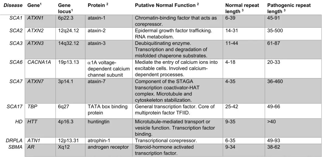
Spinocerebellar ataxia type 7 (SCA7) belongs to the family of polyglutamine (polyQ) diseases, which are neurodegenerative disorders caused by an abnormal and unstable expanded cytosine-adenine-guanine (CAG) repeat in their corresponding causative gene. The expanded CAG repeat is consequently translated into an abnormally expanded polyQ tract in the disease protein. Apart SCA7, this group is composed of five other types of SCAs (type 1-3, 6, and 17) and three non-SCA including: Huntington’s disease (HD), spinal and bulbar muscular atrophy (SBMA) and dentatorubral pallidoluysian atrophy (DRPLA) (Zoghbi and Orr, 2000) (Table 1). SCA7 and other polyQ diseases are clinically and genetically heterogeneous, however they share a number of common features. They are dominantly inherited, with the exception of SBMA, which is linked to a mutation in a gene located on the X chromosome, they are all progressive, adult onset and have a relationship between the number of CAG repeats on expanded alleles and the age of onset and severity of the disease, a prominent feature of all polyQ disorders named anticipation (Schols et al., 2004).
These diseases have their own causative gene in different chromosomes and present a variable threshold number of repeats (Table 1). Interestingly the nine proteins associated with these disorders share no sequence or structural homology except for the unstable PolyQ tract. Additionally, although the altered associated proteins are widely expressed in the central nervous system (CNS), each present specific populations of vulnerable neurons resulting in defined patterns of neurodegeneration and clinical features (Zoghbi and Orr, 2000) (Figure 1). The synthetized proteins that bear an abnormal expanded polyQ appear to take on an atypical configuration resulting in the formation and deposition of insoluble polyQ aggregates that subsequently accumulate in neurons forming nuclear or cytoplasmic inclusions, which are neuropathological hallmarks in these diseases (Perutz et al., 2002; Ross and Poirier, 2004).
Multiple common mechanisms of neurodegeneration have been identified in the polyQ disorders, including: autophagy, Ca2+ homeostasis/signaling alterations, transcription
alteration, disruption of axonal transport and vesicle trafficking, mitochondrial impairment, alteration of proteasome degradation as well as caspase activation and aggregation, amongst others, all leading to early synaptic neurotransmission deficits, that will progressively cause cell death (He et al., 2010; Matilla-Duenas et al., 2014; Shao and Diamond, 2007). Alterations in the protein configuration, formation of insoluble polyQ aggregates, as well as proteolytic cleavage of the expanded polyQ proteins have been suggested as enhancers of cell toxicity (Shao and Diamond, 2007).
Understanding how different proteins share pathological features has become one of the main interests for researchers. Identification of the steps in the pathogenic cascade leading to the onset and progression of the disease is likely to be cross-beneficial for developing effective therapies.
Disease Gene1 Gene
locus1
Protein 2 Putative Normal Function 2 Normal repeat
length 3
Pathogenic repeat length 3
SCA1 ATXN1 6p22.3 ataxin-1 Chromatin-binding factor that acts as
corepressor.
6-39 45-91
SCA2 ATXN2 12q24.12 ataxin-2 Epidermal growth factor trafficking.
RNA metabolism.
14-31 35-500
SCA3 ATXN3 14q32.12 ataxin-3 Deubiquitinating enzyme.
Transcription and degradation of misfolded chaperone substrates.
11-44 61-87
SCA6 CACNA1A 19p13.13 1A
voltage-dependent calcium channel subunit
Mediate the entry of calcium ions into excitable cells. Involved calcium-dependent processes.
4-18 20-33
SCA7 ATXN7 3p14.1 ataxin-7 Component of the STAGA
transcription coactivator-HAT complex. Microtubule and cytoskeleton stabilization.
4-35 36-460
SCA17 TBP 6q27 TATA box binding
protein
General transcription factor. Core of multiprotein factor TFIID.
25-42 49-66
HD HTT 4p16.3 huntingtin Microtubule-mediated transport or
vesicle function. Transcription factor binding.
9-35 >40
DRPLA ATN1 12p13.31 atrophin-1 Transcriptional corepressor. 6-35 49-93
SBMA AR Xq12 androgen receptor Steroid-hormone activated
transcription factor.
9-34 38-62
Table 1 Polyglutamine disorders, their associated gene, gene locus, protein and function, and the CAG repeat thresholds.
1 Gene information is in accordance with information provided by NCBI. 2 Protein abbreviation and function is in accordance with UniProt.
a. Gain vs loss of function.
The mechanisms underlying polyQ diseases are complex. Different experiments have revealed that PolyQ disorders might be acting in two different but not mutually exclusive ways: a gain of toxic function and a partial loss of the normal protein function.
It is noteworthy to mention that while PolyQ expansion is classified as a dominant mutation, there is not a pure complete or 50% loss of the normal function for the protein. Evidence for this has been shown in multiple studies performed with various levels of mutant huntingtin. A complete huntingtin Knock-Out (KO) in HD mouse led to embryonic lethality (Nasir et al., 1995; Zeitlin et al., 1995). Furthermore, neuropathological findings from heterozygote and homozygote patients for HD showed that homozygote patients present an increase in the rate of disease progression without changes in age of onset, suggesting that the mutant protein is able to execute the normal protein function at least until the disease onset (Squitieri et al., 2003). Moreover, huntingtin hemizygous inactivation does not cause HD disease symptoms despite the reduction of expression to half normal in humans and mice (Ambrose et al., 1994; Duyao et al., 1995; Persichetti et al., 1996). Additionally, deletion of one huntingtin allele does not result in HD (Housman, 1995).
The toxic gain of function is supported by the fact that expression of different expanded chains of polyQ peptides alone are intrinsically cytotoxic and cause neuronal degeneration in fly (Marsh et al., 2000) and in C.elegans (Morley et al., 2002). The concept of gain of toxic function is further supported by experiments where an expanded form of polyQ in the hypoxanthine-guanine phosphoribosyltransferase (HPRT), a protein unrelated to the known polyQ diseases-causing proteins, induces neurotoxicity and mice develop a similar phenotype to the CAG repeat disorders (Ordway et al., 1997), while HPRT knock-out mice develop a different phenotype.
However, there is growing evidence showing that the protein context is critical. In fact, in some cases, expansion of the polyQ tract by itself is not sufficient for disease (Chen et al., 2003; Emamian et al., 2003), pointing out that partial loss of normal protein function occurs alongside the pathogenesis. For example, conditional KO of normal huntingtin in a large proportion of neurons has been shown to decrease the survival and phenotypic stability of CNS cells along with, motor phenotypes and premature death (Dragatsis et al., 2000; O'Kusky et al., 1999). Moreover, overexpression of wild type form of huntingtin can lessen the mutant huntingtin effect (Leavitt et al., 2001; Van Raamsdonk et al., 2006). Furthermore, functions such as vesicular and mitochondrial trafficking or the Neuron Restrictive Factor (NRSF) regulation are
impaired in huntingtin KO mice (Dragatsis et al., 2000; Trushina et al., 2004) . Thus, deficits in trafficking observed in HD models probably represent a loss-of-function feature of polyQ huntingtin.
Similarly, studies in Drosophila, have demonstrated that neurodegeneration caused by the expanded form of ataxin-3 can be rescued by the normal ataxin-3, suggesting that at least in this model, the neurodegenerative phenotype might be due to the loss of ataxin-3 function (Warrick et al., 2005). Additionally, it was found that loss-of-function of ataxin-1 in mice is sufficient to cause many transcriptional changes common to the SCA1 knock-in mice, a model that faithfully replicates many features of the disease, suggesting that several molecular changes could be attributed to loss of ATXN1 function in SCA1(Crespo-Barreto et al., 2010). More recently, downregulation of key factors involved in calcium homeostasis were observed in the ataxin-2 KO. Interestingly, some of them also occurred in SCA2. For example, the transporter ITPR1 was depleted from soluble fractions in both mutants, suggesting a partial loss-of function in SCA2 (Halbach et al., 2017).
Nonetheless, the mechanisms underlying the loss of function are not clear. The polyQ expansion could cause the loss of function of the mutant protein and/or act negatively on the functions of the normal one.
It has been proposed that huntingtin expression levels is reduced along the pathology, as HD transgenic mice presented decreased levels of endogenous huntingtin, suggesting an alteration in the stability of the protein (Cattaneo et al., 2001).
It is known that at least two distinct large native protein complexes are associated with ATXN1: one containing the Capicua (CIC) transcription factor, and the other RNA binding motif protein17 (RBM17). In SCA1, expansion in ataxn1 causes increased interaction with RBM17 and a loss of interaction with the CIC, altering the proportion of the mutant protein participating in the formation of these complexes in vivo. This suggests that this alteration could at least partially account for SCA1 pathogenesis (Fryer et al., 2011; Lim et al., 2008). Likewise, proteins differentially interact with the normal or polyQ androgen receptor (AR). The N- terminal domain (NTD) of AR contains a transactivation domain that participates in multiple protein–protein interactions with general transcription factors and co-regulatory proteins. Thus, changes in the AR NTD induced by polyQ expansion can potentially strengthen or diminish the interaction of the polyQ AR with these proteins, implicating different pathways in SBMA pathogenesis (Beitel et al., 2013). For example, cytochrome c oxidase subunit Vb (COXVb) interacted more strongly with normal AR than polyQ AR in a hormone-dependent manner (Beauchemin et al., 2001).
Transactivation-domain-interaction Protein), a protein that functions in DNA repair, suggesting the polyQ AR may attenuate the DNA damage response (Xiao et al., 2012).
Therefore, it is becoming important to study the interplay between gain and loss of function. The full impact on cellular functions mediated by novel or altered interactions between the polyQ mutant proteins and other proteins is still being explored. Knowing the context and the normal functions of the implicated proteins for each disease can contribute to a better understanding of the pathogenesis that they cause.
b. Developmental component
PolyQ disorders are classified as adult or late onset disorders, which means symptoms appear in middle adulthood (variable for each disorder and depending on the size of the expansion). Several studies have focused on changes present right before, or during the disease onset and progression. However, studies in SCA2, SCA3, SCA6, and HD are challenging and changing this approach, showing that alterations during development could serve as the biological base for the programming and the vulnerability of adult onset disorders that they cause. Supporting this view, recent studies in non-symptomatic patients have shown subtle developmental brain differences that may account for susceptibility and neurodegeneration are present in these diseases (Lee et al., 2012; Nopoulos et al., 2011).
Mice null mutation in huntingtin leads to an early embryonic death (Nasir et al., 1995; Zeitlin et al., 1995). This can be rescued by providing wild-type extraembryonic tissue (Dragatsis et al., 1998). While low levels of huntingtin may rescue the lethality phenotype, huntingtin insufficiency causes abnormal brain development and mild movement abnormalities (Auerbach et al., 2001). Interestingly, decreased level of wild-type huntingtin followed by a later reconstitution, resulted in progressive striatal and cortical degeneration and motor coordination (Arteaga-Bracho et al., 2016). Similarly, mutant huntingtin has been shown to cause changes in the developing cortex along with defects in the proliferation of neuroprogenitors, or lead to perinatal death (Auerbach et al., 2001; Molina-Calavita et al., 2014). Furthermore, Molero et al. (2016) showed that when mice are selectively exposed to mutant HTT 97Q until postnatal day 21, they recapitulate a HD-like phenotype including neuropathology and motor deficits (Molero et al., 2016). More recently, it was shown that the loss of huntingtin during embryonic development has an impact in the dendritic morphology in young adults (Barnat et al., 2017).
All these studies have shown that variations in the levels of expression of either normal or mutant huntingtin, during the embryonic development is sufficient for generate neurological phenotype in mice. Moreover, huntingtin knockdown in zebrafish led to a variety of developmental defects, including hypochromic blood, associated to alteration in iron metabolism; loss of olfactory and lateral line sensory neurons and the presence of massive apoptosis of neuronal cells with enlarge in the ventricle area (Diekmann et al., 2009; Henshall et al., 2009; Lumsden et al., 2007), suggesting that huntingtin appears to be important for vertebrate development.
In the case of SCA3, injection of mutant ataxin-3 mRNA into the zebrafish embryos led to p53-dependent apoptosis, which occurred mainly in the central nervous system of zebrafish at early development stage (Liu et al., 2016). Additionally, using RNA interference for Ataxin-2,
C.elegans embryos showed arrest in different stages of development, indicating an essential
role of ataxin-2 for early embryonic development (Kiehl et al., 2000). On the contrary, null mutation in mouse ataxin-2 didn’t display obvious defects until adult stage, when they presented obesity and subtle rotarod defect (Kiehl et al., 2006). These differences present between both organisms during development could relate to the presence of orthologs and redundant mechanisms that may rescue the function.
Recently, it was shown that SCA6 mice presented alterations during early development (P10-13) in the maturation of Purkinje cells, which suggests impaired function. Notably, no motor deficit was detected. Moreover, the development alterations were transient and no longer observed at later stages (P21-24), showing that changes in the developing cerebellar circuit can occur without detectable motor abnormalities, and that changes in cerebellar development may not necessarily remain persistent into adulthood (Jayabal et al., 2017).
Put together, these studies provide new insight into polyQ disorders, showing that the pathogenesis present in these diseases might be more complicated and multiple approaches are needed to understand the pathological mechanisms involved in each disease. The role of loss of normal function of the polyQ proteins can be readdressed in light of their role during development as a complementary strategy to understand the pathological mechanisms involved. Recognizing the molecular changes that precede the neuronal death, as well as the alterations that provide susceptibility and predisposal to later pathophysiology could be of great value to identify putative therapeutic targets that might prevent, delay or cure determined polyQ diseases.
2. Autosomal Dominant Cerebellar Ataxias
SCA7 also belongs to the group of Autosomal dominant cerebellar ataxias (ADCAs), which are late onset heterogeneous neurodegenerative disorders. This group had been described in multiple families worldwide and in 1982 it was first proposed a classification system for them. By 1993 was refined and grouped the ADCAs into three main categories based on clinical presentation that is still a guideline in clinical practice (Harding, 1982, 1993). Characterized by progressive ataxia but also often associated with a broad spectrum of neurological or other clinical findings (Table 2). Currently, ADCAs are classified under different genetic subtypes known as SCAs. It is noteworthy that Spinocerebellar Ataxia type 7 (SCA7) is the only type of ADCA type 2, which is characterized by progressive cerebellar ataxia and retinal degeneration.
ADACA type ADCA I ADCAII ADCAIII
Clinical presentation
Cerebellar syndrome with ophtalmoplegia/pyramidal/extrapyramidal signs/cognitive impairment/peripheral neuropathy Cerebellar syndrome with pigmentary retinopathy Pure cerebellar syndrome
Neuropathology Degeneration of the cerebellum, and of
the basal ganglia/cerebral cortex/ optic nerve/pontomedullary systems/spinal tracts/peripheral nerves Cerebellar and pigmentary retinal degeneration Cerebellar degeneration SCA 1, 2, 3, 4, 8, 10, 12, 13, 17, 18, 19/22, 20, 21, 23, 24, 25, 27, 28, 32, 33, 34, 35, 36 7 5, 6, 11, 14, 15/16, 26
Table 2: Modified Harding’s classification of ADCAs (adapted from (Duenas et al., 2006)).
The SCAs are clinically and genetically heterogeneous. Clinically they are characterized by progressive loss of coordination in gait and limb movements. They are also associated with variable additional symptoms, including cerebellar dysarthria, dysphagia, extrapyramidal movement disorders, peripheral neuropathy, sphincter disturbances, cognitive impairment, epilepsy and dementia (Rub et al., 2013; Schols et al., 1997).
To date, 39 types of SCAs have been identified and are classified as SCA1 through SCA43 (no record for SCA33 or 39), most of which are autosomal dominant (except SCA24 and 28) and were categorized according to ADCA types (OMIM database 2017 https://omim.org/
(Amberger et al., 2015)). Moreover, ~80% have found and associated gene, however a large group of pedigrees with ADCA have still not found a determined loci, suggesting that the number could increase (Matilla-Duenas, 2012).
Interestingly, the increasing number of SCAs have shown multiple and different mechanisms that ultimately lead to ataxia clinical symptoms (Matilla-Duenas et al., 2014). Rising the question if there are unifying or converging pathways for these SCA.
Among all these ADCAs and polyQ disorders, SCA7 represents an interesting pathology. It present a unique retinal degeneration in addition to the cerebellar degeneration.
3. Major features of SCA7
a. Clinic-genetic
Efforts to identify the genetic cause of SCA7 began in the mid 90’s. In 1995, the causative mutation of SCA7 was identified (Trottier et al., 1995). By 1998, through positional cloning the
ATAXIN-7 gene (ATXN7) was identified and was shown to contain a polymorphic CAG repeat
making SCA7 the eighth disease to be classified as a polyglutamine disorder (David et al., 1998).
The wild-type alleles of ATXN7 have between 4-35 CAG repeats, while SCA7 alleles have typically 36-460 repeats (Table 1). Among CAG/polyQ disorders, SCA7 CAG repeats show the highest tendency to expand upon transmission, explaining the strong anticipation observed in families (mean 19 ± 13 years) (David et al., 1998; Michalik et al., 2004). Interestingly individuals with 25-35 repeats are asymptomatic, but they clearly gave rise to an SCA7 expansion in the next generation. This would explain why, despite the extreme anticipation observed in SCA7, the disease has not disappeared (Stevanin et al., 1998).
There is a significant negative correlation between the size of the CAG expansion and the age of onset and disease duration. This correlation is independent on the sex of the patient. While SCA7 alleles with ranges between 36-55 CAG repeats are responsible for the classical adult-onset form,(Michalik et al., 2004), >70 CAG repeats typically result in juvenile-adult-onset forms with accelerated disease course. The repeat length also influences the symptoms at onset:, large
cerebellar ataxia, while shorter expansions with later onset cause ataxia symptoms before visual loss (David et al., 1998; Giunti et al., 1999). Extremely large CAG expansions (>100 CAG) cause infantile forms with multisystem disorders such as failure to thrive, hypotonia, myoclonic seizures and non-central nervous systems dysfunctions like congestive heart failure, patent ductus arteriosus, renal failure, and muscle atrophy, and lead to death within few years or months (Ansorge et al., 2004; Benton et al., 1998; Giunti et al., 1999; Trang et al., 2015; van de Warrenburg et al., 2001; Whitney et al., 2007).
b. Cerebellar pathology.
SCA7 progressive cerebellar ataxia is manifested by the inability to coordinate balance, gait, and speech. Additional neurological deficits include slow saccades, opthalmoplegia, dysphagia, as well as pyramidal signs (David et al., 1998; Giunti et al., 1999). Variable levels of cerebellar and pontine atrophy are observed by magnetic resonance imaging (Alcauter et al., 2011; Bang et al., 2004; David et al., 1998; Horton et al., 2013; Michalik et al., 2004). Neuropathologically, the neuronal loss is substantial in the cerebellum (Purkinje cell layer and in the dentate nuclei), thalamus, nuclei of the basal ganglia, in the inferior olivary nuclei and brainstem (in basis pontis), which is associated with the atrophy of spinocerebellar and pyramidal tracts and milder changes are present in the granule cell layer (Horton et al., 2013; Michalik et al., 2004; Rub et al., 2005). Atrophy or loss of myelin is observed in the cerebellar white matter and extra cerebellar associated structures (Martin et al., 1999; Rub et al., 2013). In the classical adult-onset form, the disease progresses over several decades until death, due to dysphagia and other motor bulbar problems, most of the patients die of bronchopneumonia. As previously mentioned SCA7 is the only type of ADCA type 2, characterized by retinal degeneration in addition to the progressive cerebellar ataxia.
c. Retinopathy.
The retina is the neurosensory part of the eye and is a simple system of the central nervous system, and because of this a study of neural processes operant in the retina could assist in an understanding of the intricate mechanisms involved in the workings of the brain.
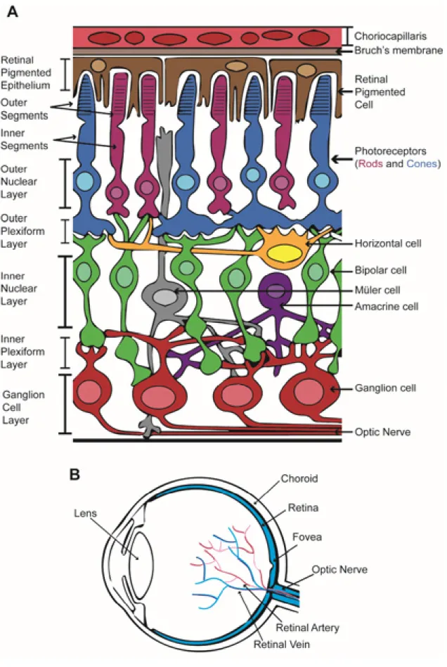
The vertebrate retina is a multilayered structure and composed of seven main cell types: rod and cone photoreceptors, horizontal cells, bipolar cells, amacrine cells, ganglion cells, and
Müller glia, which are each found in a specific layer of the organ (Thoreson, 2008) (see Figure 2).
Behind the neurosensory retina is a monolayer of pigmented epithelial cells known as the retinal pigmented epithelium (RPE) that forms part of the blood/retina barrier along with the Bruch’s membrane and the choriocapillaris. As a layer of pigmented cells the RPE absorbs the light energy focused by the lens on the retina but as well assists in support and maintenance of the neural retina, by transporting ions, water, metabolic products and recycling photo pigments generated during the visual cycle. The RPE also phagocytoses shed photoreceptor membranes to assist in constant renewal of outer segments (OS) (Strauss, 2005).
Basal to the RPE it is located the rod and cone photoreceptor (PR) layer, which is subdivided into the OS and the inner segments (IS). Rod and cone photoreceptors are light sensing cells. Cone photoreceptors are responsible for color vision and perception in bright light, and in humans are concentrated in the central macular region of the retina, decreasing in number towards the periphery of the eye. Three cone subtypes with different spectral sensitivities are found in primates: short-wavelength (S or blue-sensitive) cones, middle-wavelength (M or green-sensitive) cones, and long-wavelength (L or red sensitive) cones. This difference in spectral sensitivity arises from the presence of different cone opsins. Rod photoreceptors aid in perception in low light conditions, and outnumber cone cells 18-20:1 in humans (Swaroop et al., 2010).
The outer nuclear layer (ONL) contains the cell bodies of rod an cone photoreceptors, cone pedicles and rod spherules are synaptic upon various bipolar cell and horizontal cell types in the outer plexiform layer (OPL). The inner nuclear layer (INL), of the retina, contains cell bodies of three varieties: horizontal, bipolar and amacrine cells. Synaptic contact among bipolar, amacrine and ganglion cells are made in the inner plexiform layer (IPL). Anterior to the IPL, closer to the front surface of the retina is the ganglion cell layer (GCL), which contains cell bodies of the retinal ganglion cells (RGCs). Axons form retinal ganglion cells join together as they exit the eye to form the optic nerve, which projects to higher visual centers. Müller glia cell body is located in the INL, however, extension of the cell spans in every layer as these glial cells role is to protect and repair retinal neurons as well as act as architectural support (Thoreson, 2008) (Figure 2).
Figure 2 .Eye and retina diagram.
“In Family 3, cerebellar ataxia was associated
with retinal degeneration in 2 of the affected individuals; the others had visual
failure but no further clinical details were available. In the patient seen by the
author, there was extensive pigmentary degeneration involving most of the retina.
Saccadic eye movements were virtually absent but he could slowly follow
movements of his own hand. This particular patient had symptoms of visual
impairment before the development of ataxia, as did his relatives.”
(Harding, 1982)
This was the first classification of SCA7 as a distinct type of ADCA. The characteristic presence of retinopathy indicated that SCA7 was genetically distinct from other SCAs (Table3).
The retinal disease begins with an initial deterioration of the central vision affecting first cone photoreceptors, manifested by the loss of blue/yellow color discrimination (dyschromatopsia) and of visual acuity (due to loss of cone function); and then progresses toward a cone-rod dystrophy, this is manifested by the loss of peripheral visual fields (due to loss of rod function). This progressively spreads out to the whole retina, declining toward complete blindness. (Enevoldson et al., 1994; Gouw et al., 1994; Thurtell et al., 2009).
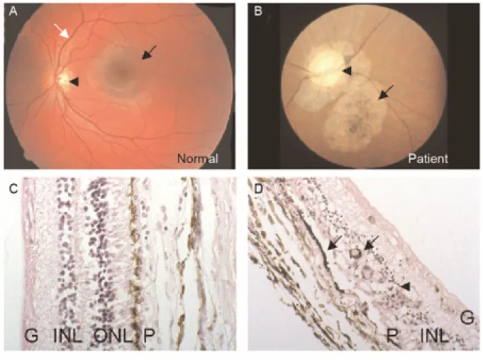
SCA7 patients may present visual impairments either before or after the onset of ataxia. Fundoscopy examination shows atrophic macula with granular pigmentation, pale areas with pigmentary atrophy and poor vasculature (Gouw et al., 1994; Michalik et al., 2004) (Figure 3A-B).
Post-mortem retinal histological examinations reveal almost complete loss of photoreceptors and substantial loss of the bipolar and ganglion neurons, associated with a severe thinning of the nuclear and plexiform layers especially in the foveal and parafoveal regions (Aleman et al., 2002; Michalik et al., 2004; Rub et al., 2008) (Figure 3C-D). In addition, damages in the Bruch’s membrane, retinal pigmentary epithelium and hypomyelinisation of the optic nerve have also been reported (Enevoldson et al., 1994; Gouw et al., 1994; Horton et al., 2013; Rub et al., 2008).
Despite the accumulated knowledge from imaging studies of SCA7 patients and the examinations of post-mortem retinal tissue, we are still lacking information regarding the temporal and spatial degeneration in the SCA7 patient retina.
Figure 3 Hallmarks of SCA7 human retinopathy.
(A, B) Fundoscopy images of a normal (A) and SCA7 patient (B) retinae. The SCA7 retina displays an extremely pale optic disc (arrowhead), atrophy of the pigmentary epithelium and choroid layer (arrow) as compared to the normal retina that shows normal macula (black arrow) and optic disc (arrowhead) and well-developed vasculature (white arrow).
(C, D) Histological sections of normal (C) and SCA7 patient (D) retinae. The normal retina (C) shows a proper organization of the retinal layers: the pigment epithelium (P), nuclei of rods and cones within the outer nuclear layer (ONL), the nuclei of bipolar, horizontal and amacrine neurons within the inner nuclear layer (INL) and the ganglion cell layer (G). In contrast, the retina of the SCA7 patient (D) is severely degenerated, and displays complete loss of photoreceptor segments and nuclei, disorganization of the INL (arrowhead) and migration of the melanin pigment (P) deep into the atrophic retina (arrows).
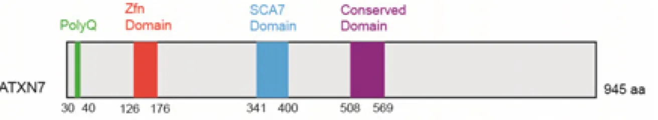
II. Ataxin-7 gene
The ATAXIN-7 (ATXN7) protein harbors a polymorphic polyQ stretch in the amino-terminus and it also presents three conserved domains that are shared with three paralogs, ATXN7L1, L2 and L3: a typical C2H2 zinc-finger (ZnF) motif, an atypical Cys-X9–10–Cys-X5–Cys-X2-His motif, known as SCA7 domain, and a third domain absent in ATXN7L3 (Figure 4) (Helmlinger et al., 2004b). ATXN7 mRNA and protein are widely expressed in neural and non-neural tissues and has been shown to be regulated by SUMOylation and acetylation (Duncan et al., 2013; Einum et al., 2001; Janer et al., 2010). ATXN7 appears predominantly in the nucleus (Kaytor et al., 1999). However, it has been reported that the intracellular distribution of ATXN7 dynamically changes and that ATXN7 distribution frequently shifts from the nucleus to the cytoplasm (Nakamura et al., 2012; Taylor et al., 2006) . Nuclear localization of ATXN7 is essential for transcription function while cytoplasmic ATXN7 has microtubule associating and stabilizing properties(Nakamura et al., 2012). There is no apparent correlation between cellular or subcellular localization and level and the vulnerability of neurons to degeneration in SCA7.
ATXN7 and its yeast ortholog sgf73 are core components of SAGA complexes (Spt-Ada-Gcn5
Acetyltransferase) involved in chromatin remodeling (also known in human as the TBP-free TAF containing complex (TFTC) and the SPT3-TAF9–GCN5 complex (STAGA) (Helmlinger et al., 2004b; Palhan et al., 2005; Sanders et al., 2002; Scheel et al., 2003).
Figure 4 Modular structure of human ATXN7 protein.
1. SAGA complex
SAGA is a multi-protein chromatin modifying complex that performs histone modifications and acts as co-factor for RNA polymerase II transcription (Pol II) (Bonnet et al., 2014). It is conserved between yeast and humans and can be subdivided in four modules (Table 3). Two
through GCN5 activity and deubiquitination module (DUB) that deubiquitinates histone H2B through USP22 activity. And two modules implicated in pre-initiation complex assembly and architecture (TBP-associated factor (TAF) and SPT modules) (Koutelou et al., 2010). ATXN7 belongs to the DUB together with the human ubiquitin protease USP22, ATXN7L3 and ENY2.
a. SAGA in Transcription
Gene expression is a complex process that requires the participation of various enzymatic complexes and is regulated at different levels. It involves two main stages: transcription and translation. Being transcription a key stage since it ensures the RNA synthesis based on a DNA template. Characterization of the yeast SAGA complex has revealed its involvement in various steps in the transcription process, including initiation and elongation; histone ubiquitination and interactions with the TATA-binding protein (TBP) (Koutelou et al., 2010; Wyce et al., 2004).
An initial step in the transcription process involve the chromatin remodeling in order to recruit the transcription machinery to the DNA. This remodeling is the consequence of histone modification. The HAT module of SAGA catalyzes the histone acetylation that ultimately will open up the chromatin allowing the formation of the pre-initiation complex (PIC) (Bhaumik, 2011). Additionally studies in yeast have revealed the complex involvement in the transcriptional activation through the recruitment of TBP by the ySpt3 subunit (Mohibullah and Hahn, 2008). Moreover, the activity of the DUB module has been shown to be necessary for switching from the initiation to the elongation stage allowing the recruitment of the Ctk1 kinase, an important catalytic subunit, which hyperphosphorylates the C-terminal heptapeptide repeat domain (CTD) of the largest RNA polymerase II subunit (Wyce et al., 2007).
Additional non transcriptional roles include mRNA export, DNA repair and telomere maintenance (Atanassov et al., 2009; Guo et al., 2011; Rodriguez-Navarro et al., 2004). Moreover SAGA complex can acetylate non-histone chromatin proteins like specific transcription activators such as c-Myc and Polo Like Kinase 4 (PLK4) (Fournier et al., 2016; Patel et al., 2004).
Component Yeast Drosophila Zebrafish Human
HAT module
(Histone acetyl transferase)
yGcn5 dGcn5 zGcn5 GCN5
yAda2b dAda2b isoform a dAda2b isoform b
zAda2b ADA2B
yAda3 dAda3 zTada3l ADA3
ySgf29 CG30390 zSgf29 SGF29
DUB module (Deubiquitination)
yUbp8 Nonstop zUsp22 USP22
ySgf73 Dmel\dAtxn7 zAtxn7 ATXN7
ySgf11 dSgf11 zAtxnL3 ATXN7L3
ySus1 dE(y)2 zEny2 ENY2
TAF module yTaf5 Wda zTaf5 TAF5L
yTaf6 dSAF6 zTaf6 TAF6L
yTaf9 dTaf9 zTaf9 TAF9
yTaf10 dTaf10b zTaf10 TAF10
yTaf12 dTaf12 zTaf12 TAF12
SPT module yTra1 Nipped-A zTrrap TRRAP
ySpt8 - - -
yAda5/Spt20 dSpt20 zSupt20 SPT20
ySpt7 CG6506 zSupt7L SUPT7L
ySpt3 dSpt3 zSupt3h SPT3
yAda1 dAda1 zTada1 TADA1
b. SAGA role in development.
Development requires the precise control of gene expression during different stages. This can range from regulation of changes of transcript levels in both space and time up to the maintenance of constant levels of expression for extended periods. Knowing that SAGA complex plays an important role in the regulation of gene expression is no surprise that components of this complex have shown involvement in events during development.
The member of the HAT module, Gcn5, was shown to be essential for the development of different organisms. Drosophila mutant revealed that while Gcn5 is not required for early larval life it does impede the metamorphosis prior to larvae death (Carre et al., 2005). This lethal phenotype is as well present in Gcn5-null mice that die as consequence of apoptosis of mesoderm cells (Xu et al., 2000). Moreover, levels of expression seem to be crucial for a proper mouse development, as mice with catalytically inactivated Gcn5 presented severe neural tube closure defects and died in mid-gastrulation (Bu et al., 2007) while reduced Gcn5 expression causes homeotic transformations in lower thoracic and lumbar vertebrae as well as exencephaly (Lin et al., 2008). Other two members of the HAT module, Ada2 and Ada3, present in Drosophila similar phenotypes to that of Gcn5 mutant larvae, with metamorphosis defect and lethality (Qi et al., 2004).
Components of the deubiquitinating module are also involved in development. usp22-null mutation is lethal in mouse due to an elevated p53 transcriptional activity (Lin et al., 2012). The
Drosophila homologues nonstop and Sgf11 mutants result in defects in neuronal connectivity
in the developing visual system as well as aberrant development of photoreceptor cells (Ma et al., 2016; Mohan et al., 2014). Finally, downregulation of the zebrafish usp22 orthologue showed the presence of hydrocephaly (Tse et al., 2009).
Thus far no members from the TAF module have been linked to a developmental phenotype, while only the drosophila homologue Nipped A from the SPT module has shown participation in the wing development acting as a coactivator of the protein mastermind (Gause et al., 2006). Up to now few subunits of the SAGA have been characterized for developmental defects. Additional studies are needed to understand how these genes function as part of the SAGA complex or independently. Understanding the role of these proteins during development may presage important implications in human diseases.
c. SAGA complex and disease.
Besides SCA7, which is caused by a member of the deubiquitinating module of SAGA, other members have been linked to disease, particularly with hallmarks of cancer, including DNA damage repair, cell cycle regulation and post translational regulation.
SAP130 and DDB1, two proteins associated to the human SAGA complex have shown to be involved in DNA repair (Brand et al., 2001; Martinez et al., 2001). Interestingly results obtained in yeast equally support the role of SAGA with DNA damage repair co-transcriptionally or independent of transcription (Ferreiro et al., 2006; Yu et al., 2005).
Three subunits of the DUB module have been linked with cancer. USP22 has been identified as a component of an 11-gene signature associated with poor prognosis of diverse types of cancer, it has been hypothesized that USP22 could regulate tumorigenesis by inducing histone modifications (Zhang et al., 2008b). Moreover, USP22 is required for the activation of target gene transcription by MYC, lack of USP22 alters MYC functions and ultimately results in a specific G1 phase cell arrest while the USP22 recruitment allows certain essential cell cycle genes to be transcriptionally activated (Zhang et al., 2008a; Zhang et al., 2008b). ATXN7, has been reported to be associated with susceptibility with breast cancer and prognosis of renal cell carcinoma (RCC) (Gotoh et al., 2014; Milne et al., 2014). Finally, the ENY2 protein member as well of the DUB module, might be defined as a diagnostic marker for primary breast cancer (Armakolas et al., 2012).
On the other hand, the HAT module member, GCN5 has been related to different types of cancer including lung, liver and colon. Alteration in the levels of expression is responsible for cell cycle progression arrest and apoptosis.GCN5 has been shown regulates the expression of cyclin D1, and cyclin E1 (cell growth and the G1/S phase transition), E2F1 (transcription factor that can induce both proliferation and apoptosis.) and p21Cip1/Waf1 (cell cycle inhibitor).
(Chen et al., 2013; Majaz et al., 2016; Yin et al., 2015).
In summary, ATXN7 is a member of the SAGA complex. This complex consist of distinct modules that participate at different levels in the transcription mediated by Pol II. The composition of these modules is now well known, however, much information regarding their biological functions remains to be elucidated. Moreover, for now multiple SAGA members are linked to disease and to multiple stages in development, rising questions of their specific roles in such biological processes.
III. Pathogenesis of SCA7 retina phenotype
Different models have been used in order to study the SCA7 disease. Current information is the result of years of biochemical, molecular and organismal studies. This section will focus on the in vivo and in vitro models that have been generated to better understand the underlying pathomechanisms of the SCA7 disease.
1.Cell-based Models
In vitro models have provided important insights about the SCA7 pathogenesis; however there
are important challenges present with their use. First, SCA7 is an adult onset neurodegenerative disorder which means long-time cultures are required in order to properly model the late onset of the disease. Additionally, there is a lack of specific affected cell types such as neurons and photoreceptors patient-derived appropriate for in vitro studies. Regarding to the latter one; while is possible to culture from post mortem tissue derived from brain (Verwer et al., 2003) or retina (Kim and Takahashi, 1988; Marita, 2011; Mayer et al., 2005) a major drawback is the limited time for establishing these cultures (less than 48 hours after patient death). Because of this, efforts have focused on using other cell types derived from SCA7 patients, such as skin derived fibroblasts (Luo et al., 2012; Scholefield et al., 2014) or lymphoblastoid cell lines (LCL) (Tsai et al., 2005). This lines give the advantage having a genetic accurate disease background, however these cells are normally unaffected by the pathology and have a limited culture life span in vitro.
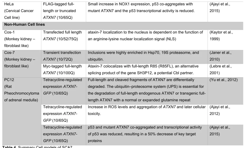
For those reasons the most common method has been to transfect established cell lines with different ATXN7 expression vectors. Providing valuable information regarding interactors, toxicity, patterns of expression, among others. Summarized in Table 4.
Patient derived cell lines
Cell line Design Contribution Reference
FIB
(patient and control fibroblast)
Individuals with 55 or 150Q
Expression of atxn7 and the antisense non-coding RNA SCAANT1. (Sopher et al., 2011)
Patient with 45 Q Generation of induced pluripotent stem (iPS) cells. (Luo et al., 2012) Individuals with 57 Q Allele-specific siRNA silences the mutant transcript more efficiently
than the wild-type.
(Scholefield et al., 2014)
LCL
(lymphoblastoid cell lines)
Individuals with 41 and 100Q
Expanded ataxin-7 significantly impaired the expression of the heat shock proteins Hsp27 and Hsp70.
(Tsai et al., 2005) Individuals with
10,51,59 or 66Q
Increased expression of ataxin-7 sense transcript levels. (Sopher et al., 2011)
Human Non-Patient derived cell lines
Hek293
(Human Embryonic Kidney 293 cells)
Transfected full length
ATXN7 (10/100Q)
shRNA decreased numbers of cells with mutant protein aggregates. (Scholefield et al., 2009) FLAG-tagged full length ATXN7 (10/60Q)
Normal and mutant ATXN7 equally interacted with the TFTC subunits TRRAP, GCN5 and TAF10.
(Helmlinger et al., 2004b)
Full length ATXN7 (10/100Q) and truncated form
Heat-shock proteins, subunits of the proteasome, transcription factors and activated caspase-3 were detected in a subset of inclusions.
(Zander et al., 2001)
HeLa (Cervical Cancer Cell line) FLAG-tagged full-length or truncated ATXN7 (10/65Q)
Small increase in NOX1 expression, p53 co-aggregates with mutant ATXN7 and the p53 transcriptional activity is reduced.
(Ajayi et al., 2015)
Non-Human Cell lines
Cos-1
(Monkey kidney – fibroblast like)
Transfected full length
ATXN7 (10/52/75Q)
ataxin-7 localization to the nucleus is dependent on the function of an arginine-lysine nuclear localization signal (NLS)
(Kaytor et al., 1999) Cos-7 (Monkey kidney – fibroblast like) Transient transfection ATXN7 (10/72Q)
Inclusions were highly enriched in Hsp70, 19S proteasome, and ubiquitin.
(Janer et al., 2010) Myc-tagged full-length
ATXN7 (10/100Q)
Ataxin-7 colocalizes with full-length R85 (R85FL), an alternative splicing product of the gene SH3P12, a potential Cbl partner.
(Lebre et al., 2001) PC12 (Rat Pheochromocytoma of adrenal medulla) Tetracycline-regulated expression ATXN7-GFP (10/65Q)
Full-length and cleaved fragments of ATXN7 are differentially degraded. The ubiquitin–proteosome system (UPS) is essential for the degradation of length endogenous ATXN7 or transgenic full-length ATXN7 with a normal or expanded glutamine repeat
(Yu et al., 2012)
Tetracycline-regulated expression
ATXN7-GFP (10/65Q)
Increase in ROS levels and aggregation of ATXN7 and later cellular toxicity. (Ajayi et al., 2012) Tetracycline-regulated expression ATXN7-GFP (10/65Q)
p53 and mutant ATXN7 co-aggregated and transcriptional activity of p53 was reduced, resulting in a 50% decrease of key target proteins
(Ajayi et al., 2015)
2.Mouse Models
Different transgenic and knock-in mouse models have been generated during the past years with the majority expressing wild-type or expanded ATXN7 under the control of a heterologous promoter specific to the brain and/or retina. These models have provided important insights into the nature of SCA7 neurodegeneration.
a. Cerebellar pathology
Analyses of the PrP-SCA7-c92Q mouse model have highlighted the importance of cell-cell interactions in the cerebellar pathology (Garden et al., 2002). These mice develop motor defects and show dark degenerating Purkinje neurons. Interestingly, Purkinje cell pathology occurs despite the fact the MoPrP promoter drives the expression of mutant ATXN7 (mATXN7) in all cerebellar neurons, except for Purkinje cells, suggesting that they are affected via a non cell-autonomous mechanism. This same model displayed as well pathological signs in Bergman glial cells (Custer et al., 2006). Given that Bergmann glia are regulators of glutamate levels in the surrounding environment of Purkinje cells and that dark degeneration often results from excitotoxicity, new transgenic mice were generated to express mATXN7only in Bergmann glia cells to assess whether the pathology would affect Purkinje cells as well. Indeed, Gfa2-SCA7-92Q mice also show Purkinje cell degeneration and motor dysfunctions. However, compared to PrP-SCA7-c92Q mice, Gfa2-SCA7-92Q mice develop a late onset and milder ataxia, suggesting that other dysfunctional neurons may account for Purkinje cell degeneration in PrP-SCA7-c92Q mice.
The contribution of different cell types and their interaction to the cerebellar pathology was further addressed using a new set of engineered mice in which mATXN7 cDNA was flanked by loxP sites at the start site of translation in the murine PrP gene in a bacterial artificial chromosome (PrPfloxed-SCA7-92Q BAC) (Furrer et al., 2011). When crossed with mice expressing Cre recombinase under Bergmann glia promoter (Gfa2) or under promoter specific to Purkinje and inferior olive neurons (Pcp2), mATXN7 was deleted specifically in these cell types. Deletion of mATXN7 from Bergmann glia has mild beneficial effects and does not prevent Bergmann glia pathology. In contrast, deletion of mATXN7 from Purkinje and inferior olive neurons improves motor performance and histopathology as well as prevents Bergmann glia pathology. Finally, deletion of mATXN7 in the three cell types is more effective to prevent the pathology. Together, these results support a complex cell-cell interaction between Bergmann glia, Purkinje and inferior olive neurons in the development of SCA7 cerebellar
The expression profile of the cerebellum of Ataxin-7-Q52 transgenic mice, which also display motor dysfunction and Purkinje cell pathology, revealed gene deregulations affecting different pathways including synaptic transmission, axonal transport, glial functions and neuronal differentiation (Chou et al., 2010). Perhaps the most interesting finding is the down regulation of a set of myelin-associated proteins (CNP, MAG, MBP, MOG, MOBP and PLP1) and of their regulators, the transcription factor Olig1 and transferrin (Chou et al., 2010). This is consistent with the loose and poorly compacted myelin sheaths observed in the cerebellar white matter of these mice, and with the myelin pallor and loss of myelinated fibers reported in the cerebellar white matter of SCA7 patients (Rub et al., 2005).
Reminiscent to the loss of photoreceptor maturation in SCA7 mouse retina, mATXN7 toxicity might compromise genetic programs controlling oligodendrocyte maturation and myelin sheath integrity and function.
b. Retinopathy
In SCA7 models, the retina develops normally before showing a progressive reduction of electroretinograph activity, thinning of the retina and repression of photoreceptor-specific genes (La Spada et al., 2001; Yoo et al., 2003). Initially, these transcriptional alterations were attributed to the dysfunction of CRX (cone-rod homeobox protein), a key transcription factor of photoreceptor genes. This is because CRX was previously shown to require interaction with ATXN7 and SAGA for its transactivation activity on photoreceptor gene promoters, and because mATXN7 was shown to suppress the transactivation activity in SCA7 retina (Chen et al., 2004; La Spada et al., 2001). Later on, analysis of SCA7266Q/5Q KI and R7E mouse retina showed that transcriptional alterations were not restricted to CRX target genes (Abou-Sleymane et al., 2006; Yoo et al., 2003). In particular, the expression profile of R7E retina showed on the one hand the downregulation of the photoreceptors specific transcription factors CRX, NRL (neural retina leucine zipper protein), and Nr2E3 (Nuclear Receptor Subfamily 2, Group E, Member 3) as well as most of their target genes, and on the other hand the re-activation of OPTX2, STAT3 and HES5 that normally inhibit the differentiation of precursor neurons into mature photoreceptors during development (Abou-Sleymane et al., 2006). SCA7 photoreceptors progressively lose their OS and cell polarity, and relapse to round cell shape (Yefimova et al., 2010). Thus, SCA7 retinopathy primarily results from the progressive regression of mature photoreceptor to an ill-defined state, which occurs long before cell death. The initial trigger leading to SCA7 photoreceptor degeneration remains to be determined. Degenerating photoreceptors in SCA7 retina ultimately die through a mechanism reminiscent
of dark neuronal cell death (Yefimova et al., 2010). Dark degeneration also occurs in SCA7 mouse cerebellum and was reported in several mouse models of polyQ disorders (Figiel et al., 2012). Different cellular responses may be triggered by different mATXN7 toxic species, since the relative amount of full-length mATXN7, proteolytic fragments, soluble and insoluble aggregates varies considerably from early to late disease stages and might influence the way individual photoreceptors respond to these different proteotoxic products.
3. Transcriptional alteration
Studies performed on cellular and mouse models of SCA7 have identified transcriptional alterations as an early pathogenic event associated with neuronal dysfunction (Abou-Sleymane et al., 2006; Chou et al., 2010; Helmlinger et al., 2006; La Spada et al., 2001; Yoo et al., 2003) This has also been reported in other polyQ disorders (Sugars and Rubinsztein, 2003).
Transcriptome analysis of SCA7 mouse retina revealed an early and progressive down-regulation of most photoreceptor-specific genes (Abou-Sleymane et al., 2006), while expression profile of SCA7 mouse cerebellum showed down-regulation of genes involved in the maintenance and function of neuronal dendrites and CNS myelin sheath (Chou et al., 2010).
Since the expanded form of ATXN7 has been shown to properly incorporate into SAGA, several studies have explored the possibility that transcriptional alterations in SCA7 could result from dysfunction of SAGA acetylation and deubiquitination activities (Helmlinger et al., 2004b; McMahon et al., 2005; Palhan et al., 2005). The outcome of these studies differs depending the model system investigated. In yeast and HEK2937 kidney cells, mATXN7-containing SAGA lacks critical subunits and leads to the reduction of GCN5 acetylation activity and gene transcription (McMahon et al., 2005; Palhan et al., 2005). In agreement with GCN5 dysfunction, promoters of photoreceptor-specific genes were shown to have histone H3 hypoacetylation, which would explain their decreased expression in SCA7 mouse retina (Prp SCA7-c92Q model) (Palhan et al., 2005). However, opposing with the above studies, mATXN7-containing SAGA purified from SCA7 mouse retina (R7E model) had normal incorporation of Gcn5, Taf12 and Spt3 and had normal acetylation activities (Helmlinger et al., 2006). In this study, promoters of photoreceptor-specific genes were found hyperacetylated, but the presence of RNA Pol II on promoters was strongly reduced, which would explain the