HAL Id: hal-01857291
https://hal.archives-ouvertes.fr/hal-01857291
Submitted on 26 May 2020
HAL is a multi-disciplinary open access
archive for the deposit and dissemination of
sci-entific research documents, whether they are
pub-lished or not. The documents may come from
teaching and research institutions in France or
abroad, or from public or private research centers.
L’archive ouverte pluridisciplinaire HAL, est
destinée au dépôt et à la diffusion de documents
scientifiques de niveau recherche, publiés ou non,
émanant des établissements d’enseignement et de
recherche français ou étrangers, des laboratoires
publics ou privés.
challenges remain
Philippe Baveye, Wilfred Otten, Alexandra Kravchenko, Maria Balseiro
Romero, Eleonore Beckers, Maha Chalhoub, Christophe Darnault, Thilo
Eickhorst, Patricia Garnier, Simona Hapca, et al.
To cite this version:
Philippe Baveye, Wilfred Otten, Alexandra Kravchenko, Maria Balseiro Romero, Eleonore Beckers,
et al.. Emergent properties of microbial activity in heterogeneous soil microenvironments: Different
research approaches are slowly converging, yet major challenges remain. Frontiers in Microbiology,
Frontiers Media, 2018, 9 (1929), pp.1929. �10.3389/fmicb.2018.01929�. �hal-01857291�
doi: 10.3389/fmicb.2018.01929
Edited by: Eoin L. Brodie, Lawrence Berkeley National Laboratory (LBNL), United States Reviewed by: James J. Moran, Pacific Northwest National Laboratory (DOE), United States Peter Nico, Lawrence Berkeley National Laboratory (LBNL), United States *Correspondence: Philippe C. Baveye baveye.rpi@gmail.com Specialty section: This article was submitted to Terrestrial Microbiology, a section of the journal Frontiers in Microbiology Received: 27 March 2018 Accepted: 30 July 2018 Published: 27 August 2018 Citation: Baveye PC, Otten W, Kravchenko A, Balseiro-Romero M, Beckers É, Chalhoub M, Darnault C, Eickhorst T, Garnier P, Hapca S, Kiranyaz S, Monga O, Mueller CW, Nunan N, Pot V, Schlüter S, Schmidt H and Vogel H-J (2018) Emergent Properties of Microbial Activity in Heterogeneous Soil Microenvironments: Different Research Approaches Are Slowly Converging, Yet Major Challenges Remain. Front. Microbiol. 9:1929. doi: 10.3389/fmicb.2018.01929
Emergent Properties of Microbial
Activity in Heterogeneous Soil
Microenvironments: Different
Research Approaches Are Slowly
Converging, Yet Major Challenges
Remain
Philippe C. Baveye1* , Wilfred Otten2, Alexandra Kravchenko3, María Balseiro-Romero1,4,
Éléonore Beckers5, Maha Chalhoub6, Christophe Darnault7, Thilo Eickhorst8,
Patricia Garnier6, Simona Hapca9, Serkan Kiranyaz10, Olivier Monga11,
Carsten W. Mueller12, Naoise Nunan13, Valérie Pot6, Steffen Schlüter14,
Hannes Schmidt15and Hans-Jörg Vogel14,16
1UMR ECOSYS, AgroParisTech, Université Paris-Saclay, Thiverval-Grignon, France,2School of Water, Energy
and Environment, Cranfield University, Cranfield, United Kingdom,3Department of Plant, Soil and Microbial Sciences,
Michigan State University, East Lansing, MI, United States,4Department of Soil Science and Agricultural Chemistry, Centre
for Research in Environmental Technologies, Universidade de Santiago de Compostela, Santiago de Compostela, Spain,
5Soil–Water–Plant Exchanges, Terra Research Centre, BIOSE, Gembloux Agro-Bio Tech, University of Liège, Gembloux,
Belgium,6UMR ECOSYS, INRA, Université Paris-Saclay, Thiverval-Grignon, France,7Laboratory of Hydrogeoscience
and Biological Engineering, L.G. Rich Environmental Laboratory, Department of Environmental Engineering and Earth Sciences, Clemson University, Clemson, SC, United States,8Faculty 2 Biology/Chemistry, University of Bremen, Bremen,
Germany,9Dundee Epidemiology and Biostatistics Unit, School of Medicine, University of Dundee, Dundee,
United Kingdom,10Department of Electrical Engineering, Qatar University, Doha, Qatar,11Institut de Recherche pour le
Développement, Bondy, France,12Lehrstuhl für Bodenkunde, Technical University of Munich, Freising, Germany,13Institute
of Ecology and Environmental Sciences – Paris, Sorbonne Universités, CNRS, IRD, INRA, P7, UPEC, Paris, France,14Soil
System Science, Helmholtz-Zentrum für Umweltforschung GmbH – UFZ, Leipzig, Germany,15Terrestrial Ecosystem
Research, Department of Microbiology and Ecosystem Science, Research Network ‘Chemistry meets Microbiology’, University of Vienna, Vienna, Austria,16Institute of Soil Science and Plant Nutrition, Martin Luther University
of Halle-Wittenberg, Halle, Germany
Over the last 60 years, soil microbiologists have accumulated a wealth of experimental data showing that the bulk, macroscopic parameters (e.g., granulometry, pH, soil organic matter, and biomass contents) commonly used to characterize soils provide insufficient information to describe quantitatively the activity of soil microorganisms and some of its outcomes, like the emission of greenhouse gasses. Clearly, new, more appropriate macroscopic parameters are needed, which reflect better the spatial heterogeneity of soils at the microscale (i.e., the pore scale) that is commensurate with the habitat of many microorganisms. For a long time, spectroscopic and microscopic tools were lacking to quantify processes at that scale, but major technological advances over the last 15 years have made suitable equipment available to researchers. In this context, the objective of the present article is to review progress achieved to date in the significant research program that has ensued. This program can be rationalized as a sequence of steps, namely the quantification and modeling of the physical-, (bio)chemical-, and microbiological properties of soils, the integration of these
different perspectives into a unified theory, its upscaling to the macroscopic scale, and, eventually, the development of new approaches to measure macroscopic soil characteristics. At this stage, significant progress has been achieved on the physical front, and to a lesser extent on the (bio)chemical one as well, both in terms of experiments and modeling. With regard to the microbial aspects, although a lot of work has been devoted to the modeling of bacterial and fungal activity in soils at the pore scale, the appropriateness of model assumptions cannot be readily assessed because of the scarcity of relevant experimental data. For significant progress to be made, it is crucial to make sure that research on the microbial components of soil systems does not keep lagging behind the work on the physical and (bio)chemical characteristics. Concerning the subsequent steps in the program, very little integration of the various disciplinary perspectives has occurred so far, and, as a result, researchers have not yet been able to tackle the scaling up to the macroscopic level. Many challenges, some of them daunting, remain on the path ahead. Fortunately, a number of these challenges may be resolved by brand new measuring equipment that will become commercially available in the very near future.
Keywords: soil microbiology, biodiversity, upscaling, tomography, X-ray computed, NanoSIMS imaging, single-cell genomics
INTRODUCTION
Over the last decade, soils have become increasingly central to a number of crucial debates on issues of great societal concern. Because they contain a huge amount of carbon, soils could lead to a dramatic acceleration of global climate change, as mean temperatures increase and rainfall patterns are altered (Baveye, 2007; Baveye et al., 2011; Hamdi et al., 2013; Crowther et al., 2016). The idea, advocated by some (Paustian et al., 1997; Lal and Bruce, 1999), that on the contrary, with proper management, soils could store even more carbon than at present, and thereby help mitigate the production of greenhouse gasses resulting from the consumption of fossil fuels, has been adopted enthusiastically by politicians in a number of countries but has stirred intense discussions among scientists (Powlson et al., 2011;Cheng et al., 2012;Dungait et al., 2012;Kowalchuk, 2012;Verbruggen et al., 2012; Minasny et al., 2017, 2018; van Groenigen et al., 2017;
Baveye et al., 2018; White et al., 2018). At the same time, humanity is faced with the prospect of having to significantly increase food production to feed the world population, which is expected to rise to 9 or 10 billion people by 2050 (Godfray et al., 2010). Since soil and water resources are already used at the maximum level of what some consider ecologically safe, a consensus seems to be emerging that as long as the focus is kept on land-based agricultural production, the best option to insure food security lies in exploiting plant-microbe partnerships to improve biomass production (Weyens et al., 2009;
Glick, 2012, 2014; Blaser et al., 2016), or in stimulating so-called plant-soil feedback processes, whereby plants induce soil microbial communities to release nutrients and store water in the rhizosphere (Sposito, 2013;Baveye, 2015). In addition, even though the issue of soil contamination does not appear at the moment to be at the forefront of environmental concerns in many countries, the question remains of what to do with millions
of severely polluted sites around the globe, especially given the fact that this number is ever increasing, as a result of practices like shale gas production (Baveye, 2013c; Meckenstock et al., 2015). Given the prospect of a progressive warming of soils in decades to come, renewed threats caused by soil contamination will undoubtedly need to be addressed at some point in the near future.
The intimately connected microbial and physico-chemical processes at the core of all these soil-related issues have posed daunting challenges to researchers. Until a decade ago, in spite of sustained research efforts, progress was very slow or even non-existent, and in several cases serious hurdles arose, which no one had anticipated. Kirschbaum (2006)admitted that in the 10 years prior to the publication of his review of the field, no real advance had been made in understanding and predicting quantitatively the effect of temperature on the decomposition of soil organic matter (OM). Available models also routinely underestimated the pulses of CO2 flux occurring when large
rainfall events follow drafts (Blagodatsky and Smith, 2012;Evans et al., 2016). Recent work byRabot et al. (2015) suggests that many of the previous measurements of the production by soil bacteria and fungi of nitrous oxide, a very potent greenhouse gas (Laughlin and Stevens, 2002;Crenshaw et al., 2008; Hu et al., 2015), probably missed very short emission bursts that occur at the onset of drying of soils, and therefore underestimated total N2O production by soils. Concomitantly, research on carbon
sequestration in soils provided evidence of the problematic “priming” effect, identified early on (Macura et al., 1965;Arsjad and Giddens, 1966), but routinely overlooked until a decade ago (Fontaine et al., 2007; Kuzyakov, 2010; Tian et al., 2015) and still poorly understood (Nunan et al., 2015;van der Wal and de Boer, 2017). Through this effect, the addition of fresh OM to soils can lead to the mineralization of very old humic substances, previously thought to be utterly stable and recalcitrant to further
degradation. In a similar fashion, in polluted soils, experiments showed that a slight change, for example brought about by the addition of a source of nutrients for microorganisms, could easily make supposedly “sequestered” contaminants once again bioavailable (Li et al., 2005). Some of these areas of ignorance remain “terra incognita” at this point, even with regard to the much ballyhooed biodiversity of soils (Baveye et al., 2016a,b). There is still no satisfactory explanation for the observation, made more than 60 years ago, that the mineralization of soil OM continues at the same rate even if 90% of soil microorganisms are wiped out by CHCl3 fumigation (Jenkinson, 1966;Powlson et al., 2017;Baveye, 2018). A final example of a situation where our understanding of soil systems is still insufficient is related to the links between the diversity of soil microbial communities and various soil parameters. Some authors have found a close correlation between this diversity and specific parameters, like soil pH (Fierer and Jackson, 2006), but more detailed statistical analyses sometimes present a different picture. In a recent study,
Terrat et al. (2017)use some of the most sophisticated molecular techniques currently available to analyze the biodiversity of soil samples across France, and try to relate it to various parameters of soils and of their environment. The results are systematically underwhelming. They find that less than half (48.2%) of the observed variance of the biodiversity could be accounted for by using soil parameters that are routinely measured. Clearly, at least in this particular study, something fundamental about soils is being missed.
In virtually all these instances, a common observation is that soil samples that appear alike in most of their overall measured characteristics can behave very differently, making replicated observations and good correlations difficult to achieve. Obviously, it is not sufficient to describe soils solely on the basis of traditional macroscopic measurements, such as the volumetric water content, microbial density, or contaminant concentration. Quantitative information on the spatial heterogeneity manifested at the micron scale, at which microorganisms operate, is also absolutely required.
In some respects, this is not as novel a perspective as it may appear. In another era, in literature that unfortunately seems to have become largely ignored since, soil microbiologists already reached the same conclusion. Sixty years ago, Rovira and Greacen (1957)subjected moist soil samples to compression and shearing to simulate tillage, and concluded, after ruling out other possible explanations, that the enhanced oxygen consumption observed in the soils after disruption was due to exposure of organisms to OM that was previously inaccessible to them. These and a number of other early observations pointing in the same direction prompted Alexander (1964, p. 219) to conclude that “microorganisms apparently in the same habitat are, in fact, often exposed to entirely different environmental influences and population pressures. To understand the forces actually affecting the organisms, a microenvironmental concept rather than the gross macroscopic view of interactions must be adopted.” The review by Griffith (1965) of the extensive work carried out in the 40s and 50s on the opposite effect of microorganisms on their physical environment, and in particular on the development of soil architecture, also raises many
questions that could be addressed only from a microscopic perspective. Experimental evidence obtained since the mid-sixties has provided steadily strengthening support for this perspective (Hattori, 1973; Cheshire, 1977; Elliott et al., 1980;
Tiedje et al., 1984;Stotzky, 1986;Crozat et al., 1987;Darrah et al., 1987;Parkin et al., 1987;Postma and Altemuller, 1990;Postma and van Veen, 1990; Killham et al., 1993;Renault and Stengel, 1993; Strong et al., 1997; Wachinger et al., 2000; Chenu and Stotzky, 2001;Attard et al., 2011;Chapman et al., 2012;Johnson et al., 2013;Vos et al., 2013;Uroz et al., 2015;Xun et al., 2015;
Barcenas-Moreno et al., 2016;Keiluweit et al., 2017, 2018). In the 50s and 60s, very little could be done to come up with better measurements, unfortunately. Alexander (1964, p. 219), again, observed that “because of inherent technical difficulties in biochemical experimentation at the microscopic level, progress in understanding of the microenvironment has been painfully slow.” Even though more and more experiments over the years confirmed the significance of microenvironments, for a long time it was not feasible practically to characterize them in quantitative terms. The advent of transmission or scanning electron microscopes, and later of confocal laser microscopes as well, provided a wealth of qualitative information about microbial habitats in the form of micrographs of increasingly high quality (Foster, 1988;Vandevivere and Baveye, 1992a,b,c,d;
DeLeo et al., 1997;Baveye et al., 1998), but the lack of related quantitative data prevented for several decades the development of satisfactory predictive models of soil microbial processes, accounting explicitly for the microheterogeneity of soils.
This situation has changed dramatically in the last decade and is continuing to evolve at a rapid pace. Significant technological advances have provided soil researchers, for example, with routine access to X-ray computed tomography (CT) systems, which provide increasingly reliable information about the geometry of pores and solids in soils at resolutions as small as 0.05 µm. Progress in near-edge X-ray spectromicroscopy (NEXAFS), scanning transmission X-ray microscopy (STXM), X-ray absorption spectroscopy, micro-fluorescence spectroscopy, and Nano-SIMS, applied to soil thin sections, has led to observations of sharp spatial heterogeneity in the chemical make-up of soils over minute distances, and in the accumulation of trace metals. Significant advances related to biological markers now allow specific bacteria to be identified in soils, and their spatial distribution at the micrometer scale to be determined in thin sections. This information can be translated into 3-dimensional distributions using recently developed statistical algorithms. In addition, very efficient modeling tools, like the Lattice-Boltzmann approach, allow the description of transport and physico-chemical processes occurring in soil pores at scales that are directly relevant to microorganisms, whereas individual-based or agent-individual-based models, also developing rapidly, can describe the dynamics of microorganisms inhabiting the pore space (Gras et al., 2010, 2011;Muci et al., 2012;Hellweger et al., 2016;Kim and Or, 2016).
In the last few years, the application of each of these technologies and modeling methods to soils has been the object of a sizeable literature. Progress achieved in the use of each technology has already been expertly reviewed
(O’Donnell et al., 2007; Taina et al., 2008; Young et al., 2008;
Behrens et al., 2012; Rennert et al., 2012; Helliwell et al., 2013; Tuller et al., 2013; Wildenschild and Sheppard, 2013;
Schlüter et al., 2014;Calistru and Jitareanu, 2015;Kuzyakov and Blagodatskaya, 2015; Prosser, 2015; Roose et al., 2016; Xiong et al., 2016;Totsche et al., 2017). For some technologies, since advances are extremely rapid, it would be useful, conceivably, to provide an updated coverage of recent work, and no doubt new reviews will fill the gaps in the near future. Yet, a different type of critical overview might be even more fruitful at this stage, one that keeps sight firmly on what started out as the ultimate goal of the research: A thorough understanding of what one needs to measure at the macroscale in order to adequately describe emergent microbial processes. Instead of surveying the increasingly widespread application of specific technologies to soils, it is worth taking a step back and analyzing how the use of these technologies and their continual improvements help us, or are expected to help us, move steadily on paths leading to the goal we seek. For each path, we can try to assess how far along we are at present and, to the best of our knowledge, to estimate how much distance remains to be covered. Also, since at the scale of bacterial and archaeal cells, it is virtually impossible to dissociate physical, (bio)chemical, and biological aspects of soils, another key point of interest is the extent to which the combined uses of different technologies, meant to access information on these complementary aspects, make us now, or at least promise to make us soon, converge consistently toward meaningful insights. In this reflection on what remains to be done, it makes sense to try to gauge as well how much assistance we could derive from measurement technologies that are barely emerging at the moment but will in all likelihood become routinely available to us in the next few years. It is to scrutiny along these different directions that the present review article is devoted.
KEEPING ONE’S EYES ON THE
ULTIMATE GOAL
First things first. As a famous microbiologist once wrote, “without the proper technological advances the road ahead is blocked. Without a proper vision, there is no road ahead” (Woese, 2004). So, it is vital to start from a clear perception of the goal that is being pursued, and then outline what paths lead to it. As pointed out above, it has been known for half a century at least that the type of macroscopic measurements that are carried out routinely on soils and sediments at the moment do not inform in a satisfactory way about the parameters that appear to be controlling the activity of microorganisms in these systems. Experience has shown clearly that knowledge of, e.g., the total microbial biomass and the total amount of OM present in a given volume of soil or sediment does not allow us to make reliable predictions about the activity of microorganisms or the fate of OM. Somehow, our usual measurements do not capture enough of the huge complexity that soils manifest at the microscopic scale to enable us to predict accurately various properties of soils, like the activity of microorganisms, at the macroscopic scale.
To describe the process by which microscale heterogeneity influences and generates macroscopic behaviors, researchers have used alternatively the terms of “emergence” (Holland, 1990;
Addiscott, 2011) or “self-organization” (Smagin, 1989; Hallet, 1990;Phillips, 1995, 2000;Manson, 2001;Young and Crawford, 2004; Barot et al., 2007; Lavelle et al., 2007, 2016; Ebrahimi and Or, 2016; Tecon and Or, 2017a,b). For a number of reasons, explained in detail in Appendix 1 (Supplementary Information), “emergence,” implying a reality that isless than the sum of its parts and is therefore much simpler to describe, is far more appropriate than the term of “self-organization” to describe the type of soil-borne processes on which this review article focuses. In the following, we shall therefore refer consistently to “emergence.”
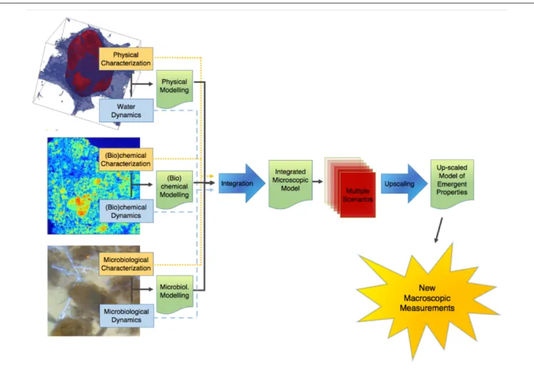
This point of terminology being resolved, the crux of the matter is that information of an entirely different nature than that currently available is needed to describe soil microbial processes adequately. We clearly need new macroscopic measurements. There are probably different ways to envisage the paths that will lead us eventually to this “Holy Grail.” Figure 1 proposes one of these perspectives, which has served as a general strategy map to a number of us in our research efforts. It starts on the left with information about basic soil features. What we understand at this point of emergent processes in soils indicates that this topic has (at least) three clear, resolutely interdependent facets, associated, respectively, with physical-, (bio)chemical-, and microbiological aspects of soils. For each of them, it is crucial to gather experimental information, either on static properties (dealt with in the boxes “physical characterization,” etc.), or on their dynamics. Alongside this evidence gathering, it is also important to develop theoretical and modeling frameworks that encapsulate experimental information and allow predictions to be made. In each case, experimental data should serve to refine theories and models, which in turn (e.g., through sensitivity analyses) can provide guidance in the procurement of additional data. The outcome of this type of iterative approach, hopefully, is a satisfactory description of each dynamic, which can then be integrated at first pairwise, and eventually all together, into a comprehensive model of soil processes at the microscale.
At that point, we are still somewhat far from the goal. Indeed, when this integrated model becomes available, running it on any given soil sample will require a tremendous amount of microscale information, which may take weeks or even months to gather, not to mention that the integrated model itself will likely take quite some time to run, which means that with this integrated microscale description of soils, only very few soil samples will ever be characterized and modeled. What we need instead is to come up with simplemacroscopic measurements that can be carried outroutinely.
One way to find out what these macroscopic measurements should be is suggested in Figure 1. It consists of expanding the available experimental database by simulating many different scenarios under different conditions of microscale heterogeneity of the soils, and of their properties. From these multiple scenarios, one can try to find out how one can simplify the description, in other words upscale the microscale model to the macroscale, while making sure that, in the process, the
FIGURE 1 | Schematic representation of the sequence of steps in the research on the emergent properties of soils, leading from a characterization of the various properties and dynamics at the microscale, onward to an upscaled macroscopic model, and finally to the ultimate goal of identifying macroscopic measurements that can be carried out routinely.
macroscopic parameters that appear in the resulting upscaled description are amenable to routine measurement in practice. This prospect of course rests on the assumption, at this stage very much open, that the simplification implied by the notion of “emergence” indeed occurs in soils. As discussed later, there is fortunately some circumstantial evidence that such simplification can be expected.
The research program, depicted in Figure 1, comprises a number of clear paths, which are discussed in the following. To the extent that some of the steps along these paths involve advanced technologies and elaborate methods of analysis or simulation, there is a definite risk of drift, i.e., to focus excessively on tools, perfect them, and progressively forget over time the reason for doing all this work in the first place, as one could argue has unfortunately happened occasionally in the past in other contexts in soil science (Baveye and Laba, 2015). One might argue that switching progressively from actual soils to very coarse sands or collections of clean 500 µm (or bigger) glass beads constitutes an example of such a drift. These systems admittedly pose far fewer technological challenges, which enable researchers to identify and isolate various microscale mechanisms, but, as
experience acquired in the past (e.g., in the sixties, when glass beads were used to research several soil processes, like water retention hysteresis) has shown, the relevance of the information acquired in these idealized systems for the functioning of real soils is unclear, at best. To avoid such drift, as much as possible, the descriptions of the various paths of Figure 1 will focus exclusively on progress made to date with actual, livingsoils, in all their wonderful complexity and messiness.
One last comment that needs to be made before we embark in the description of the program of Figure 1 is that there is no reason to be so wedded to it as not to be open to alternatives that may surface. If tomorrow, an experimentalist comes up with a robust empirical relationship among novel macroscopic measurements, similar to what is envisaged as the ultimate outcome of the program of Figure 1, every researcher interested in the field should probably rejoice, change gear, and adopt an entirely different perspective, for example to try to understand why the solution works. This is reminiscent of the debate about top-down vs. bottom-up approaches in hydrology (Basu et al., 2011;Baveye and Laba, 2015). Regardless of how strongly held one’s philosophical beliefs are, what matters most is to find a
satisfactory answer to a number of questions, not necessarily the manner in which the answers are obtained. This being said, no experimentalist has stepped forward yet with a ready answer, and the stepwise plan of Figure 1 appears to be our best bet at this point to ever obtain one in the limited time we have to do so.
PROGRESS ON THE PHYSICAL FRONT
Computed Tomography and Image
Processing
Any overview of the quantitative research of the past 10 years on microscale processes in soils needs to start with their physical characterization. Indeed, soil physicists have undeniably led the charge. The pioneering work carried out in the early 1980s with medical and custom-made X-ray and gamma-ray computed tomography systems brought to the attention of the soil physics community the potential of this technology, then still in its infancy (Petrovic et al., 1982;Hainsworth and Aylmore, 1983;
Crestana et al., 1985; Pires et al., 2010). The low (millimeter) resolution of scanners available at the time enabled researchers to characterize the geometry of macropores (e.g., earthworm burrows) in soils (Warner et al., 1989;Joschko et al., 1991;Heijs et al., 1995;Capowiez et al., 1998;Rogasik et al., 2003;Luo et al., 2010), but was much too coarse to provide information relevant to microorganisms. In the mid-1990s, various synchrotron facilities around the world began to devote beam time to soils, and researchers immediately took advantage of the significantly higher spatial resolution (down to a few µm) these facilities afforded, as well as the fact that the synchrotron X-ray beams are monochromatic (single-energy) (Anderson and Hopmans, 1994;Spanne et al., 1994;Garnier et al., 1998;Wildenschild et al., 2002;Feeney et al., 2006). However, access to synchrotron beam time was, and still is to a large extent, somewhat scarce and difficult to obtain, so that the extent of adoption of synchrotron-X-ray tomography has remained limited. The commercialization, around 2002, of the first tabletop, non-medical X-ray tomography systems, which were not excessively onerous and could therefore be entirely dedicated to soil science research, marked the beginning of a new era. The X-rays produced by these machines are polychromatic (i.e., are a mixture of X-rays of different energies), which in a number of ways is a disadvantage compared to the monochromatic X-ray produced by synchrotrons, but the resolution of these tabletop scanners has steadily improved since 2002 and several machines now allow resolutions that, in small soil samples of a few cm3, can be as low as 0.3 µm, i.e., commensurate with the resolution afforded by synchrotrons (Voltolini et al., 2017) and with the size of some of the “ultra-small” bacteria and archaea found in soils. The very high resolution of X-ray CT has for a time at least made other types of measuring instruments, like dual-energy gamma-ray scanners, neutron radiography, or nuclear magnetic resonance micro-imaging systems, fall off the radar screen, at least in applications to soils. Nevertheless, as we shall see later, these instruments afford advantages over X-ray CT, and are therefore likely to play a more significant role in the future.
The “3-dimensional” soil images that CT scanners provide are in fact stacks of 2-dimensional, grayscale images associated with virtual slices within the soil sample. Very early on in the use of these images, researchers came to the conclusion that these grayscale images would not be very useful to quantify the geometry of the soil pore space and that it was necessary to derive binary (black and white) images from the original grayscale ones, a process alternatively referred to as “thresholding” or “segmentation.” Significant progress has occurred over the years in how this thresholding is approached. Initially, it was carried out slice by slice, either manually by simple visual inspection (“eye-balling”) or with the assistance of one of a number of available 2D algorithms (e.g., Nunan et al., 2006). The first improvement consisted of thresholding the whole 3-d image at once, using an algorithm to calculate a unique, global threshold value. Then, various researchers showed that in the presence of textural heterogeneities (e.g., stones) within the samples, it was preferable to instead use local thresholds, which can vary from location to location within a sample (Iassonov et al., 2009;
Schlüter et al., 2014). Up to that point, all thresholding algorithms required operator input, to adjust one or more parameters. This introduced unavoidable subjectivity in the process, which in principle would make it improbable for different individuals to threshold a given soil sample the same way, or even for a single individual to threshold different soil samples (e.g., associated with different agricultural practices or with successive times) in a consistent manner (Baveye et al., 2010).
The question of objectivity in the generation of X-ray CT images of soils is in fact much broader than just this issue regarding thresholding/segmentation. Indeed, as a number of authors have pointed out (Vaz et al., 2011; Houston et al., 2013b), the process of obtaining CT images of soils requires many decisions to be made by operators, concerning in particular the value of scanning parameters (e.g., energy level, choice of filter, scanning resolution), the selection of one among a number of alternative image reconstruction and artifact correction algorithms, the format (8- or 16 bit) used to store the images, and the use of a method to increase image sharpness or reduce the noise that is unavoidably present in the images after reconstruction. As with thresholding 10 years ago, different groups, and sometimes even different individuals within a group, adopt alternative perspectives with respect to the various decisions that need to be made by operators, which can lead to sometimes significant differences in some of the metrics that are associated eventually with CT images (see, e.g., Houston et al., 2013b). Nevertheless, at this point, there appears to be no effort underway to develop a set of materials that could be used as “scanning standards,” as suggested by
Baveye et al. (2010), or simply to standardize analyzes. One way out of the difficulty would be to document exhaustively the parameter values used at each and every step of the image acquisition process, as well as, through detailed sensitivity analyses, the extent to which conclusions that are reached on the basis of CT images are affected by these parameter values.
Nevertheless, recognition a few years ago that the subjectivity in thresholding operations and in the manipulation of CT images
could be substantial, prompted the development of a number of automated thresholding algorithms requiring no operator input (Schlüter et al., 2010;Hapca et al., 2013;Houston et al., 2013a), regardless of the level of “supervision” (learning from training data) adopted. These objective algorithms have been used in a number of investigations (e.g.,Beckers et al., 2014a,b;Houston et al., 2017), and new algorithms are appearing that do not require any parameter tuning (e.g.,West et al., 2018), but so far they have not stopped the development of operator-dependent approaches (Kulkarni et al., 2012;Hashemi et al., 2014;Ojeda-Magana et al., 2014; Martin-Sotoca et al., 2017). Therefore, further progress is needed in this area, especially in order to segment images containing multiple distinct populations of voxels.
BIB- and FIB-SEM
Another approach that has recently been explored to obtain basically the same physical information as with X-ray CT consists of using broad- or focused ion beam scanning electron microscopy (BIB- or FIB-SEM). The ion beam can directly modify or “mill” a specimen surface, and this milling can be controlled with nanometer precision. By carefully controlling the energy and intensity of the ion beam, it is possible to perform very precise nano-machining to remove very thin layers of material, for example in a block of soil impregnated with resin. BIB milling produces cross-sections of a few mm2to cm2, whereas FIB deals with surfaces that at most are a few hundredµm2. Once a new surface has been exposed, it can be imaged via SEM, at resolutions typically between 10 and 500 nm (Cantoni and Holzer, 2014). The sequence of images obtained in successive layers can be assembled into a 3D image, similar to those resulting from X-ray CT tomography, and subsequently segmented (Salzer et al., 2015;
Liu et al., 2017). In the last few years, this approach has been used extensively to investigate the morphological characteristics of dolomite rocks, shales, and clays using BIB alone (Houben et al., 2013), a combination of BIB- and FIB-SEM (Hemes et al., 2015), or the joint use of micro-CT and FIB-SEM (Devarapalli et al., 2017). In soils, FIB-SEM presents a tremendous potential, but its use appears to have been limited so far to observations of microbially induced calcite precipitation in sandy soils (Li et al., 2017) and to obtain high-resolution images of the colonization of soil–root interfaces (Vidal et al., 2018).
Soil Structure Versus Architecture
Early in the use of CT scanners to characterize the physical properties of soils, it became apparent that this technology afforded a convenient response to the age-old question of how to best quantify soil “structure,” this term being understood either as “the arrangement or organization of the particles in the soil” (Hillel, 2004), or, followingDexter (1988), as “the spatial heterogeneity of the different components or properties of soils.” For many decades, the vast majority of the research on the topic has viewed soil structure as intimately linked with the fact that it is possible to fragment soils into distinct aggregates upon the application of mechanical stress (Rabot et al., 2018). Undoubtedly this perspective has its roots in the soil surveyors’ traditional poking of exposed soil profiles with knives, leading to the detachment of chunks of soils, called “aggregates,” whose size
and shape is used to diagnose the types of pedogenetic processes that might have taken place at that location, to classify soils, and to evaluate their agronomic potential. Since the 1940s, an extensive body of literature has been devoted to the assessment of the stability of soil aggregates under a variety of operational conditions, for example under dry or wet sieving. AsYoung et al. (2001) point out, “the ease and seeming reproducibility of the many standard stability tests are the main drivers behind the prevalence of this type of research.”
A common criticism of the concept of aggregate in soils is that it is little more than an artifact. The hierarchical organization of aggregates, identified and described in detail byTisdall and Oades (1982), suggests that the distribution of sizes of aggregates one obtains might depend on the amount of energy that is applied to take soils apart. This operational issue, discussed byAmézketa (1999), is particularly well illustrated by the experimental results ofDíaz-Zorita et al. (2002), who show that the size of fragments obtained by sieving soils is inversely related to the mechanical stress applied.Hallett et al. (2013)also point out that breakdown of soils by dynamic or static mechanical loading yields different fragmentations of soil aggregates. This dependence of the aggregate size distribution on the operational conditions under which it is measured raises the question of whether aggregates exist in soils in their natural state (Young et al., 2001), calling into question the extensive literature that tries to analyze the influence of aggregate size on various processes, e.g., in terms of the sequestration of OM, the distribution of bacteria, a wide range of geochemical processes, or the release of greenhouse gasses (Ranjard and Richaume, 2001;Jasinska et al., 2006;Nunan et al., 2006;Razafimbelo et al., 2008;Goebel et al., 2009;Pallud et al., 2010;Chivenge et al., 2011;Masue-Slowey et al., 2011, 2013;
Blaud et al., 2014;Rabbi et al., 2014, 2016;Ebrahimi and Or, 2015;
Jiang et al., 2015;San José Martínez et al., 2015; Sheehy et al., 2015;Hausladen and Fendorf, 2017;Rillig et al., 2017;Zhao et al., 2017;Bocking and Blyth, 2018; Li et al., 2018), and explaining perhaps why some authors have failed to observe anticipated correlations between OM content and aggregation (Razafimbelo et al., 2013). Nevertheless, one might argue that this dependence problem can be alleviated somewhat by standardizing methods, and that, in any event, it does not particularly affect attempts to understand at a very local scale in soils the interactions between pore geometry, chemical composition, and microbial activity. As long as aggregates are viewed as chunks of soil that are convenient to manipulate because they do not fall apart too easily, e.g., when they are rotated on the stage of a CT scanner, and to the extent that no particular significance is associated with their external surfaces, which might just have been failure planes in some larger aggregate, no harm is done in using aggregates to gain insight into microscale processes, as various authors have done successfully (Remusat et al., 2012;Ananyeva et al., 2013;Kravchenko et al., 2013, 2015;Voltolini et al., 2017;Yu et al., 2017).
One could also consider that there is no problem either with repacking aggregates extracted from a soil, and trying to find out experimentally or through simulation how this now entirely artificial system behaves (e.g.,Daly and Roose, 2014; Ebrahimi and Or, 2016). We are often forced by journals to use repacked soil columns in order to have actual replicates, and be able to
calculate statistics, which some reviewers view as sacred and indispensable. However, it is entirely unclear at this point to what extent the conclusions that one reaches from this kind of exercise relate to the behavior of real soils, including the very soil from which the aggregates that are used originated. The reason for this has to do fundamentally with the absence of any theoretical framework or set of procedures to, as it were, put the pieces of the puzzle back together, once a soil has been disaggregated and its aggregates have been characterized, e.g., relative to their size distribution and individual geometries. In the process of disaggregating a soil sample, as long as no information is obtained about the geometry and topology of the interstices that may have existed originally between what eventually becomes distinct aggregates, there is no way practically to “reconstruct” the original soil, even for computational purpose, and in particular to guarantee that the pores between aggregates in the repacked system be similar in shape to those that existed originally1. One
could draw parallels here with architecture (Letey, 1991;Baveye, 2006) or even with card games: Indeed, one cannot say anything about the size and shape of a house of cards after it has been torn down, simply by looking at the pile of cards that is left.
Aware of these obstacles already many years ago, a number of authors argued for a different way to approach the structure of soils. Dexter (1988), in a thorough review of the then available methodology in this field, recommends that preference be given to methods involving direct observation of structural features by scanning electron microscopy and by optical scanning of resin-impregnated sections and fracture surfaces. A few years later,
Letey (1991) vents his frustration in the face of many failed attempts to link soil structure, defined in terms of aggregates, to functionality within the soil system. He suggests that instead of focusing on the solid components of soil structure, as had been the tradition for decades, one should emphasize instead the arrangement of voids, and the properties that these voids confer to soils, just as to describe a building, it is not primarily the shape of the bricks or stones that matters, or the thickness of the walls, but the size of the rooms and openings (windows, door frames). Reiterating these same messages, Young et al. (2001)
argue that “an investigation of discrete aggregates or distributions of aggregates does not offer any spatial information. Functional traits of soil structure, at all scales, rely on the connectivity, tortuosity, and heterogeneity of pore space in 3D.” The same message is echoed in the recent thorough review of the literature byRabot et al. (2018), who conclude that “although appealing, the aggregate perspective does not seem to be the most appropriate to link soil structure with soil functions and processes.” Because of the historically close connection between “soil structure” and aggregates,Young et al. (2001)propose to drop the expression of “soil structure” in favor of that, less history-laden, of “soil architecture.” This terminology has been routinely adopted since (e.g., Baveye, 2006; Lin et al., 2010; de Jonge et al., 2012; Lin, 2012; Bouckaert et al., 2013a,b; Cazelles et al., 2013; Helliwell
1There is one exception to this general statement, in the case of some oxisols in
tropical areas, such as the La Selva soil investigated byRadulovich et al. (1992). This soil, containing 95% kaolinite clay, is made of “pseudo-sand” microaggregates that are very regular in shape and size, so that it is possible in practice to repack the soil in a state that is very close to what it was originally.
et al., 2013;Kravchenko and Guber, 2017;San José Martínez et al., 2017) and will be used consistently in the following.
In principle, it is feasible to analyze this architecture by taking 2D images of sequences of thin sections in resin-impregnated blocks of undisturbed soil, and then using dedicated software to reconstruct from these images a full 3D picture of the geometry of soil pores. This tedious, time-consuming approach has been adopted with success byCousin et al. (1996, 1999),Vogel (1997), andVogel and Roth (2001). However, access to X-ray beams at various synchrotron facilities, and especially the availability of table-top X-ray CT scanners, have allowed the work in this area to experience a quantum leap around the turn of the century. The new technology has made it possible to obtain 3D images of the pore space in intact soil cores much more rapidly, and at resolutions that have gradually improved over time (Mooney, 2002;Rozenbaum et al., 2012; Bouckaert et al., 2013a;Calistru and Jitareanu, 2015;Rabot et al., 2015).
The gradual conceptual shift from the aggregate-based “structure” to the “architecture” of soils has been accompanied by a refocus of the discourse on the voids within this architecture, following in that respect the suggestion ofLetey (1991). Another conceptual shift as well is occurring in that respect. Conditioned to think in terms of a traditional analogy between the pore space of soils and a bundle or network of capillaries, soil physicists used to be concerned about the size of “pores” in soils. It is clear from CT images that there are no identifiable pores in soils, and that the delineation of individual pores is necessarily somewhat subjective. Some authors have tried to make the concept of pore size distribution less arbitrary by using automatic algorithms to determine locally the radii of maximum balls that are fully inside the pore space. Partly because of the historical weight of the capillary analogy and partly with the help of these “inscribed balls” algorithms, pore size distributions are still being computed (e.g.,Kuka et al., 2007;Papadopoulos et al., 2009;Ostadi et al., 2010;Bouckaert et al., 2013a;Peng et al., 2014; Houston et al., 2017;Meira Cassaro et al., 2017). Yet, clearly, researchers have increasingly turned in recent years to other approaches to describe quantitatively the make-up of soils. Indeed, a whole panoply of mathematical tools is now available, and is steadily expanded, to characterize a number of aspects of the pore space. These tools include various algorithms to calculate the tortuosity and connectivity of the pore space on the basis of grayscale or binary 3D CT images (Gommes et al., 2009;Houston et al., 2017;
Meira Cassaro et al., 2017). Another approach to describe the pore space quantitatively is provided by the fundamental set of Minkowski functional measures (Lehmann et al., 2006; Vogel et al., 2010; Falconer et al., 2012). These functional measures comprise the volume, surface area, integral mean curvature, and the Euler-Poincaré characteristic (or topological measure Chi).Karsanina et al. (2015)propose another set of descriptors, including two-point probability functions, linear functions, and two-point cluster functions, and they used the first two in simulated annealing optimization procedures to reconstruct soil architecture artificially, based on original images of soil thin sections. For a number of years, fractal geometry was thought to be an ideal tool to characterize the inner space of soils, since according to the way the theory was interpreted, a single
parameter, the fractal dimension, could inform about the make-up of soils over a range of scales. This vision of fractal geometry has since been confronted with the reality that a single dimension, whose value turns out to be itself scale- and resolution-dependent, does not suffice. As explained by Mandelbrot from the start, at least one other parameter, either the lacunarity (e.g.,
Pendleton et al., 2005; San José Martínez et al., 2017) or the succolarity (de Melo and Conci, 2013) is required to obtain an accurate description. Since the lacunarity (Pendleton et al., 2005) and likely also the succolarity are affected by the resolution of images, it is not clear at this point whether fractal geometry still offers much interest. Another approach that might (but has not yet) provide a solution to the resolution-dependence derives from the application of the theory of multifractal measures to CT images of soils (Lafond et al., 2012; Wang et al., 2016;
Torre et al., 2018). From a different perspective, various authors have also tried to extract networks from 3D images of soils, to which graph theory principles can be applied to characterize the connectivity and topology of the pore space (Vogel and Roth, 2001;Perez-Reche et al., 2012).
Can We “See” Water and Organic Matter
in Soils?
Since the ultimate objective of the research reviewed here is to eventually be able to predict the activity of microorganisms in soils, for whom the presence of water and readily biodegradable OM is crucial, it is important to be able to detect in what portion of the pore space they are located. In this respect, researchers have been confronted with the difficulty that, typically, if one places a wet soil sample in a table-top X-ray CT scanner, the outcome is a 3D grayscale image characterized by a histogram with a single, broad peak that is not suitable at all to tease apart the water from the solids without resorting to arbitrary assumptions (Tracy et al., 2015), nor to identify in the solid phase the portion that corresponds to OM. So far, to avoid this obstacle, researchers have either shifted their attention toward artificial media, or they have worked with actual soils but under special conditions that allow the identification of water and OM.
In terms of artificial porous media, researchers have used glass beads (Culligan et al., 2006;Schaap et al., 2007) and coarse sands (Brusseau et al., 2007) to quantify the 3-dimensional distribution of water in the pore space. If one scans these systems under partially saturated conditions, as these researchers did, evidence suggests that it is not very difficult to locate air-water interfaces. Another way to proceed, made possible by the low reactivity of glass beads or sands compared to soils, is to increase the contrast between the attenuation of X-rays in the solid phase and the liquid phase by using a contrast agent that increases X-ray attenuation in the liquid. Although the results obtained with glass beads and sands are definitely interesting and probably applicable to coarse aquifer materials, it is not clear at this stage how they help us identify water and OM in actual soils, which as a rule tend to be tremendously more heterogeneous, and have much smaller pores. There is no real answer at this point to the question of how one can transition from glass beads to actual soils. This is a perennial problem, also faced by researchers who for a time
carried out extensive work in the 1960s on the hysteresis of water retention in glass beads systems (Topp and Miller, 1966;Topp, 1971).
Another approach that can conceivably work in some soils, consists of scanning a soil sample when it is dry, and then re-scan it when it has been brought to the desired moisture content (e.g.,
Tracy et al., 2015). Comparison between the non-air phases in the “before” and “after” images yields the distribution of water in the system. In principle, this approach could work very well if the soil does not swell at all when its moisture content is increased. This apparently was the case in the experiments carried out by
Tracy et al. (2015), who report that “no significant evidence of shrinkage was observed.” Yet the problem is that most soils in the world do shrink/swell to some extent (Garnier et al., 1997), including soils like those described by Radulovich et al. (1992)
whose kaolinitic mineralogy one does not traditionally associate with this phenomenon. The question remains at this point of what is significant enough evidence of shrinkage or swelling in a soil sample to prevent this “subtraction” method to be used to visualize the distribution of water.
Yet another strategy is to carry out CT measurements on real soils under conditions where water and OM are not intimately mixed with the solid constituents at very fine scales. This approach has been adopted by a number of researchers in the last few years who worked on plant residues within soils (De Gryze et al., 2006;Negassa et al., 2015;Kravchenko et al., 2017) or attempted to directly visualize soil moisture (Carminati et al., 2008;Tippkötter et al., 2009;Pot et al., 2015). Working with a real clay-loam soil material near water saturation,Carminati et al. (2008) focused on the water that occupies part of the volume in the larger pores. They were able under these conditions to clearly observe pendular rings of water in images at a resolution close to 6µm.Tippkötter et al. (2009)adopted a similar focus, in undisturbed soil samples, and were able with a table-top X-ray CT scanner to visualize the presence of water films coating the inner surfaces of meso- and macropores. Similarly, Pot et al. (2015), working with synchrotron X-rays, were able to generate CT images of repacked aggregates in whose histograms there was a good separation of voxels associated with the air, liquid, and solid phases (Figure 2).
A last approach that could work in principle to see the moisture in soils consists of adding various contrast agents to the water, to modify its X-ray attenuation (Van Loo et al., 2014). However, in practice, contrast agents need to diffuse sufficiently for the method to work, which again, in many cases, in undisturbed soil samples, might be operationally workable only to image the largest pores near saturation.
It might thus be tempting to look elsewhere for a possible answer. Indeed, over the last decade, the resolution associated with 3D nuclear magnetic resonance (NMR) micro-imaging has become steadily better.Lee and Lee (2017), for example, managed to obtain images of 1.2 mm by 1.2 mm columns of glass beads and crushed silica gels particles, respectively, with a spatial resolution of 46.875 µm, which is still coarser than the resolution of CT scanners for this column width, but is not as far from it as it used to be. As encouraging as these results are, however, NMR micro-imaging as currently implemented still suffers from a major
FIGURE 2 | (A) two dimensional section of synchrotron X-ray computed tomography image of a soil cube equilibrated at –1 kPa and (B) histogram of the corresponding 3D SR-µCT image. In the tomographic sections, black is the air phase, dark gray is the water phase and light gray to white is the matrix phase. The scale bars represent 500µm. (Reprinted with permission fromPot et al., 2015).
obstacle when it comes to real soils, and therefore is not a real solution in that context. Because of the very powerful magnets that are used to generate the signal, only soils that do not contain paramagnetic elements can be imaged. Since many if not most soils contain some iron, at least, this limits tremendously the conditions under which NMR is a viable alternative to X-ray CT to generate 3D images. An alternative to NMR would be to use neutron computed tomography (Tumlinson et al., 2008) to observe the distribution of water in soils, but at this stage the resolution of images that can be generated is still relatively low, comparable to that obtainable with medical or table-top X-ray CT equipment 15 or 20 years ago (Perfect et al., 2014).
The best option to “see” water at this point, even though it has not been implemented very much of late, appears to be
the use of dual energy X-rays in CT scanners. With gamma rays of two different energies, typically produced by241Am and 137Cs sources, it has been possible for a while to simultaneous
assess the moisture content and bulk density of soils (Soane, 1967;Hopmans and Dane, 1986; Biassusi et al., 1999), but the measurements are extremely slow, and their spatial resolution is low. Garnier et al. (1998) applied dual-energy synchrotron X-rays for the first time to soils, to assess rapid vertical soil density and water content changes in swelling soils during infiltration. Shortly thereafter, Rogasik et al. (1999) used a medical scanner that allowed them to scan silt loam subsoil samples at two energy levels (80 and 120 kV) to evaluate the distributions of water, air, and solids, as well as the voxel dry bulk density. The spatial resolution during scanning was 0.25 mm in the horizontal and 1 mm in the vertical direction, which was (and still is) standard for scanners routinely used in hospital settings. Since this work almost 20 years ago, there has been to our knowledge no application of dual-energy X-ray tomography to soils. Several table-top X-ray scanners currently commercialized offer the possibility to carry out dual-energy scanning sequentially on soil samples. The fact that nobody so far has reported on the use of this feature with table-top scanners suggests that polychromatic X-rays are not suitable for dual-energy scanning to work in the case of soil samples. Further research is needed to determine if with monochromatic X-ray beams, at synchrotron facilities, dual-energy scanning produces promising results.
Part of the reason for the limited use of dual-energy scanning − and it would be true as well for attempts to scan soils rich in OM with dual-energy gamma-rays − is that until not too long ago, it would have been difficult to tease apart water from soil OM in CT images (Taina et al., 2008). The problem is not OM per se. Kettridge and Binley (2011)demonstrate that X-ray CT can image beautifully the structure of peat samples of various compositions. The difficulty has to do with the fact that at the high X-ray energies required to penetrate through soil materials, there is very little difference in X-ray attenuation between water and water-filled OM, whose peaks in grayscale image histograms are often not clearly distinguishable from a broad peak associated with mineral constituents. This problem was resolved, at least in part, in 2014, when Van Loo et al. (2014) tested 52 different chemical compounds. They perfused aqueous solutions saturated with the compounds through undisturbed soil samples under partial vacuum and found that 4 of these chemicals [phosphomolybdic acid (PMA), silver nitrate, lead nitrate and lead acetate] successfully enhance the X-ray attenuation contrast of OM relative to soil minerals and allow particulate organic matter (POM) to be easily detected.
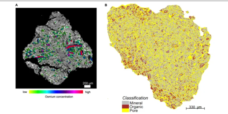
Peth et al. (2014)tried to take advantage of the fact that osmium has a marked absorption K-edge2at a photon energy ∼74 keV.
They exposed air-dry soil aggregates to a 25 w/w OsO4solution
for 48 h at room temperature in a closed vial under a fume hood (because of the very high toxicity of OsO4), and scanned
2K-edge is the binding energy of the K shell electron of an atom. There is a sudden
increase in the attenuation coefficient of photons occurring at a photon energy just above the binding energy of the K shell electron of the atoms interacting with the photons.
FIGURE 3 | Illustrative two-dimensional spatial distributions of osmium-stained OM mapped onto the reconstructed image cross-sections of aggregates. In (A), red colors are typically associated with particulate OM. Green colors reflect lower concentrations (Modified fromPeth et al., 2014. Reprinted with permission). In (B), the red patches correspond to OM (Modified fromRawlins et al., 2016. Reprinted with permission).
these aggregates at a synchrotron facility below and above the absorption K-edge, respectively. Preliminary results, obtained by
Peth et al. (2014) andRawlins et al. (2016), suggest that this technique makes it possible to visualize the distribution of OM in soils, and to distinguish between POM and OM that is distributed more diffusely throughout the soil architecture (Figure 3). One promising approach to identify POM in CT images consists of building on both the attenuation, thus gray scale, values of the organic materials and on the spatial distribution patterns of POM grayscale values, which uniquely separate it from the rest of the soil solids (Kravchenko et al., 2014a). Indeed, even from a “naked eye” examination, POM often stands out on CT images due to much greater uniformity of its grayscale values.Kravchenko et al. (2014a)successfully used geostatistical parameters of POM fragments as indicators of the presence of POM in intact soil samples. This approach has advantages over POM identification via Os staining, since, unlike Os staining, CT scanning has minimal effect on soil microorganisms (Bouckaert et al., 2013b;Kravchenko et al., 2014b;Schmidt et al., 2015). Thus, the samples can be used for exploring the decomposition of the identified POM fragments in a sequence of initial CT scanning, incubation, and post-incubation CT scanning activities, as done byKravchenko et al. (2015). However, as of now the process of POM identification using this approach is time consuming and requires a substantial user input.
Sub-Resolution Pores
Another issue that, at the moment, is still awaiting a definite solution, is related to the soil pores having dimensions smaller than the resolution of CT images. From the mid-1980s to the
early 2000s, the resolution of X-ray CT scanners accessible to soil scientists improved by about 3 orders of magnitude, from a few hundred microns to a fraction of a micron in the best of cases. However, no matter how small this resolution is, a portion of the pore space unavoidably remains invisible to scanners. The practical significance of sub-resolution pores depends strongly on the resolution of CT images, of course, but also, critically, on the type of soil considered. For a coarse sandy soil, it is possible that at a 20 or 30 µm resolution, only a minute portion of the total porosity would not be visible in thresholded CT images. Contrastedly, in other types of soil, the portion of sub-resolution pores can be substantial. In microaggregated tropical soils like those investigated bySollins and Radulovich (1988), pores with a diameter less than 5 µm make up approximately 70% of the pore space, and are key to understanding the unique physical and chemical properties of these systems (e.g.,Radulovich et al., 1992). In the silty soils investigated by Piccoli et al. (2017), 82% of the pores a dimensions smaller than the resolution of 30 µm at which the CT scans were made. In clayey or clay-loam soils, like those whose pore size distribution was determined via mercury intrusion porosimetry by Churchman and Payne (1983), a resolution of 10 µm would be too large to identify any pore at all, and it would be of paramount importance to be able to somehow characterize the sub-resolution porosity in some fashion.
In some very special cases, grayscale CT images, before any thresholding or segmentation is carried out, may contain some information about sub-resolution features. In porous media like sandstone samples or columns filled with glass beads, which consist solely of a homogeneous mineral phase, grayscale values
intermediate between those of the pores and solids in CT images result from partial-volume effect if the solid particles are not porous. If, on the contrary, the solid particles are themselves porous, intermediate grayscale values are associated both with partial-volume effects and with sub-resolution pores, and blind application of a ternary segmentation approach, like that used by
Scheibe et al. (2015), may yield meaningless results if interpreted solely as sub-resolution porosity. However, in these same simple systems, as long as partial-volume effects are negligible, grayscale values contain unambiguous information about sub-resolution pores, information that is lost entirely when the images are thresholded (Gommes et al., 2009;Baveye et al., 2010). To refine the analysis, insight obtained from images of these systems at different resolutions can be combined, as was done recently by various authors (Sok et al., 2010;Pini and Madonna, 2016;Shah et al., 2016) and applied to soils by others (Vogel et al., 2010;Dal Ferro et al., 2014), to try to get a better handle on predicting the properties of sub-resolution voids. Unfortunately, in real soils, in most cases of practical interest, both these single- and multiple-resolution approaches run into as yet insurmountable hurdles, related to the intimate, fine-scale mixing of minerals, OM, and water. The contribution of these different phases and constituents to the grayscale value of voxels in reconstructed CT images cannot at this stage be differentiated, and as a result, it is not possible in general to correlate this grayscale level with the porosity of the volume of soil to which it corresponds. It might be necessary to wait until tunable X-ray and gamma-ray scanners become routinely available, to resolve this issue.
Moving From 3D to 4D: Dynamical
Measurements
In order to get a dynamical picture of physical processes in soils, one needs to transition from 3D to 4D, the fourth dimension of course being time. In many disciplines outside soil science, this transition has captured the attention of researchers over the last few years, and very interesting results have been obtained, in particular for very coarse-textured porous media (Berg et al., 2013; Dobson et al., 2016). Yet, as far as soils are concerned, forays along these lines have been timid. At the mesoscale, very interesting work, starting already 25 years ago, describing how earthworm burrow systems evolve over time (Joschko et al., 1991;
Capowiez et al., 1998), how the geometry of macropores in paddy soils evolves during soil shrinkage (Bottinelli et al., 2016), or how loamy soils are compacted during centrifugation (Schlüter et al., 2016). Also, various researchers have used MRI systems to monitor the infiltration of water in clay and coarse sandy loam columns (Amin et al., 1994, 1996;Preston et al., 2001;Votrubová et al., 2003), γ-ray CT equipment to quantify the swelling of vertisols over time (Biassusi et al., 1999), or neutron CT systems to investigate the dynamics of water flows in soil, especially in the rhizosphere (Badorreck et al., 2010;Perfect et al., 2014;Tötzke et al., 2017). However, virtually all of this work has been carried out at relatively low resolutions, at best of 15µm but more often than not of several tens or even hundreds of microns.
At the micron scale sensu stricto, very little 4D work has been carried out so far. None of this research includes water
movement, which is not very surprising, given the difficulties mentioned earlier concerning the detection of water at a sufficiently high resolution to be relevant to the microscale. Even under the various conditions where this detection is possible, water movement tends to be too fast to be monitored by X-ray CT, even at the fastest scanning times (of the order to 10–15 min, typically) available with table-top scanners. Ultrafast scanning techniques have been used recently with columns filled with gravel (Dobson et al., 2016), but similar research has yet to be conducted with soils. Because of these constraints, dynamic microscale measurements have been limited to situations that involve ice formation, or slow changes in the architecture of soils. Using a table-top X-ray CT scanner,Torrance et al. (2008)
investigated the changes in structure and the redistribution of water to form ice lenses in saturated samples of an Aurora silt loam frost-susceptible soil that were thoroughly mixed to produce an initially homogeneous material, and of a Honeywood silt loam that was deliberately contaminated with motor oil. The soils were subjected to relatively rapid, downward freezing, with access to water at their base. The results indicate that CT can produce excellent images of the ice lens distribution within a frozen silt loam soil, the consolidation of soil between the ice lenses, and the effects of hydrocarbon contamination on ice formation. Also using X-ray CT in freezing soils,Starkloff et al. (2017)assessed the impact of a succession of freezing-thawing cycles on the pore network of a silty clay loam and a loamy sand topsoil. Also recently,Schlüter and Vogel (2016)quantified soil architecture turnover by labeling soil constituents in place with small garnet particles and tracking their fate in successive CT images. The particles adhere to pore boundaries at the beginning of the experiment but gradually change their position relative to the nearest pore as structure formation progresses and pores are destructed or newly formed.
Modeling the Physics
Over the last 2 decades, a significant body of literature has been devoted to the mathematical modeling of water retention and transport within the complex geometry of soil pores, revealed with increasing resolution by X-ray CT scanners. The bulk of this literature has dealt with the development and application of the Lattice-Boltzmann method (Martys and Chen, 1996;Genty and Pot, 2013, 2014; Liu et al., 2016; Cruz et al., 2017; Zhou et al., 2018), but in recent years, other methods have also been adopted, based on finite element or finite difference schemes, or on geometric primitives.
Most of the work in this area has involved a number of variants of the Lattice-Boltzmann method, in which a fluid is viewed as a collection of fictitious particles that, alternatively, propagate from node to node on a regularly spaced grid (lattice mesh), then collide with the particles that end up on the same nodes. In the modeling of soils, the nodes correspond to the centers of voxels in 3D CT images. The method originates from a molecular description of a fluid and can directly incorporate physical terms stemming from a knowledge of the interaction between molecules. Hence, in principle, it keeps the cycle between the elaboration of a theory and the formulation of a corresponding numerical model short, which undoubtedly