HAL Id: hal-03180884
https://hal.inrae.fr/hal-03180884
Submitted on 25 Mar 2021
HAL is a multi-disciplinary open access
archive for the deposit and dissemination of
sci-entific research documents, whether they are
pub-lished or not. The documents may come from
teaching and research institutions in France or
abroad, or from public or private research centers.
L’archive ouverte pluridisciplinaire HAL, est
destinée au dépôt et à la diffusion de documents
scientifiques de niveau recherche, publiés ou non,
émanant des établissements d’enseignement et de
recherche français ou étrangers, des laboratoires
publics ou privés.
viruses in rodents and shrews in context of
forest-savannah-urban areas interface in the city of
Franceville (Gabon)
Joa Mangombi, Nadine N’dilimabaka, Jean-Bernard Lékana-Douki, Octavie
Banga, Sydney Maghendji-Nzondo, Mathieu Bourgarel, Eric Leroy, Florence
Fenollar, Oleg Mediannikov
To cite this version:
Joa Mangombi, Nadine N’dilimabaka, Jean-Bernard Lékana-Douki, Octavie Banga, Sydney
Maghendji-Nzondo, et al.. First investigation of pathogenic bacteria, protozoa and viruses in rodents
and shrews in context of forest-savannah-urban areas interface in the city of Franceville (Gabon).
PLoS ONE, Public Library of Science, 2021, 16 (3), pp.e0248244. �10.1371/journal.pone.0248244�.
�hal-03180884�
RESEARCH ARTICLE
First investigation of pathogenic bacteria,
protozoa and viruses in rodents and shrews in
context of forest-savannah-urban areas
interface in the city of Franceville (Gabon)
Joa Bra
ï
the Mangombi
ID1,2,3, Nadine N’dilimabaka
1,4*
, Jean-Bernard Lekana-Douki
1,5,
Octavie Banga
1, Sydney Maghendji-Nzondo
6, Mathieu Bourgarel
ID7,8
, Eric Leroy
1,9,
Florence Fenollar
2,3, Oleg Mediannikov
3,101 Centre Interdisciplinaire de Recherches Me´dicales de Franceville (CIRMF), Franceville, Gabon, 2 Aix
Marseille Univ, IRD, AP-HM, Microbes, VITROME, Marseille, France, 3 IHU Me´diterrane´e Infection, Marseille, France, 4 De´partement de Biologie, Faculte´ des sciences, Universite´ des Sciences et Techniques de Masuku (USTM), Franceville, Gabon, 5 De´partement de Parasitologie, Universite´ des Sciences de la Sante´ (USS), Owendo, Libreville, 6 De´partement Epide´miologie-Biostatistique et Informatique Me´dicale (DEBIM), Universite´ des Sciences de la Sante´ (USS), Owendo, Libreville, 7 CIRAD, UMR ASTRE, Harare, Zimbabwe, 8 ASTRE, Univ Montpellier, CIRAD, INRA, Montpellier, France, 9 UMR MIVEGEC IRD-CNRS-UM, IRD, Montpellier, France, 10 Aix Marseille Univ, IRD, AP-HM, Microbes, MEPHI, Marseille, France
*nadinendilimabaka@yahoo.fr
Abstract
Rodents are reservoirs of numerous zoonotic diseases caused by bacteria, protozoans, or
viruses. In Gabon, the circulation and maintenance of rodent-borne zoonotic infectious
agents are poorly studied and are often limited to one type of pathogen. Among the three
existing studies on this topic, two are focused on a zoonotic virus, and the third is focused on
rodent Plasmodium. In this study, we searched for a wide range of bacteria, protozoa and
viruses in different organs of rodents from the town of Franceville in Gabon. Samples from
one hundred and ninety-eight (198) small mammals captured, including two invasive rodent
species, five native rodent species and 19 shrews belonging to the Soricidae family, were
screened. The investigated pathogens were bacteria from the Rickettsiaceae and
Anaplas-mataceae families, Mycoplasma spp., Bartonella spp., Borrelia spp., Orientia spp.,
Occiden-tia spp., Leptospira spp., Streptobacillus moniliformis, Coxiella burnetii, and Yersinia pestis;
parasites from class Kinetoplastida spp. (Leishmania spp., Trypanosoma spp.),
Piroplasmi-dae spp., and Toxoplasma gondii; and viruses from ParamyxoviriPiroplasmi-dae, HantaviriPiroplasmi-dae,
Flavivir-idae and Mammarenavirus spp. We identified the following pathogenic bacteria: Anaplasma
spp. (8.1%; 16/198), Bartonella spp. (6.6%; 13/198), Coxiella spp. (5.1%; 10/198) and
Lep-tospira spp. (3.5%; 7/198); and protozoans: Piroplasma sp. (1%; 2/198), Toxoplasma gondii
(0.5%; 1/198), and Trypanosoma sp. (7%; 14/198). None of the targeted viral genes were
detected. These pathogens were found in Gabonese rodents, mainly Lophuromys sp.,
Lem-niscomys striatus and Praomys sp. We also identified new genotypes: Candidatus
Barto-nella gabonensis and Uncultured Anaplasma spp. This study shows that rodents in Gabon
harbor some human pathogenic bacteria and protozoans. It is necessary to determine
whether the identified microorganisms are capable of undergoing zoonotic transmission
a1111111111
a1111111111
a1111111111
a1111111111
a1111111111
OPEN ACCESSCitation: Mangombi JB, N’dilimabaka N,
Lekana-Douki J-B, Banga O, Maghendji-Nzondo S, Bourgarel M, et al. (2021) First investigation of pathogenic bacteria, protozoa and viruses in rodents and shrews in context of forest-savannah-urban areas interface in the city of Franceville (Gabon). PLoS ONE 16(3): e0248244.https://doi. org/10.1371/journal.pone.0248244
Editor: Wanda Markotter, University of Pretoria,
SOUTH AFRICA
Received: August 27, 2020 Accepted: February 23, 2021 Published: March 8, 2021
Copyright:© 2021 Mangombi et al. This is an open access article distributed under the terms of the
Creative Commons Attribution License, which permits unrestricted use, distribution, and reproduction in any medium, provided the original author and source are credited.
Data Availability Statement: All relevant data are
within the paper and itsSupporting Information
files.
Funding: JBM 1: AUF- Agence universitaire de la
Francophonie. https://www.auf.org/afrique- centrale-grands-lacs/nouvelles/actualites/liste- beneficiaires-college-doctoral-regional- mathematiques-informatique-biosciences-geosciences-de-lenvironnement-publiee/orhttps://
from rodents to humans and if they may be responsible for human cases of febrile disease
of unknown etiology in Gabon.
Introduction
For several decades, rodents have been recognized as reservoirs or hosts carrying zoonotic
patho-gens [
1
–
4
] that can have very dramatic impacts on the economy and public health [
2
]. These
zoo-noses include the plague [
5
,
6
], Lassa hemorrhagic fever (LHF) [
7
], and hemorrhagic fever with
renal syndrome [
8
]. Even today, zoonotic diseases involving rodents may cause hundreds or even
thousands of deaths worldwide [
9
–
11
]. Many of these rodent-borne diseases are often
misdiag-nosed. For example, leptospirosis cases can easily be misdiagnosed as dengue or malaria infection
because of the similarity of the initial symptoms [
12
]. Such misdiagnosis is especially frequent in
countries of sub-Saharan Africa, where access to the necessary diagnostic tools is limited [
13
,
14
].
Countries of sub-Saharan Africa are experiencing a remarkable expansion of their urban
agglomerations [
15
–
18
]. This growth of cities is so dramatic that it may exceed the absorption
and management capacity of municipal environmental services, leading to the development of
large informal areas characterized by particularly degraded socioenvironmental conditions
(high human density, waste accumulation, precarious dwellings, etc.) [
18
–
20
]. Such living
con-ditions are reported risk factors favorable to rodent infestations [
21
], leading to the assertion
that the level of infestation of cities by rodents is correlated with the rapid growth of these cities
[
22
]. Finally, when the density of a rodent population increases, contacts (direct or indirect)
between rodents and humans will become more common, and the likelihood of disease
trans-mission will increase [
20
,
23
,
24
].
In developing countries in sub-Saharan Africa, the contribution of rodents to human
dis-ease is very poorly understood [
13
,
14
]. The relevant studies that have been reported to date
have often focused on specific major pathogen agents such as Lassa virus, which causes
thou-sands of cases and deaths each year in West Africa [
25
], or plague, which is widely studied in
Madagascar, where new cases are still recorded every year [
26
]. However, West Africa is an
exception to this situation. Indeed, numerous studies have been conducted in Senegalese
rodents addressing topics ranging from rodent ecology to invasive rodents as well as the
bacte-rial, parasitic and virus communities carried by these rodents [
13
,
27
–
35
]. Similarly, studies on
the ecology of rodents and rodent-borne diseases in urban areas are emerging in Niger [
14
,
36
–
38
], Mali [
39
] and Benin [
40
–
44
].
In Gabon, located at the equator in the western portion of the Central Africa, there are
many circulating pathogens. These pathogens include the etiologic agents responsible for viral
hemorrhagic fever, examples Chikungunya fever, Dengue fever and Ebola hemorrhagic fever,
reviewed by Bourgarel et
al, [
45
]. Several diseases of parasitic origin are also reported in the
region, such as toxoplasmosis [
46
] and malaria, which is the most common parasitosis in
trop-ical Africa, [
47
–
50
] and shows no improvement compared to other countries in the region
according to the most recent data [
51
]. Many such pathogens may be carried by rodents, but
in Gabon, very few studies have focused on inventories or the identification of potentially
zoo-notic infectious agents in these animals. The existing studies have focused on one virus at a
time [
52
,
53
] and one plasmodium parasite [
54
] in rodents. They do not provide sufficient
data to reveal the diversity and abundance of infectious agents carried by rodents in Gabon.
Franceville is the third largest city in Gabon. It is located in the southeast of the country and
is characterized by a spatial structure in which constructed, forest and savannah areas come
into contact, referred to as a forest mosaic and savannah [
55
]. This heterogeneity of habitats
www.auf.org/wp-content/uploads/2018/09/Liste-des-doctorants-MATHINBIO.pdf. Number: CDMATHINBIO 17872. The funders had no role in study design, data collection and analysis, decision to publish, or preparation of the manuscript.
Competing interests: The authors have declared
makes Franceville city an excellent model for the study of zoonoses since the human
popula-tion is in close contact with both domestic and wild animals in this area.
In this study, we sought to identify a wide range of potential zoonotic bacteria, protozoans
and viruses hosted by rodents in the city of Franceville. The aims were to (i) identify and
char-acterize these pathogens and (ii) compare their distribution according to the different types of
habitats encountered within the city. This is the first time such a study has been conducted on
rodents in Gabon.
Materials and methods
Franceville, study area
Franceville is a Gabonese city located 500 km southeast of the capital, Libreville. Its population
increased dramatically between 1993 and 2013, from 31,193 to 110,568 inhabitants [
56
,
57
]. It
continues to grow at a moderate rate; the current population is approximately 129,000 [
58
].
Franceville is an atypical city characterized by the presence of buildings and vegetation
referred to as mosaic forest and savannah.
Rodents were captured during four trapping sessions in houses and small savannah and
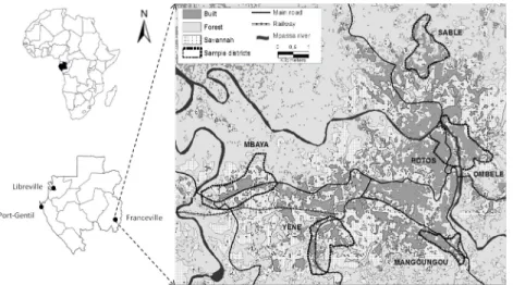
for-est islands in 2014. Trapping took place in houses in six (6) districts of the city of Franceville
(
Fig 1
), including four peripheral districts (Mangoungou, Mbaya Sable and Ye´ne´) and two
cen-tral districts (Ombe´le´ and Potos). It should also be noted that these districts are located along
the main access routes to the city (roads and railways). These districts display many of the
dominant characteristics of the city as a whole in terms of the level of connectivity and the
aggregation of buildings as well as the presence or absence of vegetation. Mbaya and Ye´ne´ are
the two main entry points of the city, by road and railway, respectively. Sable and
Mangoun-gou are more isolated districts. Mbaya is mainly industrial. Potos is the central trade district,
including large storehouses and the main open market [
59
].
Rodent and organ sampling
Rodents were sampled according to a standardized live-trapping protocol as previously
described [
59
]. Live-trapped rodents were brought back to our laboratory, euthanized,
weighed, sexed, measured and autopsied. During autopsy, various organs and tissues,
includ-ing the kidney, liver, brain, lungs, and spleen, were collected. All of these samples were stored
Fig 1. Map of Franceville. Study area and location of the six districts sampled for rodents. https://doi.org/10.1371/journal.pone.0248244.g001
at -80˚C. In this study, all bacteria and protozoa were screened on the liver, except
Leptospira
on the kidney and
Toxoplasma on the brain. While, all viruses were screened in the spleen.
Ethics statements
Trapping campaigns were performed with prior (oral) agreement from local authorities (city
Mayor, district chief and family heads). All sampling procedures were approved by the Ethics
Committee named “Comite´ Nationale d’Ethique pour la Recherche” under the number: Prot
n˚ 0020/2013/SG/CNE. Live-trapped rodents were brought back to our laboratory, euthanized
with a halothane solution and autopsied in accordance with guidelines of the American Society
of Mammalogists [
60
]. None of the rodent species captured in the present study had a
pro-tected status (CITES lists and IUCN).
Specific identification of rodent species
Specific species identification was carried out according to the identification keys provided by
Jean-Marc DUPLANTIER and Violaine NICOLAS following their studies on rodents in
Gabon [
61
–
69
].
Rattus rattus was the only rat species identified in our study. Morphologically, it is easily
distinguishable from
Rattus norvegicus. In addition, the Rattus sample collected in this study
was the subject of a population genetic study validating the identification of
R. rattus [
59
].
Sim-ilarly, the
Mus musculus domesticus specimens included in this study were genetically
identi-fied previously [
53
].
Lemniscomys striatus and Lophuromys sikapusi were identified to the
species level by using morphological identification keys.
Praomys and Mus (Nannomys) were
identified to the subgenus level. Nevertheless, the determination of host species was performed
only in pathogen-positive samples. A fragment of the 16S ribosomal RNA gene (16S rRNA)
was amplified as previously described [
61
] and sequenced under BigDyeTM terminator
cycling conditions. All sequences from this study have been deposited in GenBank under
accession numbers MT256376 to MT256385 for rodent host species and MT677677 to
MT677695 for shrews host species.
DNA and RNA extraction from organs and tissues
Small pieces of the liver, kidney, spleen and brain of rodents were collected and placed
individ-ually in Eppendorf tubes. Total DNA or RNA was extracted with a BioRobot EZ1 system
(Qia-gen, Courtaboeuf, France) using a commercial EZ1 DNA/RNA Tissue Kit (Qiagen) (Qia(Qia-gen,
Courtaboeuf, France) following the manufacturer’s instructions. DNA and RNA were eluted
in 100
μl of TE buffer. DNA was stored at 4˚C until being used for PCR amplification, while
RNA was stored at -20˚C.
Molecular detection of virus, bacterial and protozoan DNA in rodents and
shrews
Virus molecular detection. The following virus families or species were screened by
one-step RT-PCR in RNA extracts from rodent spleens:
Hantavirus spp., Mammarenavirus spp.,
Flavivirus, Paramyxovirus, Lymphocytic choriomeningitis mammarenavirus (LCMV) and Zika
virus (ZIKV). Methodological details and primer sequences are provided in
Table 1
.
Bacteria and protozoan molecular detection. The real-time PCR (qPCR) was performed
to screen all rodent samples using previously reported primers and probes for
Bartonella spp.,
Anaplasmataceae, Coxiella burnetii, Borrelia spp., Rickettsiaceae, Mycoplasma spp., Orientia
Table 1. Method of investigat ion of target ed viruses. Virus familly Target virus (group) Techniqu e Target gene Primer names Sequences (5’-3’) Amplific ation Amplic on References Arenaviridae Mammarenavirus �Nested PCR S ARS16V GGCATWGANCCAAACTGATT 95˚C for 2min, then 40 cycles of 95˚C—30 S, 55˚C-30 S and 72˚C -1 min. Extension 72˚C -5min. Same for the both round 640 bp [ 70 ] ARS1 CGCACCGGGGATCCTAGGC ARS3V CATGACKMTGAATTYTGTGACA 460 bp ARS7C (modified) ATRTGYCKRTGWGTTGG Arenaviridae LCMV qRT-PCR GPC LCMVS GGGATCCTAGGCTTTTTGGAT 95˚C -20 sec, then 45cycles of 95˚C-3 sec 57˚C-30 sec [ 53 ] LCMVAS GCACAATAATGACAATGTTGAT LCMVP- FAM CCTCAAACATTGTCACAATCTGACCCAT Hantaviridae Hantavirus �Convention nal PCR S UHanta F2 GGVCARACWGCHGAYTGG 95˚C 2 m, then 45 cycles of 95˚C -15 S, 52˚C—30 S and 72˚C -1m. 72˚C -1 m 236 bp [ 71 ] UHantaR2 TCITCWGCYTTCATDSTDGA L HAN-L-F2 TGCWGATGCHACIAARTGGTC 95˚C-5 m, then 45 cycles of 96˚C-30 S, 60˚C-35 S and 72˚C -50 S. finish with 72˚C-5min. 388 bp [ 72 ] HAN-L-R2 GCRTCRTCWGARTGRTGDGCAA Paramyxoviridae Paramyxovirus (Respirovirus , Morbillivirus , Henipavirus) One step RT-PCR / semi-nested PCR L RMH-F1 TCITTCTTTAGAACITTYGGNCAYCC 60˚C-1 min for denaturing, 44 to 50˚C-30 min (for RT), 94˚C-2 min, and then 40 cycles of 94˚C-15 s, 48 to 50˚C—30 s, 72˚C-30 s, and final extension 72˚C-7 min For semi-nested : 94˚C-2 min, and then 40 cycles of 94˚C-15 s, 48 to 50˚C—30 s, 72˚C-30 s, and final extension 72˚C-7 min 497 bp [ 73 ] RMH-F2 GCCATATTTTGTGGAATAATHATHAAYGG RMH-R CTCATTTTGTAIGTCATYTTNGCRAA Paramyxovirus (Avulavirus , Rubulavirus) One step RT-PCR / semi-nested PCR L AR-F1 GGTTATCCTCATTTITTYGARTGGATHCA 250 bp [ 73 ] AR-F2 ACACTCTATGTIGGIGAICCNTTYAAYCC AR-R GCAATTGCTTGATTITCICCYTGNAC Pneumoviridae Paramyxovirus (Pneumovirinae) One step RT-PCR / semi-nested PCR L PNE-F1 GTGTAGGTAGIATGTTYGCNATGCARCC 300 bp [ 73 ] PNE-F2 ACTGATCTIAGYAARTTYAAYCARGC PNE-R GTCCCACAAITTTTGRCACCANCCYTC Flaviviridae Zika virus One step RT-qPCR NS5 ZIKV 1086 CCGCTGCCCAACACAAG 95˚C -20 sec, then 45cycles of 95˚C-3 sec 57˚C-30 sec 160 bp [ 74 ] ZIKV 1162c CCACTAACGTTCTTTTGCAGACAT ZIKV 1107-FAM AGCCTACCTTGACAAGCAGTCAGACACTCAA Flaviviridae Flavivirus One step RT-PCR / semi-nested PCR NS5 PF1S TGYRTBTAYAACATGATGGG 45˚C-20 min, 95˚C-2 min, then45 cycles of 95˚C-25 sec, 51˚C-30 sec, 68˚C-30 sec. End 68˚C-5 min Semi-nested : 94˚C-2 min,then45 cycles of 94˚C-25 sec, 5˚C-30 sec, 72˚C-30 sec. End 72˚C-5 min 280 bp 210 bp [ 75 ] PF3S ATHTGGTWYATGTGGYTDGG PF2Rbis GTGTCCCAiCCNGCNGTRTC Oligonucle otide sequences of the prime rs and probes used for virus detection in rodent spleens in this study. �Analys es were performed with cDNA using Superscri pt III following the manufact urer’s instruction s. https://doi. org/10.1371/j ournal.pone .0248244.t001
Piroplasmida., Toxoplasma gondii., and Kinetoplastida (including the Trypanosoma and
Leish-mania genera). The sequences of the primers and probes are shown in
Table 2
. For all systems,
any sample with a cycle threshold (Ct) value of less than 40 Ct was considered positive.
Con-ventional PCR analysis was performed for all qPCR-positive samples using the primers and
conditions described in
Table 2
. The amplification reaction was conducted in a final volume of
25
μl containing 12.5 μl of AmpliTaq Gold master mix, 0.75 μl of each primer [20 μM], 5 μl of
DNA template, and 6
μl of water. The thermal cycling profile consisted of one incubation step
at 95˚C for 15 min, 45 cycles of 30 s at 95˚C, 30 s to 1 min at the annealing temperature
(
Table 2
) and 1 min at 72˚C, and a final extension step of 5 min at 72˚C. Successful
amplifica-tion was confirmed by electrophoresis in a 1.5% agarose gel, and the amplicons were
completely sequenced on both strands.
Quantitative real-time PCR was performed on the CFX96 Real-Time system (Bio-Rad) with
the Roche LightCycler 480 Probes Master Mix PCR kit (Roche Applied Science, Mannheim,
Germany). For each assay, DNA extracts of the targeted bacteria or parasites were used as
posi-tive controls and distilled water as negaposi-tive control (
S1 Table
). For the viral families
Bunyaviri-dae and ArenaviriBunyaviri-dae, the positive controls used were plasmids, designed during the
PREDICT project. For
Flaviviridae, we used the Yellow fever virus RNA (vaccinal strain 17D)
and RNA transcripts from mumps, measles, and respiratory syncytial viruses, for
Paramyxo-viridae. Conventional PCR was performed in an automated DNA thermal cycler (GeneAmp
PCR Systems Applied Biosystems, Courtaboeuf, France). Sequencing analyses were performed
on the ABI Prism 3130XL Genetic Analyzer (Applied Biosystems, Thermo Fisher Scientific,
France) using the DNA sequencing BigDye Terminator V3.1 Cycle Sequencing Kit (Applied
Biosystems, Foster City, CA, USA, Perkin-Elmer) according to the manufacturer’s
instruc-tions. The BigDye products were purified on Sefadex G-50 Superfine gel filtration resin
(Cytiva, Formerly GE Healthcare Life Science, Sweden).
The sequences were compared to sequences available in the GenBank database using the
BLAST algorithm (
http://blast.ncbi.nlm.nih.gov/Blast.cgi
).
Multispacer sequence typing (MST) genotyping of
Coxiella burnetii. The multispacer
sequence typing (MST) method was used for
Coxiella burnetii genotyping. For this purpose,
five (5) different spacers, which were previously described [
80
], were selected and amplified
(Cox 2, 5, 18, 22, 37). Conventional PCR was performed as described below with a
hybridiza-tion temperature of 59˚C. Then, the web-based MST database (
https://ifr48.timone.univ-mrs.
fr/mst/coxiella_burnetii/groups.html
) was used for MST identification.
Phylogenetic and statistical analyses
Phylogenetic analysis. The obtained sequences were analyzed using ChromasPro version
1.3 (Technelysium Pty, Ltd., Tewantin, Queensland, Australia) for assembly and were aligned
with other sequences of targeted bacteria or parasite species from GenBank using
CLUS-TALW, implemented in BioEdit v7.2 [
89
]. Phylogenetic trees were constructed with MEGA
software v.7 [
90
]. The maximum likelihood method based on the Hasegawa-Kishino-Yano
model (HKY) was used to infer the phylogenetic analysis with 500 bootstrap replicates.
Statistical analysis. Statistical analysis was performed with R software V3.2.5 [
91
] using
chi-square/Fisher’s exact tests for data comparisons between the prevalence of infected rodents
for all parasites according to habitat type. A
p-value� 0.05 was considered to be significant.
General linear mixed models (GLMMs) run using the lme4 package [
92
] were also
employed to examine the potential determinants of parasite richness (number of parasite
spe-cies in a host), as reported in a previous similar study [
78
]. We assumed a Poisson distribution
for the parasite richness data. The sampling site was considered a random factor, and other
Table 2. Method of investigation of targeted bacteria and protozoa. Target Organis m Target gene Technique Primer names SEQUENC ES (5’-3’) Annealing Tempera ture Amplicon Reference Anaplasmatacae 23S Broad-range qPCR TtAna_F TGACAGCGTACCTTTTGCAT 55 190 bp [ 76 ] TtAna_R GTAACAGGTTCGGTCCTCCA TtAna_P 6FAM-GGATTAGACCCGAAACCAAG Broad-ran ge conventiona l PCR Ana23S-212F ATAAGCTGCGGGGAATTGTC 58 650 bp [ 76 ] Ana23S-7 53R TGCAAAAGGTACGCTGTCAC(for sequencing only) Ana23S-908r GTAACAGGTTCGGTCCTCCA Bartonella sp ITS (Intergenic 16S-23S) Broad-range qPCR Barto_ITS 3_F GATGCCGGGGAAGGTTTTC 60 104 bp [ 77 ] Barto_ITS3_ R GCCTGGGAGGACTTGAACCT Barto_ITS 3_P 6FAM-GCGCGCGCTTGATAAGCGTG Broad-ran ge conventiona l PCR Urbarto1 CTTCGTTTCTCTTTCTTCA 50 733 bp [ 78 ] Urbarto2 CTTCTCTTCACAATTTCAAT Coxiella burnetii IS1111A Broad-range qPCR CB_IS1111 _0706F CAAGAAACGTATCGCTGTGGC 60 154 bp [ 79 ] CB_IS1111_ 0706R CACAGAGCCACCGTATGAATC CB_IS11 11_0706P 6FAM-CCGAGTTCGAAACAATGAGGGCTG IS30A Broad-range qPCR CB_IS30A _3F CAAGAAACGTATCGCTGTGGC 60 154 bp [ 77 ] CB_IS30A_3R CACAGAGCCACCGTATGAATC CB_IS30 A_3P 6FAM-CCGAGTTCGAAACAATGAGGGCTG Spacer 2 Species-spec ific PCR Cox2 F CAACCCTGAATACCCAAGGA 59 358 bp [ 80 ] Cox2 R GAAGCTTCTGATAGGCGGGA Spacer 5 Species-spec ific PCR Cox5 F CAGGAGCAAGCTTGAATGCG 59 344 bp [ 80 ] Cox5 R TGGTATGACAACCCGTCATG Spacer 18 Species-spec ific PCR Cox18 F CGCAGACGAATTAGCCAATC 59 556 bp [ 80 ] Cox18 R TTCGATGATCCGATGGCCTT Spacer 22 Species-spec ific PCR Cox22 F GGGAATAAGAGAGTTAGCTCA 59 340 bp [ 80 ] Cox22 R CGCAAATTTCGGCACAGACC Spacer 37 Species-spec ific PCR Cox37 F GGCTTGTCTGGTGTAACTGT 59 322 bp [ 80 ] Cox37 R ATTCCGGGACCTTCGTTAAC Leptospira sp 16S Broad-range qPCR Lepto_F CCCGCGTCCGATTAG 58 88 bp [ 81 ] Lepto_R TCCATTGTGGCCGRACAC Lepto_P 6FAM-CTCACCAAGGCGACGATCGGTAGC LipL32 Broad-ran ge conventiona l PCR LipL32 F ATCTCCGTTGCACTCTTTGC 58 474 bp [ 82 ] LipL32 R ACCATCATCATCATCGTCCA Borrelia sp 23S Broad-range qPCR TTB23s F CGATACCAGGGAAGTGAAC 60 148 bp [ 78 , 83 ] TTB23sR ACAACCCYMTAAATGCAACG TTB23SP 6-FAM-TTTGATTTCTTTTCCTCAGGG-TAMRA Mycoplasma sp ITS Broad-range qPCR Mycop_ITS _F GGGAGCTGGTAATACCCAAAGT 60 114 bp [ 84 ] Mycop_IT S_R CCATCCCCACGTTCTCGTAG Mycop_ITS _P 6FAM-GCCTAAGGTAGGACTGGTGACTGGGG (Continued )
Table 2. (Continued ) Target Organis m Target gene Technique Primer names SEQUENC ES (5’-3’) Annealing Tempera ture Amplicon Reference Rickettsia sp gltA (CS) Broad-range qPCR RKND03_F GTGAATGAAAGATTACACTATTTAT 60 166 bp [ 77 , 79 ] RKND0 3_R GTATCTTAGCAATCATTCTAATAGC RKND03 P 6-FAM-CTATTATGCTTGCGGCTGTCGGTTC Orientia_Occidentia sp 23S Broad-range qPCR OcOr23 S-F TGGGTGTTGGAGATTTGAGA 55 140 This study OcOr23S-R TGGACGTACCTATGGTGTACCA OcOr23S-P FAM-GCTTAGATGCATTCAGCAGTT Occidentia sp sca Broad-range qPCR OMscaA-F AGTTTAAAATTCCCTGAACCACAATT 55 240 [ 78 ] OMscaA-R ACTTCCAAACACTCCTGAAACTATACTTG OMscaA-P FAM-TGAAGTTGAAGATGTCCCTAATAGT Streptobacillus moniliformis gyrB Broad-range qPCR Smoni-g yrB-F AGTTTAAAATTCCCTGAACCACAATT 60 96 bp [ 85 ] Smoni-gyrB-R ACTTCCAAACACTCCTGAAACTATACTTG Smoni-g yrB-P 6FAM-TCACAAACTAAGGCAAAACTTGGTTCATCTGAG Yersina pestis caf1 species-speci fic qPCR YPcaf-S TACGGTTACGGTTACAGCAT 45 240bp [ 86 ] YPcaf-A GGTGATCCCATGTACTTAACA YPcaf-1 6-FAM-ACCTGCTGCAAGTTTACCGCCTTTGG Toxoplasma gondii ITS1 Broad-range qPCR Tgon_ITS1_F GATTTGCATTCAAGAAGCGTGATAGTA 60 333 bp [ 87 ] Tgon_ITS 1_R AGTTTAGGAAGCAATCTGAAAGCACATC Tgon_ITS1_ P 6-FAM-CTGCGCTGCTTCCAATATTGG Piroplasma sp 5 ,8S Broad-range qPCR 5,8s-F5 AYYKTYAGCGRTGGATGTC 60 40 bp [ 78 ] 5,8s-R TCGCAGRAGTCTKCAAGTC 5,8s-S 6-FAM-TTYGCTGCGTCCTTCATCGTTGT 18S Broad-ran ge conventiona l PCR piro18S-F3 GTAGGGTATTGGCCTACCG 58 969 bp piro18S-R3 AGGACTACGACGGTATCTGA Leishmania sp 18S SSU Broad-range qPCR F GGTTTAGTGCGTCCGGTG 60 75 bp [ 88 ] R ACGCCCCAGTACGTTCTCC Probe leish S FAM-CGGCCGTAACGCCTTTTCAACTCA Kinetoplastidea 28S LSU (24 alpha) Broad-range qPCR F LSU 24a AGTATTGAGCCAAAGAAGG 60 200 bp [ 88 ] R LSU 24a TTGTCACGACTTCAGGTTCTAT P LSU 24a 6FAM-TAGGAAGACCGATAGCGAACAAGTAG Broad-ran ge conventiona l PCR F2 28S ACCAAGGAGTCAAACAGACG 58 ~ 550 bp [ 88 ] R1 28S ACGCCACATATCCCTAAG Trypanosoma sp 5 .8 S rRNA Broad-range qPCR F 5,8S CAACGTGTCGCGATGGATGA 60 83 bp [ 88 ] R 5,8S ATTCTGCAATTGATACCACTTATC P 5,8S 6-FAM-GTTGAAGAACGCAGCAAAGGCGAT 28S Broad-ran ge conventiona l PCR F2 28S ACCAAGGAGTCAAACAGACG 58 ~ 550 bp [ 88 ] R1 28S ACGCCACATATCCCTAAG Oligonu cleotide sequences of primers and probes used for real-tim e PCR and conven tional PCR to screen bacteria and protozoans in this study. https:// doi.org/10.1371 /journal.pone. 0248244.t0 02
factors, including host factors (species, sex, weight, body mass), status (native vs. invasive),
trap location (inside vs. outside the door), habitat type (central districts, peripheral districts,
vegetal areas) and seasons (dry season and rainy season), were considered fixed effects (
S2
and
S3
Tables). The significance of the interactions of different effects was estimated by using the
Akaike information criterion (AIC) for model selection. AIC changes were evaluated when
model parameters were modified (added or removed). Full-model averages, available in the
MuMIn package [
93
], were used for AIC estimation. The best model showed a null
ΔAICC.
Results
Rodents sampled for this study
A total of 198 small mammals were captured including 49 in Mbaya, 19 in Mangoungou, 19 in
Ombe´le´, 15 in Potos, 25 in Sable, 18 in Ye´ne´ and 53 in vegetative areas (
S2
and
S3
Tables). The
captured animals included two (2) invasive species of rodents,
Rattus rattus (N = 54) and Mus
musculus domesticus (N = 29), five (5) native rodent taxa, Lophuromys sp. (N = 27),
Lemnisc-omys striatus (N = 27), PraLemnisc-omys sp. (N = 17), Mus (NannLemnisc-omys) sp. (N = 22) and CricetLemnisc-omys sp.
(N = 3), and shrews (N = 19).
According to the three types of established habitats, small mammals were distributed as
fol-lows 29 rodents (3
Lemniscomys striatus, 2 M. m. domesticus, 1 Praomys sp., 3 Mus (Nannomys)
sp. and 20
R. rattus) and 5 shrews in central districts; 102 rodents (3 Cricetomys sp, 8 Le.
stria-tus, 7 Lophuromys sp, 27 M. m. domesticus, 17 Mus (Nannomys) sp., 6 Praomys sp., 34 R.
rat-tus) and 9 shrews in peripheral districts and 48 rodents (16 Le. striatus, 20 Lo. sikapusis, 2 Mus
(
Nannomys) sp., 10 Praomys sp.) and 5 shrews in forest-savannah areas (
Table 3
).
Bacterial, protozoan and viral nucleic acids detected in rodents and shrews
All rodents were found negative for all viral pathogens screened in the spleen by conventional
PCR and qPCR, including
Hantavirus spp., Mammarenavirus spp., Flavivirus, Paramyxovirus,
Lymphocytic choriomeningitis mammarenavirus (LCMV) and Zika virus (ZIKV). Similarly, all
rodents were found negative by qPCR on tissues for several bacteria and protozoa, specifically
Borrelia sp, Leishmania sp, Mycoplasma sp, Orientia sp, Occidentia sp, Streptobacillus
monili-formis, Rickettsia sp and Yersinia pestis. In contrast, 49/198 (24,7%) rodents were positive for 8
of the 16 pathogens (bacteria and protozoans) tested via qPCR. In total, 7 genera of pathogenic
microorganisms were identified, including bacterial
Anaplasma spp. (8.1%; 16/198), Bartonella
spp. (6.6%; 13/198),
Coxiella burnetii (5.1%; 10/198), and Leptospira spp. (3.5%; 7/198). The
protozoans that we identified included
Trypanosoma sp (7%; 14/198), Piroplasma sp (1%; 2/
198) and
Toxoplasma gondii (0.5%; 1/198) (
Table 3
). All microorganisms were detected in the
liver samples except for
Leptospira spp. and Toxoplasma gondii, which were only detected in
the kidney and brain, respectively.
Multiple infections (i.e., many infectious agents in the same organ of an individual rodent)
were found in 11 rodents (5.5%), including 10 double infections (5%) and one triple infection
(0.5%).
Rattus rattus and Le. striatus presented the highest carriage rate for all of the identified
pathogens, including 5 out of 7 infectious agents, while
M. m. domesticus appeared to harbor
the fewest pathogens (1/7). Other species carried between 3 and 4 pathogens (
Table 3
).
Phylogenetic analysis for the taxonomic description of detected pathogens
Bartonella. The sequencing of 733 bp of the ITS gene from the DNA extracts of 13
qPCR-positive individuals revealed five sequences of
Bartonella ranging from 690 to 722 bp
Table 3. Bacteria and protozoa identified in Franceville rodents. Pathogen screening (qPCR positive individual number)
Genotype founded Rodent species
Cricetomys sp. (N = 3) Lemniscomys striatus (N = 27) Lophuromys sp. (N = 27) Mus m. domesticusŦ (N = 29) Mus Nannomys sp. (N = 22) Praomys sp. (N = 17) Rattus rattusŦ (N = 54) Shrews (N = 19) Central districts (Potos and Ombe´le´ districts) N1 = 34 Bartonella spp. (1) Bartonella elizabethae 0 0 0 0 0 1/54 (1.8%) 0 Anaplasma spp. (1) Candidatus Anaplasma gabonensis 0 0 0 0 0 0 1/54 (1.8%) 0 Coxiella burnetii - 0 0 0 0 0 0 0 0 Leptospira spp. - 0 0 0 0 0 0 0 0 Piroplasma - 0 0 0 0 0 0 0 0 Trypanosoma spp. (8) Trypanosoma congolensis riverine forest /Trypanosoma brucei brucei / Trypanosoma otospermophili 0 2/27 (7.4%) 0 0 1/22 (4.55%) 0 5/54 (9.3%) 0 Toxoplasma gondi (1) Toxoplasma gondi 0 1/27 (3.7%) 0 0 0 0 0 0 Peripheral districts (Mang� -Mbaya-Ye´ne´ and Sable
districts) N2 = 111 Bartonella spp. (3) Bartonella massiliensis 2/3 (67%) 0 1/27 (3.7%) 0 0 0 0 0 Anaplasma spp. (8) Candidatus Anaplasma gabonense 0 4/27 (14.8%) 0 0 0 1/17 (5.88%) 2/54 (3.7%) 1/19 (5.3%) Coxiella burnetii (3) Coxiella burnetii MST group 20 0 1/27 (3.7%) 0 0 1/22 (4.55%) 0 1/54 (1.8%) 0 Leptospira spp. (3) Lepstospira kirschneri 0 0 0 0 0 0 1/54 (1.8%) 2/19 (10.6%) Piroplasma (1) Theileria sp. 0 1/27 (3.7%) 0 0 0 0 0 0 Trypanosoma spp. (6) Trypanosoma congolensis riverine forest 1/3 (33%) 0 0 1/29 (3.45%) 1/22 (4.55%) 0 3/54 (5.6%) 0 Toxoplasma gondi - 0 0 0 0 0 0 0 0 Vegetation areas (Forest and savannah fragments) N3 = 53 Bartonella spp. (9) Candidatus Bartonella gabonensis 0 0 9/27 (33.3%) 0 0 0 0 0 Anaplasma spp. (7) Candidatus Anaplasma gabonense 0 4/27 (14.8%) 1/27 (3.7%) 0 0 2/17 (11.8%) 0 0 Coxiella burnetii (7) Coxiella burnetii 0 4/27 (14.8%) 2/27 (7.4%) 0 0 1/17 (5.88%) 0 0 Leptospira spp. (4) L. borgpetersenii 0 0 4/27 (14.8%) 0 0 0 0 0 Piroplasma (1) Theileria sp. 0 0 0 0 0 1/17 (5.88%) 0 0 Trypanosoma spp. - 0 0 0 0 0 0 0 0 Toxoplasma gondi - 0 0 0 0 0 0 0 0
The infectious agents identified and described in this study and the rodents associated with them. One hundred and ninety-eight (198) rodents, collected in N1, N2 and
N3 were analyzed by qPCR.
Ŧindicates invasive rodents.
�Mang represents the Mangoungou district.
Lophuromys sp. hosts showed that the most closely related species was Bartonella
queenslan-densis (GenBank:
EU111769.1
), which presented the highest score and a percentage of identity
of 84–86% (611/721, 546/634, 550/635). This percentage of identity below 95% and the fact
that all these three sequences were grouped in the same cluster (with 99% of identity between
each other) in the phylogenetic tree suggested that the obtained
Bartonella pathogen
repre-sented a new species, an undescribed species. We provisionally named this probable new
geno-type
Candidatus Bartonella gabonensis. The fourth sequence obtained from a Cricetomys sp.
rodent matched the
B. massiliensis OS23 and OS09 strains (
HM636450
and
HM636449
) with
96.7% (699/723) and 96.1% (700/728) identity, respectively. The last sequence, obtained from
R. rattus, matched Bartonella sp. ’Tel Aviv’ of the Bartonella elizabethae complex (GenBank:
CP031843.2
) with 100% (690/690) identity. It is referred to here as
B. elizabethae (
Fig 2
).
Anaplasma. Among 16 qPCR-positive individuals, six sequences ranging from 623 to 683
bp (GenBank: MT269268 to MT269273) were obtained after the sequencing of the
Anaplasma
23S rRNA gene [
76
,
94
]. BLAST analysis of these sequences showed identities with
A.
phagocy-tophilum (
KM021418
) ranging from 91% to 92% (578/633, 606/659, 607/659, 606/660, 607/
659, 607/659). The percentage of identity below 95% suggests that the obtained pathogen is a
new or undescribed species, with similarity to
Anaplasma phagocytophilum. However, the
dis-similarity between the
Anaplasma sequences, as shown in our data (
Fig 3
), could also suggest
Fig 2. Taxonomic tree and description of the identifiedBartonella. Phylogenetic tree of Bartonella spp. identified in
rodents in Franceville. The evolutionary history was inferred by using the Maximum Likelihood method based on the Hasegawa-Kishino-Yano model. The analysis involved 36 nucleotide sequences. All positions containing gaps and missing data were eliminated. There were a total of 189 positions in the final dataset. Evolutionary analyses were conducted in MEGA7. Sequences obtained in this study are indicated in bold. The hosts are indicated after the underscore.
that the amplified genetic material would come from organisms of a different genus. Finally,
not being able to conclude on the basis of our results, we refer to it here as Uncultured
Ana-plasma sp.
Coxiella burnetti. The genotyping of
Coxiella burnetii from 10 qPCR-positive individuals
via MST analysis showed the following profile allele codes: 3–2–6–5–4, corresponding to Cox
2—Cox5—Cox 18- Cox22—Cox37, respectively. This profile identified MST group 20. This
genotype has been found in Europe and the United States [
80
] and is associated with human
and animal disease. The same genotype, MST20 was also found on domestic animal ticks in
Ethiopia in Africa [
95
]. In Franceville,
C. burnetii MST group 20 was found in all the samples
tested from the five rodent species mentioned
Praomys sp, R. rattus, Mus Nannomys sp, Le.
striatus and Lo. sikapusi.
Leptospira. Seven samples were positive in qPCR screening of the
16S rRNA gene of
Lep-tospira sp. The sequencing of a portion of the LipL 32 gene from the DNA extracts of 7
qPCR-positive individuals revealed two
Leptospira sequences (MT274303 and MT274304). Sequence
MT274303 was 99.1% (442/446; 444/448) similar to both
Leptospira borgpetersenii serovar
Hardjo (
CP033440.1
) and
Leptospira weilii (
AY461930.1
), identified from
Lophuromys sp.
Sequence MT274304 was 100% (474/474) similar to both
Leptospira kirshneri (
JN683896.1
)
and
Leptospira interrogans (
KC800991.1
), identified in the
Crocidura goliath shrew
(
MT256384.1
).
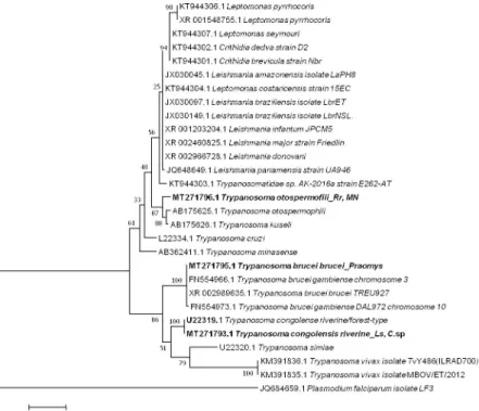
Trypanosoma. Five sequences of
Trypanosoma ranging from 464 to 571 bp were obtained
(GenBank: MT271793 to MT271797) after the sequencing of 550bp of the
28 S gene from 14
qPCR-positive individuals identified when screening for
Trypanosoma spp. (10 positive
indi-viduals) and the
Kinetoplastidae class (4 positive individuals). Two presented as T. congolense
riverine/forest-type (
U22319
) (2/198) with 99% (570/571) of identity from
Le. striatus and
Fig 3. Taxonomic tree and description of the identifiedCandidatus Anaplasma gabonense. Phylogenetic tree of
Anaplasma species identified in rodents of Franceville. The evolutionary history was inferred by using the Maximum
Likelihood method based on the Hasegawa-Kishino-Yano model. The analysis involved 24 nucleotide sequences. All positions containing gaps and missing data were eliminated. There were a total of 420 positions in the final dataset. Sequences obtained in this study are indicated in bold. Evolutionary analyses were conducted in MEGA7. The hosts are indicated after the underscore.Ls, Lemniscomys striatus; Rr, Rattus rattus.
Cricetomys sp; one, from Praomys sp (1/198), showed 100% (522/522) identity to both T. brucei
brucei (
XR_002989635
) and
T. brucei gambiense (
FN554966.1
); and two others from
R. rattus
and Mus Nannomys (
AB190228
) (2/198) were identified as
T. otospermophili, with 97% (453/
467) identity (
Fig 4
).
Theileria. The two samples that were positive according to the pan-
Piroplasma 5.8S qPCR
analysis and were sequenced (GenBank: MT269266 and MT269267) were shown 98% (869/
883, 868/881) identity to
Theileria sp strain HaD-2019a (
MK484070.1
) found in Senegalese
rodents (
Fig 5
). These two infected rodents were
Le. striatus and Praomys sp.
Toxoplasma gondii. Of the tested brain samples, only one (0.5%, N = 198) was positive in
the qPCR screening of
T. gondii according to the ITS1 gene, with a Ct value of 35.1. However,
it could not be identified; the sample came from an
Le. striatus rodent.
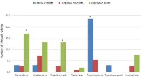
Habitats and pathogens in rodents
We categorized the sampling areas into three groups as follows: central districts (Ombe´le´,
Potos), peripheral districts (Mbaya, Ye´ne´, Sable, and Mangoungou) and non urban areas of
vegetation (savannah-forest) (
Fig 6
). The prevalence of infection by the pathogens in each
group was 32.3% (11/32), 21.6% (24/111) and 52.8% (28/53) for the central districts, peripheral
districts and vegetated areas, respectively.
In terms of overall prevalence, a significant difference was found between the average
prev-alence in the infected rodents in the three habitat types (X-squared = 16.659, df = 2,
P < 0.0002413). The residual value of the X-squared test showed that the difference was
attrib-uted to the vegetated areas. The rodents from vegetated areas showed the highest infection
Fig 4. Taxonomic tree and description of the identifiedTrypanosoma sp. Maximum likelihood tree of Trypanosoma
species identified in rodents in Franceville. Sequences obtained in this study are indicated in bold. The evolutionary history was inferred by using the Maximum Likelihood method based on the Hasegawa-Kishino-Yano model. The analysis involved 30 nucleotide sequences. All positions containing gaps and missing data were eliminated. There were a total of 415 positions in the final dataset. Evolutionary analyses were conducted in MEGA7. The hosts are indicated after the underscore.Ls, Lemniscomys striatus; C. sp., Cricetomys sp.; MN, Mus Nannomys sp.
prevalence in Franceville. Similarly, at the pathogen group scale (only for pathogens found in
more than 5 rodents), the rodents from vegetated areas showed the highest prevalence of
Fig 5. Taxonomic tree and description of the identifiedTheileria sp. Phylogenetic tree of Theileria species identified
in rodents in Franceville. Sequences obtained in this study are indicated in bold. The evolutionary history was inferred by using the Maximum Likelihood method based on the Hasegawa-Kishino-Yano model. The analysis involved 36 nucleotide sequences. All positions containing gaps and missing data were eliminated. There were a total of 749 positions in the final dataset. Evolutionary analyses were conducted in MEGA7.Ls, Lemniscomys striatus.
https://doi.org/10.1371/journal.pone.0248244.g005
Fig 6. Histogram of the prevalence of infections. Richness of pathogens in Franceville rodents by habitat type. Three
habitat types were identified in this study: the central district with little vegetation, peripheral districts with abundant vegetation around dwellings and vegetation areas (mixture of only savannah and forest).�indicates significantly
different richness at p <0,05.
infection by
Bartonella sp (P <0.001) and C. burneti (P <0.004) pathogens (
Fig 6
). However, a
different result was obtained in rodents from central districts, which showed a significantly
higher rate of infection by
Trypanosoma pathogens than the rodents coming from the 2 other
habitats (
P< 0.002).
Host factors and pathogens in rodents
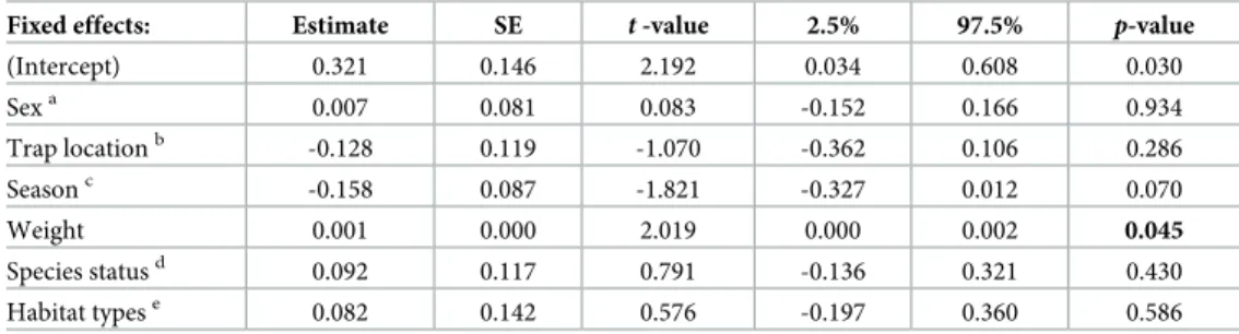
GLMM analysis revealed a strong association effect between the parasite richness and body
mass of host rodents (
t-value = 0.791, P< 0.0449) (
Table 4
). Parasitic richness was positively
correlated with the weight of the rodents. Conversely, no association could be identified
between parasitic richness and the other factors tested among the rodents in Franceville.
Discussion
Rodents are hosts of numerous zoonotic diseases caused by bacteria, protozoans, or viruses all
around the world [
96
]. Luis et
al., 2013 [
1
] noted the importance of paying serious attention to
rodents because they are the most diverse mammals and carry many pathogens responsible for
emerging viral zoonoses. In Gabon, over the last five years, some studies have focused
specifi-cally on rodent viruses [
52
,
53
] and plasmodium parasites [
54
]. Herein, we broaden the
spec-trum of these studies by investigating a wide range of bacteria, protozoa and viruses in the
rodents of Franceville in Gabon.
We did not identify the presence of viruses in these rodents. Several hypotheses can be put
forth to explain this result. We suggest that a low viral load could explain the failure to detect
the targeted viral fragments. In such instances, the use of high-throughput sequencing,
partic-ularly next generation sequencing methods [
97
], could be more effective and efficient, as
reported by Diagne et
al., 2017 [
27
]. Another hypothesis is that the failure to detect pathogens
may be due to the absence in our sample of rodent species that are reservoirs for the targeted
pathogens. For example,
Mastomys natalensis and Mastomys erythroleucus, which are LHF
res-ervoirs whose distribution area includes Gabon, were missing from our sample. Nevertheless,
R. rattus and M. m. domesticus are reservoirs of many pathogens, including Hantavirus and
Lymphocytic choromeningite virus (LCMV), respectively, but we did not succeed in identifying
Table 4. Factors and parasitic richness association.
Fixed effects: Estimate SE t -value 2.5% 97.5% p-value
(Intercept) 0.321 0.146 2.192 0.034 0.608 0.030 Sexa 0.007 0.081 0.083 -0.152 0.166 0.934 Trap locationb -0.128 0.119 -1.070 -0.362 0.106 0.286 Seasonc -0.158 0.087 -1.821 -0.327 0.012 0.070 Weight 0.001 0.000 2.019 0.000 0.002 0.045 Species statusd 0.092 0.117 0.791 -0.136 0.321 0.430 Habitat typese 0.082 0.142 0.576 -0.197 0.360 0.586
Evaluation of the Poisson generalized linear mixed models fitted to estimate the host factors and parasitic richness association.
The reference categories corresponding to: a male, b indoor, c rainy season, d native and e vegetal areas. https://doi.org/10.1371/journal.pone.0248244.t004