Advances in molecular probe-based labeling tools and their
application to multiscale multimodal correlated microscopies
The MIT Faculty has made this article openly available.
Please share
how this access benefits you. Your story matters.
Citation
Ellisman, M. H. et al. “Advances in Molecular Probe-Based Labeling
Tools and Their Application to Multiscale Multimodal Correlated
Microscopies.” Journal of Chemical Biology 8.4 (2015): 143–151.
As Published
http://dx.doi.org/10.1007/s12154-015-0132-6
Publisher
Springer Berlin Heidelberg
Version
Author's final manuscript
Citable link
http://hdl.handle.net/1721.1/105352
Terms of Use
Creative Commons Attribution-Noncommercial-Share Alike
REVIEW
Advances in molecular probe-based labeling tools
and their application to multiscale multimodal correlated
microscopies
M. H. Ellisman1,2,3&T. J. Deerinck1&K. Y. Kim1&E. A. Bushong1&S. Phan1&
A. Y. Ting4&D. Boassa1
Received: 19 February 2015 / Accepted: 29 March 2015 / Published online: 22 May 2015 # Springer-Verlag Berlin Heidelberg 2015
Abstract The need to determine the precise subcellular dis-tribution of specific proteins and macromolecular complexes in cells and tissues has been the major driving force behind the development of new molecular-genetic and chemical-labeling approaches applicable to high-resolution, correlated, multidi-mensional microscopy. This short review is intended to pro-vide an overview of recently developed and widely used elec-tron microscopy (EM)-compatible probes, including tetracysteine tags, mini singlet oxygen generator (MiniSOG), time-specific tag for the age measurement of proteins (TimeSTAMP) with MiniSOG, and enhanced ascorbate peroxidase (APEX). We describe how these highly specific and genetically introduced EM probes are now used, in con-junction with lower resolution light microscopic methods, to obtain wide field or dynamic records of live preparation or of large maps in 3D using recently developed laboratory-scale X-ray microscopes. The article is intended to enable researchers through a high-level view of the toolbox of labels available today for studies aiming to analyze dynamic subcellular and molecular processes in cell culture systems as well as in ani-mal tissues—and ultimately allow investigators to determine the precise location of macromolecular complexes by EM.
Keywords CLEM . MiniSOG . APEX . Tetracysteine . Photooxidation . TimeSTAMP
Introduction
Microscopy has played a central role in biomedical research, due in large part to the vast range of scales over which it is capable of delivering information. It is arguably the most im-portant tool in biomedical science. Even today, microscopy continues to experience a tremendous expansion in its appli-cation to research, catalyzed in large part by the introduction of genetically encodable fluorescent proteins (FPs) to live-cell imaging [11,21] and the synergistic development of new in-strumentation and imaging modalities, leading to a wealth of new discoveries in cell biology and medicine. Key to the suc-cess and widespread adoption of these technologies is their ease of use, robustness, and consistency of results obtained. Efforts to expand, improve, and simplify the use of FP re-porters have spawned rapid growth of research and develop-ment of new probes systems with broadening impact on basic and biomedical research. A related undertaking involves ex-tending these types of molecular-genetic probes to other forms of microscopy, particularly electron microscopy (EM) (for review, see [8]). The ultimate goal is that, with the greatly improved resolution afforded by EM, these new types of molecular-genetic labels will support advances in quantitative spatial proteomics in small sub-volumes by enabling direct in situ visualization of molecules and macromolecular com-plexes in cells and tissues. These new types of genetic labels promise to deliver unprecedented in situ spatio-proteomic in-formation in cells and tissues.
All these efforts are meant to impact bioscience, as there is a general need to improve the resolution and fidelity of protein labeling and macromolecular recognition achievable by * D. Boassa
1 National Center for Microscopy and Imaging Research and Center
for Research on Biological Systems, University of California, San Diego, La Jolla, San Diego, CA 92093, USA
2
Department of Neurosciences, University of California, San Diego, La Jolla, San Diego, CA 92093, USA
3
Salk Institute for Biological Studies, San Diego, CA, USA
4 Department of Chemistry, Massachusetts Institute of Technology,
electron microscopy and improve and streamline methods available for multiscale, multicolor, multimicroscope modali-ty imaging. A current focus in the biology field is to continue to develop and apply molecular probes for correlated imaging to propel multiresolution analysis of the structure and function of proteins in supramolecular complexes underlying cellular processes. The expectation is to enable biomedical researchers to follow small molecular components, requiring a resolution of <10 nm, and support their ability to do supramolecular imaging throughout a continuous field of view as great as 100μm wide. This range is required to observe and map molecular constituents in their cellular and tissue context.
Despite research efforts to decode entire genomes and re-cent advances in genomic-screening technologies for uncovering mutations in various genes linked to different forms of diseases, we are often left with a profound gap in understanding their final product of regulation and expression. Particularly in complex organisms, we often lack knowledge of the function and distribution of many cellular proteins. Furthermore, knowing the unique expression dynamics and distribution of each protein from a systems biology point of view will help understand their roles in normal physiology and/or pathological states associated with diseases. A success-ful approach we are using to try to fill this gap is genetically encoded markers for proteins to follow dynamics in light mi-croscope systems, then high-resolution imaging by electron microscopy. We can count on an expanding molecular probes toolbox to assess protein location and function, considering, for every target protein, the appropriate tag and methodology. For example, while most proteins tolerate tags, some proteins require specific or smaller tags resistant to proteases or differ-ent subcellular microenvironmdiffer-ents. Importantly, the probes reviewed here (Fig.1) are appropriate to the capabilities of sophisticated multiscale imaging tools that can be used in a complementary manner to examine the entirety of events as-sociated with complex biological questions. The ultimate goal is to visualize macromolecules of interest with high spatial and temporal resolution, potentially generating the 4D maps of their distribution and interactions within a given tissue or organism.
Common challenges
EM-level mapping of the distribution of specific proteins in situ has proven vital to the understanding of many cellular functions, but most of the methods previously developed have fallen short of the resolution afforded by electron microscopy and suffer from a range of seemingly intractable limitations. These limitations include procedures that do not adequately preserve cellular and supramolecular ultrastructure and native protein distribution, lack the capability to deliver uniform 3D labeling, and generally provide suboptimal resolution of the
contrasting agent. All of these limitations greatly limit the ability to interpret the imaging data. Compounding this is the fact that many of these approaches involve complex pro-cedures and specialized instrumentation that made their use challenging technically. For example, antibody-based protein detection, the most widely used approach, usually requires a compromise in chemical fixation to maintain antibody-epitope binding and the use of permeabilizing detergents to facilitate access of the labeling reagents, both of which signif-icantly perturb cellular ultrastructure. The introduction and refinements of molecular-genetic approaches for EM-level protein localization allowed incorporation of tags and labels directly into the target protein and offered the ability to over-come many of the aforementioned limitations, in addition to facilitating excellent correlated light and EM. Below, we re-view the probes currently available to the research community and examples of their application in addressing biomedical challenges.
Tetracysteine tags
Fluorescent labeling of tetracysteine-tagged (4Cys) proteins followed by photooxidation of diaminobenzidine (DAB) was one of the first examples of a genetically encoded light microscopy (LM)/EM-compatible approach [9] and has been very successful in a host of diverse applications. Tetracysteine tags are short encodable peptides having the general sequence Cys-Cys-Xaa-Xaa-Cys-Cys and can be fused to either the amino or carboxyl terminus of the protein of interest or can be incorporated into internal sites. Tetracysteine tags exhibit sub-picomolar affinity for a variety of membrane-permeable biarsenicals such as the green fluorescent Fluorescein Arsenic Hairpin-binder (FlAsH) (ex 508, 528 em) and the red fluores-cent Resorufin-based AsH (ReAsH) (ex 593, 608 em), and the latter is capable of photooxidizing DAB to create an EM-visible reaction product. Exhaustive screening of a clone library of internal and flanking sequences around the tetracysteine motif in living mammalian cells revealed the consensus sequence FLNCCPGCCMP exhibits the highest affinity, stability, and quantum efficiency of the 4Cys-biarsenical complex thus far. One powerful application of the 4Cys labeling methodology is to temporally and spatially distinguish new vs. old proteins using spectrally distinct fluorescent biarsenical ligands that on-ly fluoresce when bound to the geneticalon-ly appended tetracysteine protein tag. We refer to this as in vivo optical pulse-chase labeling. Numerous researchers employed the small 4Cys tag as it is especially effective in situations that require minimally perturbing modifications to proteins such as labeling of small viral proteins [22]—or situations that re-quire live-cell imaging of in vitro preparations by light micros-copy [15]. Use of these 4Cys constructs to follow the dynamics of beta-actin is a good example of the usefulness of a small
probe [8]. With this system, it is possible to label the 4Cys-tagged actin with FlAsH with high specificity for live-cell ob-servations, and to subsequently fix the cells and then counter-stain to mark other structures with a variety of other labeling methods (Fig.2). Living cells labeled with ReAsH can also be used for time-lapse imaging followed by glutaraldehyde fixa-tion to arrest cellular activity, and then ReAsH can be used to photooxidize DAB for subsequent highly correlated EM (Fig.3). Another powerful application is to incorporate 4Cys motifs into fluorescent proteins such as green fluorescent pro-tein (GFP) in order to allow them to be used for both light and electron microscopy [10]. Labeling of GFP-4Cys with ReAsH allows a DAB reaction product to be generated either by direct excitation of the ReAsH or by exciting the ReAsH by Förster resonance energy transfer (FRET) from the GFP (Fig.4). The main drawbacks to 4Cys tags are that (1) they require the ad-dition of the exogenous biarsenical-labeling reagent ReAsH to enable DAB photooxidation and (2) control of background staining due to the lipophilic nature of this biarsenical requires
careful work to determine labeling and washout conditions [12]. Furthermore, our effort to adapt this labeling technology to complex tissues and whole organisms has proven difficult due to the requisite stringent labeling conditions and disruptive oxidative effects of aldehyde fixation on the tetracysteine motif [12]. Finally, due to the modest reactive oxygen produced by ReAsH, it is not the tool of choice today for imaging low-abundance proteins by EM, when compared to mini singlet oxygen generator (MiniSOG) and enhanced ascorbate peroxidase (APEX), described in the following paragraphs.
MiniSOG
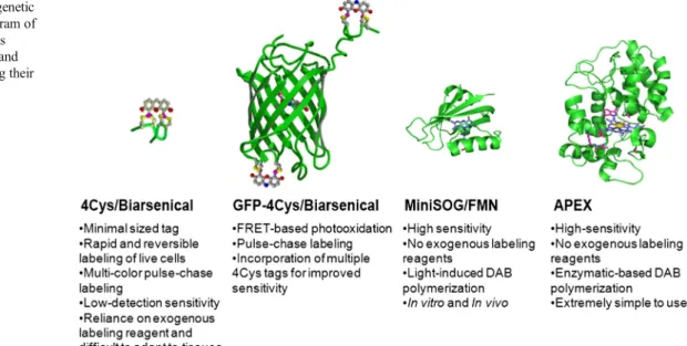
Since its introduction [20], MiniSOG, the first fluorescent protein genetically engineered specifically for correlated light and electron microscopy (CLEM), overcame the aforemen-tioned limitations by having the intrinsic ability to efficiently photooxidize DAB, thereby providing EM contrast without Fig. 1 Comparison of genetic
probes for CLEM. Diagram of different tagging systems described in this article and important facts regarding their application
Fig. 2 Imaging of actin dynamics using the 4Cys technology. a Live-cell confocal imaging of tetracysteine-tagged beta-actin in cultured cells using the green fluorescent biarsenical FlAsH. The lower cell is expressing the recombinant protein while the upper cell is not. b, c Following fixation,
cells counterstained with phalloidin-rhodamine show excellent co-staining in the transfected cell (yellow). Residual green fluorescence in the non-transfected cell is typical of background non-specific biarsenical cellular labeling (a). Bar=5μm
the need for an extraneously applied labeling agent. Photoox-idation using MiniSOG fusion proteins requires only the small molecules of DAB and dissolved molecular oxygen, which can readily penetrate fixed cells and tissues. This technique has been employed with great success in a wide variety of research activities [1–3,6,17,19]. For example, it was used to visualize, for the first time, the dynamic organization of the small p53-silencing adenoviral protein E4-ORF3 in intact and well-preserved nuclei (Fig.5; [19]).
TimeSTAMP-MiniSOG
MiniSOG has also been combined recently with the time-spe-cific tag for the age measurement of proteins (TimeSTAMP) [16] and a split-fluorescent protein, creating the first fully
genetically encoded, time-resolvable protein tag for correlated light and EM [5]. The use of this new type of molecular Bpulse-chase^ tool allows tracking of specific subpopulations of a target protein with high spatial and temporal resolutions. Specifically, this tool provides the ability to screen samples during a live imaging study using light microscopy as labeled copies of a target protein are being positioned in their cellular subdomains, and to visualize them both by fluorescence and at high resolution by EM. Applying the TimeSTAMP-specific protease inhibitor (BILN-2061) for a short incubation time (1– 3 h) allows for the marking of new proteins made during the labeling pulse. In this way, one can visualize in living neurons newly synthesized tagged proteins in time-lapse LM imaging, and follow them as they trafficked through the expressing cell. EM analysis, including 3D electron tomography of labeled proteins, can reveal their localization at high resolution. Using Fig. 3 CLEM imaging of
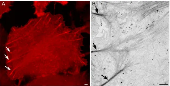
cytoskeleton arrangement. a Following live-cell time-lapse multiphoton imaging of cells expressing tetracysteine-tagged beta-actin labeled with ReAsH, the cells are fixed and the ReAsH is used to photooxidize DAB into an electron-dense polymer visible by EM (b). Arrows indicate the same bundles of actin visible by both LM and EM. Bar=1μm
Fig. 4 FRET-driven photooxidation. a Small tetracysteine tags can be incorporated into fluorescent proteins such as GFP to enable direct or FRET-based photooxidation to enable EM visualization. b–e CLEM of cells expressing connexin43-GFP-4Cys labeled with ReAsH. Bar=200 nm
TimeSTAMP for pulse-chase labeling has revealed previously uncharacterized aspects of the life cycle of the synaptic protein PSD95 [5]. Similarly, the application of this tagging system allowed the visualization ofBlonger lived^ alpha-synuclein proteins as they traffic and accumulate at presynaptic termi-nals (Fig.6).
APEX
APEX (for enhanced Ascorbate PEroXidase) [18] is a comple-mentary probe utilized for protein labeling at the EM level. It is a small (40 % smaller than horseradish peroxidase, HRP), genetically encodable peroxidase engineered to express as a monomer with a high degree of enzymatic activity towards DAB. It also has the remarkable property, unlike HRP, of exhibiting high enzymatic activity in all cellular compart-ments, including cytoplasm, and does so after strong aldehyde fixation. While APEX is not fluorescent, it is very attractive as
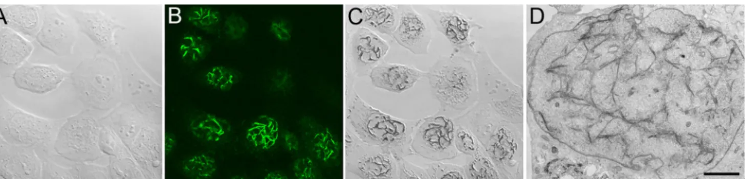
a probe because of its sheer simplicity and ease of use for labeling of proteins and making them visible to both light and electron microscopy. The protocol for labeling APEX-expressing fusion proteins in cells and tissues for EM is just one additional step in an otherwise conventional protocol to optimally preserve ultrastructure using glutaraldehyde prima-ry fixation and osmium tetroxide post-fixation. In cells or tissues expressing an APEX-fusion protein, following glutar-aldehyde fixation, the sample is incubated in a DAB solution containing 0.03 % hydrogen peroxide for as short as a few seconds to minutes. The formation of the reaction product can be monitored readily by ordinary transmitted light microscopy and the labeling intensity easily controlled. Since the reaction takes place in highly cross-linked glutaraldehyde-fixed speci-mens at 4 °C, diffusion of the reaction product can be mini-mized. Furthermore, since the labeling can be observed by ordinary light microscopy, many specimens can be screened rapidly prior to more time-consuming preparation for electron microscopy (Fig. 7). APEX has been used successfully to Fig. 5 CLEM imaging of cultured cells expressing the adenoviral protein
E4-ORF3 fused to MiniSOG. a Transmitted light image prior to fluorescence photooxidation. b Confocal image of MiniSOG fluorescence showing an intranuclear filamentous network formed by E4ORF3. c Transmitted light image following fluorescence
photooxidation showing DAB labeling corresponding to the fluorescence. d Electron microscopy of a reacted cell showing staining of the otherwise refractory elaborate filamentous network formed by E4ORF3. Bar=2μm
Fig. 6 Distribution ofBolder^ AS-TimeSTAMP-YFP-MiniSOG proteins at presynaptic terminals in neurons by CLEM. YFP fluorescence map allows the visualization of labeled proteins. a Fluorescence image superimposed to transmitted light image. Bar = 10μm. Following photooxidation and EM processing, a correlated EM map (b) is used to identify target labeled areas (green arrow) to perform 3D electron
tomographic analysis. c Representative slice of the electron tomogram corresponding to the presynaptic terminal indicated by the green arrow in a, b. The darker intensity of the signal reflects the AS labeling associated with various presynaptic endomembrane systems. Bar= 200 nm
localize a large number of cellular proteins, including actin, tubulin, vimentin, histone 2B, connexin43, PSD-95, and var-ious mitochondrial proteins, in a wide variety of cell types with excellent results. Like MiniSOG, the ultrastructural pres-ervation is excellent since no permeabilizing detergents or compromises to chemical fixation are required for labeling. However, in our work with APEX, we observed a limitation in its detection sensitivity, as some low-level expressing pro-teins were not always detectable. We hypothesized that the limited sensitivity of APEX may result from suboptimal fold-ing or stability, poor heme bindfold-ing, or some combination of these factors. These shortcomings helped to motivate an effort to identify key residues that could be modified in order to further improve these properties. This work led to the develop-ment of APEX2 via yeast-display-based directed evolution. APEX2 is a soybean-based ascorbate peroxidase that contains the original three APEX mutations (K14D, W41F, and E112K) plus an additional key modification (A134P) [14]. The APEX2 probe demonstrates significantly improved sensitivity when compared to the original APEX probe. This improvement allowed us to successfully localize a number of low-level ex-pressing proteins including MICU1, a mitochondrial calcium uptake protein that was not possible to visualize using APEX. APEX probes can also be used for live-cell time-lapse imaging
and CLEM when combined with GFP or other fluorescent proteins. We have successfully done this using a connexin43-GFP-APEX construct that, when expressed in living cells, ex-hibits the brightness, photostability, and low phototoxicity of GFP and, after chemical fixation and the addition of DAB and H2O2, is able to create a reaction product visible by EM
(Fig.8). Notably, we also observed a sensitivity of APEX to light; particularly intense blue light can deteriorate its enzymat-ic activity. To avoid this problem, fusions with yellow or red fluorescent proteins are recommended.
The APEX2 probe is likely to be adopted widely due to its ease of use and the fact that it overcomes so many obstacles encountered with other EM-level protein mapping approaches.
Tracking genetic labels across imaging modalities
A powerful application of these genetically encoded CLEM probes is to combine them with volume imaging by LM using confocal or multiphoton microscopy, followed by 3D EM imaging to enhance the ultrastructural reconstruction of spe-cific cells and tissues. Since the labeling is done prior to the physical sectioning of the sample, these probes are perfectly suited to conventional serial section EM, electron Fig. 7 Correlated light (a–c) and EM (d–f) imaging of a variety of APEX fusions in cultured cells. a Histone 2B-APEX in a dividing cell showing chromosome labeling. b Mitochondria matrix targeted APEX. c Cytoplasmic staining of vimentin-APEX. Scale bars=1μm
tomography, and serial block-face scanning EM (SBEM). For example, we have used MiniSOG to enable the targeted label-ing of retinal ganglion cell subtypes and subsequent 3D EM reconstruction. Mice expressing Cre recombinase in retinal ganglion cells were subjected to intravitreal injection with AAV virus carrying stop-floxed farnesylated MiniSOG. Fol-lowing primary fixation with aldehydes, the retina was photooxidized (Fig.9a) and then stained for SBEM imaging. However, the staining protocol for SBEM imaging involves intense labeling with heavy metals, which completely
obscures the location of labeled ganglion cells in the specimen and renders the specimen completely opaque to light. There-fore, in order to track the cells of interest, X-ray microtomography was performed on the tissue to reveal the photooxidized cells in the stained tissue (Fig.9b) [4]. The X-ray tomographic data allows for precise trimming and mount-ing of the specimen for serial block-face SBEM imagmount-ing. SBEM involves the iterative process of removing an ultrathin section of tissue from a specimen in the SEM, and then imag-ing the resultimag-ing block-face [7]. When this process is repeated Fig. 8 CLEM imaging of
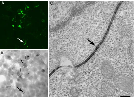
connexin43-GFP-APEX. Labeling of the same gap junction (arrows) is visualized by a confocal, b transmitted light, and c electron microscopy. Bar=200 nm
Fig. 9 Multimodal multiscale reconstruction of neurons using MiniSOG. The retinal ganglion cell subpopulation is specifically targeted for MiniSOG expression. a Cell bodies and neurites are clearly visible in transmitted LM following photooxidation of DAB with MiniSOG. b The same region of interest is tracked by X-ray microscopy following
infiltration of the tissue with heavy metals. The DAB-labeled neurons and tissue landmarks such as blood vessels are visible in the X-ray tomogram. c Following SBEM imaging, 3D segmentation of dendrites from ganglion cells is facilitated by the presence of DAB. Scale bars=10μm
hundreds or thousands of times, a large 3D volume is gener-ated with nanometer resolution. The labeled cells are electron-dense due to DAB deposition and are consequently signifi-cantly easier to track and segment for large-scale cellular re-construction (Fig.9c).
Conclusions
The development of the next-generation molecular-genetic and chemical-labeling approaches will advance the applica-tion of high-resoluapplica-tion, correlated, multiscale, and multimodal microscopy to biomedical research. With these expanding methods, we will elevate our ability to determine the locations of specific proteins in situ, a capability that has proven impor-tant in biology where localization of molecular constituents in cell and tissue contexts has provided important new under-standing. The EM-compatible probes presented here consist of complementary tools, each presenting different strengths as well as limitations, and are not meant to serve as a compre-hensive list of those available for CLEM, but rather ones we helped to develop. Indeed, new molecular-genetic imaging probes and techniques are continually under development with new capabilities [13], and one can expect this trend to continue into the future. Ultimately, the end-user will need to match the tag that best fits the desired goal in the context of the specific biological question, taking into account the target protein and their subcellular environment, the spatial and/or temporal resolution desired, the size of the tag, and the ap-proach utilized to make them visible by EM. Whether one chooses to combine the probes or to apply different ones in parallel, their application by CLEM allows visualizing in 3D a desired molecule in the context of extremely complex cell morphology and with respect to other key cellular structures, which is a prerogative of CLEM.
Acknowledgments The work presented here was conducted at the Na-tional Center for Microscopy and Imaging Research at San Diego, which is supported by NIH Grant GM103412 awarded to Dr. Mark Ellisman. National Institutes of Health grants P41RR004050 (Mark Ellisman) and GM086197 (Roger Y. Tsien and Mark Ellisman) and AHA Grant 10SDG2610281 (Daniela Boassa) provided funding for this research. Compliance with ethical standards
Conflict of interest The authors declare that they have no conflict of interest.
References
1. Boassa D, Berlanga ML, Yang MA, Terada M, Hu J, Bushong EA, Hwang M, Masliah E, George JM, Ellisman MH (2013) Mapping the subcellular distribution of alpha-synuclein in neurons using ge-netically encoded probes for correlated light and electron
microscopy: implications for Parkinson’s disease pathogenesis. J Neurosci 33(6):2605–2615
2. Boassa D, Nguyen P, Hu J, Ellisman MH, Sosinsky GE (2015) Pannexin2 oligomers localize in the membranes of endosomal ves-icles in mammalian cells while Pannexin1 channels traffic to the plasma membrane. Front Cell Neurosci 8:468
3. Burgers PP, Ma Y, Margarucci L, Mackey M, van der Heyden MA, Ellisman M, Scholten A, Taylor SS, Heck AJ (2012) A small novel A-kinase anchoring protein (AKAP) that localizes specifically pro-tein kinase A-regulatory subunit I (PKA-RI) to the plasma mem-brane. J Biol Chem 287(52):43789–43797
4. Bushong EA, Johnson DD, Kim KY, Terada M, Hatori M, Peltier ST, Panda S, Merkle A, Ellisman MH (2014) X-ray microscopy as an approach to increasing accuracy and efficiency of serial block-face imaging for correlated light and electron microscopy of bio-logical specimens. Microsc Microanal 13:1–8
5. Butko MT, Yang J, Geng Y, Kim HJ, Jeon NL, Shu X, Mackey MR, Ellisman MH, Tsien RY, Lin MZ (2012) Fluorescent and photo-oxidizing TimeSTAMP tags track protein fates in light and electron microscopy. Nat Neurosci 15(12):1742–51
6. Cleyrat C, Darehshouri A, Steinkamp MP, Vilaine M, Boassa D, Ellisman MH, Hermouet S, Wilson BS (2014) Mpl traffics to the cell surface through conventional and unconventional routes. Traffic 15(9):961–982
7. Denk W, Horstmann H (2004) Serial block-face scanning electron microscopy to reconstruct three-dimensional tissue nanostructure. PLoS Biol 2(11), e329
8. Ellisman MH, Deerinck TJ, Shu X, Sosinsky GE (2012) Picking faces out of a crowd: genetic labels for identification of proteins in correlated light and electron microscopy imaging. Methods Cell Biol 111:139–155
9. Gaietta G, Deerinck TJ, Adams SR, Bouwer J, Tour O, Laird DW, Sosinsky GE, Tsien RY, Ellisman MH (2002) Multicolor and elec-tron microscopic imaging of connexin trafficking. Science 296: 503–507
10. Gaietta GM, Giepmans BN, Deerinck TJ, Smith WB, Ngan L, Llopis J, Adams SR, Tsien RY, Ellisman MH (2006) Golgi twins in late mitosis revealed by genetically encoded tags for live cell imaging and correlated electron microscopy. Proc Natl Acad Sci U S A 103:17777–17782
11. Heim R, Tsien RY (1996) Engineering green fluorescent protein for improved brightness, longer wavelengths and fluorescence reso-nance energy transfer. Curr Biol 6(2):178–182
12. Hoffmann C, Gaietta G, Zürn A, Adams SR, Terrillon S, Ellisman MH, Tsien RY, Lohse MJ (2010) Fluorescent labeling of tetracysteine-tagged proteins in intact cells. Nat Protoc 5(10): 1666–1677
13. Kuipers J, van Ham TJ, Kalicharan RD, Veenstra-Algra A, Sjollema KA, Dijk F, Schnell U, Giepmans BN (2015) FLIPPER, a combinatorial probe for correlated live imaging and electron mi-croscopy, allows identification and quantitative analysis of various cells and organelles. Cell Tissue Res. 360(1):61–70
14. Lam SS, Martell JD, Kamer KJ, Deerinck TJ, Ellisman MH, Mootha VK, Ting AY (2015) Directed evolution of APEX2 for electron microscopy and proximity labeling. Nat Methods 12(1): 51–54
15. Lelek M, Di Nunzio F, Henriques R, Charneau P, Arhel N, Zimmer C (2012) Superresolution imaging of HIV in infected cells with FlAsH-PALM. Proc Natl Acad Sci U S A 109(22):8564–8569 16. Lin MZ, Tsien RY (2010) TimeSTAMP tagging of newly
synthe-sized proteins. Curr Protoc Protein Sci Chapter 26:Unit 26.5 17. Ludwig A, Howard G, Mendoza-Topaz C, Deerinck T, Mackey M,
Sandin S, Ellisman MH, Nichols BJ (2013) Molecular composition and ultrastructure of the caveolar coat complex. PLoS Biol 11(8), e1001640
18. Martell JD, Deerinck TJ, Sancak Y, Poulos TL, Mootha VK, Sosinsky GE, Ellisman MH, Ting AY (2012) Engineered ascorbate peroxidase as a genetically encoded reporter for electron microsco-py. Nat Biotechnol 30:1143–1150
19. Ou HD, Kwiatkowski W, Deerinck TJ, Noske A, Blain KY, Land HS, Soria C, Powers CJ, May AP, Shu X, Tsien RY, Fitzpatrick JA, Long JA, Ellisman MH, Choe S, O’Shea CC (2012) A structural basis for the assembly and functions of a viral polymer that inacti-vates multiple tumor suppressors. Cell 151(2):304–319
20. Shu X, Lev-Ram V, Deerinck TJ, Qi Y, Ramko EB, Davidson MW, Jin Y, Ellisman MH, Tsien RY (2011) A genetically encoded tag for correlated light and electron microscopy of intact cells, tissues, and organisms. PLoS Biol 9(4):e1001041
21. Tsien RY (1998) The green fluorescent protein. Annu Rev Biochem 67:509–544, Review
22. Whitt MA, Mire CE (2011) Utilization of fluorescently-labeled tetracysteine-tagged proteins to study virus entry by live cell mi-croscopy. Methods 55(2):127–136