See discussions, stats, and author profiles for this publication at: https://www.researchgate.net/publication/233538136
Comparative study of nitric oxide (NO) production during human hydatidosis:
Relationship with cystic fluid fertility
Article in Parasitology Research · November 2012
DOI: 10.1007/s00436-012-3181-6 · Source: PubMed
CITATIONS
8
READS
192
5 authors, including:
Some of the authors of this publication are also working on these related projects:
study of the immunomodulatory effects of Echinococcus granulosus on allergic process View project Study of the polymorphism of antioxidant enzymes in the pathophysiology of type 1 diabetes View project Razika Zeghir-Bouteldja
AKLI MOHAND OULHADJ UNIVERSITY. BOUIRAH 11PUBLICATIONS 66CITATIONS
SEE PROFILE
Manel Amri
University of Science and Technology Houari Boumediene 39PUBLICATIONS 272CITATIONS SEE PROFILE Samia Bouaziz Université de M'sila 9PUBLICATIONS 58CITATIONS SEE PROFILE Touilboukoffa Chafia
University of Science and Technology Houari Boumediene 197PUBLICATIONS 1,332CITATIONS
SEE PROFILE
All content following this page was uploaded by Touilboukoffa Chafia on 16 May 2014. The user has requested enhancement of the downloaded file.
ORIGINAL PAPER
Comparative study of nitric oxide (NO) production during human
hydatidosis: relationship with cystic fluid fertility
Razika Zeghir-Bouteldja&Manel Amri&
Samia Bouaziz&Dalila Mezioug&Chafia Touil-Boukoffa
Received: 12 July 2011 / Accepted: 31 October 2012 # Springer-Verlag Berlin Heidelberg 2012
Abstract Human hydatidosis is characterized by a pro-longed coexistence of Echinococcus granulosus and its host without effective rejection of the parasite. This parasitic infection constitutes a major health problem in Algeria. In this study, we investigated in vivo production of nitrite
(NO2−+ NO3−) in sera of Algerian patients carrying
differ-ent cyst locations. Nitrite (NO2−+ NO3−) levels were
eval-uated by the Griess method. Our results indicated that the levels of nitrite were significantly higher in the sera of hydatic patients than those of healthy controls supporting the involvement of nitric oxide (NO) in antihydatic action. The levels of nitrite in sera of the patients with hepatic hydatidosis were significantly higher than those with
pul-monary infection. The lower serum (NO2− + NO3−) levels
were observed in the relapsing cases. In addition, (NO2−+
NO3−) levels of fertile hydatic fluids were significantly
higher compared to infertile fluids. Our results suggest that the presence of NO products in hydatic fluids seems to be related to the location and the fertility of hydatic cysts. The assessment of protein concentration in hydatic fluids showed that the concentration of proteins was not exclu-sively dependent on the fertility but on the cyst locations.
The assessment of (NO2− + NO3−) production in hydatic
patients may be a useful tool to evaluate effector mecha-nisms of NO and clinical manifestations.
Abbreviations
NO Nitric oxide
NOs2 Nitric oxide synthase 2
IFN-γ Interferon gamma
PBMCs Peripheral blood mononuclear cells
NO3− Nitrate
NO2− Nitrite
E. granulosus Echinococcus granulosus
Introduction
Human hydatidosis is an endemic parasitic disease caused by the larval stage of the tapeworm of Echinococcus gran-ulosus. It occurs practically worldwide, including countries of Central and South America, Western and Southern/South-eastern Europe, the Middle East and North Africa, some sub-Saharan countries, Russia and adjacent countries
(Eckert and Deplazes 2004), and China (Wen and Yang
1997). It constitutes a major health problem in North
Africa, particularly in Algeria. It is considered as a disease for obligatory declaration by the National Insti-tute of Public Health. The annual incidence is 1.47–2.3/ 100,000 Algerian population. The transmission of E. granulosus in the dog–sheep cycle is known to occur most frequently in rural and grazing areas. Larval infec-tion is characterized by long-term growth of hydatic
cysts in the intermediate host (Zhang et al. 2003).
Variability and severity of clinical expression of hydatidosis are not only associated with the duration and intensity of infection but also with the variety of human immunological responses to the antigens of larval hydatic cyst with detectable
R. Zeghir-Bouteldja
:
M. Amri:
S. Bouaziz:
D. Mezioug:
C. Touil-Boukoffa (*)
Laboratory of Cellular and Molecular Biology, FSB-USTHB, University of Bab-Ezzouar, BP 32, Algiers, Algeria e-mail: touilboukoffa@yahoo.fr R. Zeghir-Bouteldja e-mail: bouteldja_raz@yahoo.fr M. Amri e-mail: manelamri@yahoo.fr S. Bouaziz e-mail: bouaziz_samia@yahoo.fr D. Mezioug e-mail: mezioug_dalila@yahoo.fr DOI 10.1007/s00436-012-3181-6
humoral and cellular responses against E. granulosus.
Touil-Boukoffa et al. (1998) have reported a correlation between in
vivo and ex vivo interferon-γ and nitric oxide (NO) production
during human hydatidosis. NO is an important regulator and mediator in many physiological and pathophysiological events. It has been implicated in neurotransmission,
vasodila-tation, and immune regulation (Moncada and Higgs1991).
Although small amounts of NO are constitutively released by endothelial and neuronal nitric oxide enzymes (eNOs and nNOs, respectively), inducible NO synthase, located mainly in immune cells, produces NO in considerable amounts after
exposure to cytokines (Nussler et al.1992). NO and its stable
metabolites (NO2−and NO3−) have been identified as major
effector molecules during the majority of parasitic infections
(Liew1992; Ascenzi et al.2003). Production of NO has been
shown to be induced by interferon gamma (IFN-γ) during
human hydatidosis, suggesting the relevant role of NO in the
host defense (Touil-Boukoffa et al.1998; Ait Aissa et al.2006;
Amri et al.2007). Our proposal was supported by in vitro
observations that hydatic cysts are susceptible to the products
of NO such as NO2−and ONOO−. These metabolites showed
in vitro scolicidal activity and degenerative effects on germinal and laminated layers of human hydatid of E. granulosus
(Zeghir-Bouteldja et al.2009).
The present study was designed to assess the
produc-tion of (NO2− + NO3−) in sera of Algerian hydatic
patients in relation with clinical status (before and after surgery), cystic location, and cystic status. We also
examined (NO2− + NO3−) and protein levels in fertile
and infertile hydatic fluids collected after surgery from cysts of different anatomical sites.
Materials and methods Patients
The group of Algerian patients (33±2, 5 years old, and 55 % men) is composed of hydatic patients with different cyst locations. Regions of origin of patients are the provinces of high plateau, the northeastern, north-central provinces, and center of Algeria. They were tested for circulating
(NO2−+ NO3−) levels. Patients were tested before and after
surgery (1 week before and 24–72 h after surgery). All patients were admitted to the Department of Surgery (Mus-tapha Bacha Hospital, Algiers, Algeria). None of the patients had received pharmacological treatment. Subjects with other acute or chronic diseases, smoking, and alcohol-ism were not included in this study. Serological reaction against parasitic antigen (antigen 5) was tested in each case by immunoelectrophoresis. The diagnosis was radiologically and surgically confirmed for all patients. Indirect hemaggluti-nation test was used for detecting hydatidosis antibodies.
Stool examination was made on both patients and controls groups to exclude intestinal parasitic infections.
Healthy controls (n020, mean age 37±6 years, 50 %
males) from the same Algerian region (Algiers) are com-posed of adult volunteer blood donors who presented no inflammatory disease nor sign of infection at the time of blood sample collection. In addition, none of the subjects had ever received blood transfusion or any medication. All subjects were informed on the study and have signed an official consent, which was conducted according to the guidelines of the local Ethics Working Group.
Serum collection
Blood samples collected from healthy donors and hydatic patients were centrifuged at 2,000 × g for 10 min to separate the serum. Hemolysis serum was excluded from this study.
The serum samples were classified into several groups
(Table 1). This classification was based on clinical stage
(before and after surgery, n060), patients who relapsed after
18 months of surgery (n015), cyst location (lung n024; liver
n032), cyst states (intact cysts n056; calcified cysts n07;
broken cysts n04), and double location (liver+lung n06). Hydatic fluids collection
Forty-six hydatic fluid samples were obtained after surgery. Hydatic fluids (HFs) were collected after aseptic dissection of the intact hydatic cysts. They were centrifuged at 3,000× g for 30 min at +4 °C, and the sediment was checked for the presence of protoscolices. Cyst fertility was determined by the presence of viable free protoscolices in hydatic fluid.
Table 1 Serum levels of (NO2−+ NO3−) in Algerian hydatic patients
Groups Population (NO2−+ NO3−)
concentration
(n) (μM)
Healthy controls 20 30.01±1.96
Patients (before surgery) 60 77.37±7.24
Patients (after surgery) 60 49.70±6.89
Patients with intact cysts 56 74.31±3.87
Double location (lung and liver) 6 83.53±14.48
Patients with lung cysts 24 59.90±4.14
Patients with liver cysts 32 85.12±5.31
Patients with relapsed infection (liver and lung)
15 20.17±1.62
Patients with calcified (liver and lung cysts)
7 28.64±2.55
Patients with broken lung cysts 4 105.81±14.74
Data are means of the (NO2− + NO3−) levels ± standard errors
n number of patients
The viability of the protoscolices was assessed by staining with 0.1 % aqueous eosin solution. Other parameters of cyst fertility were considered such as a whitish color of the laminated layer and limpidity of hydatic fluid. The fertility was variable for the cysts located in the same organ. The fertile fluid (+) was characterized by the presence of a high number of protoscolices and daughter vesicles. The hydatic fluid had a milky aspect and whitish color. The number of protoscolices was more than 1,000 parasites/ml. Less fertile fluid (+/−) was characterized by the presence of reduced number of protoscolices. The hydatic fluid was clear and had whitish color, and the number of protoscolices was less than 500 parasites/ml. Infertile fluid (−) was characterized by total absence of protoscolices and daughter vesicles and was limpid and clear. The hydatic fluids were classified into several groups. This classification is based on fertility and
cyst location [liver HFs (Table2) (+) HF n013, (+/−) HFs
n05, and (−) HFs n07; lung HFs (+) HFs n010, (+/−) HFs
n04, (−) HFs n04), and (−) muscular HF n01; (+) splenic
HF n01; and (−) pancreatic HF n01]. Nitric oxide assay
NO production was assessed by determination of the end
products of NO oxidation. Total concentration of (NO2−+
NO3−) was quantified by the Griess reaction as described in
1879 (Sun et al.2003). Briefly, the Griess Reagent System
was based on a reaction which uses sulfanilamide and N-1-napthylethylenediamine dihydrochloride under acidic
con-ditions. NO2− was assayed after reduction of NO3− by
nitrate reductase containing Pseudomonas oleovorans bac-teria (ATCC 8062). One hundred microliters of each serum
or hydatic fluid to be tested was mixed with 50μl of 1:10
dilution of pelleted bacteria cells. In parallel, the NO2−
standard for calibration curves was treated similarly as the
samples. More than 90 % of NO3− was converted to NO2−
under these conditions. After incubation at 37 °C for 90 min,
the sample was centrifuged and mixed with 100μl of the
Griess reagent (0.5 %N-1-naphtyl-ethylenediamine in 20 %
HCl, 5 % sulphanilamide in 20 % HCl (1v/v). After 20 min of incubation at 22 °C, the formation of chromogenic deriv-ative was detected by spectrophotometry at 540 nm as
described by Touil-Boukoffa et al. (1998).
Protein assay
The protein concentration in hydatic fluids was determined by
the Bradford method (Bradford1976). A convenient standard
curve was made using a series of dilutions of bovine serum albumin (BSA) with the concentrations of 10–100 μg/μl in phosphate-buffered saline. The concentration of protein was
assessed by the addition of 100μl of each sample to 3 ml of
Bradford reagent. When mixed with a protein solution, the acidic Coomassie dye reagent changes color from brown to blue in proportion to the amount of protein present in the sample (Coomassie stains; Sigma-Aldrich). After incubation for 5 min at 22 °C, the absorbance was read at 595 nm. The average absorbance value of each concentration of the BSA was plotted. For this study, the hydatic fluid samples were
treated similarly, and 100μl of hydatic fluid was mixed with
3 ml of Bradford reagent. The concentration of proteins was determined by extrapolation of the value of optic density on the BSA standard curve.
Statistical analysis
All values are expressed as means ± standard error. Data analysis was performed using the Origin 7.5 software
(OriginLab2003, USA). Differences between means were
analyzed by student’s t test. Values for P<0.05 were considered statistically significant.
Results
Production of nitrite in sera of patients
As shown in the Table 1, sera (NO2−+ NO3−) levels were
higher in the presurgical hydatic patients group (77.37 ±
7.24μM) than those of healthy subjects (30.01±1.96 μM).
The (NO2−+ NO3−) levels were significantly higher in the
patients group before surgery (77.37 ±7.24μM) than in the
patients group after surgery (49.70 ±6.89μM); P00.01023),
whereas the relapsing patients group showed lower nitrite
levels (20.17 ±1.62μM) in comparison to healthy
individu-als. There are no significant differences between relapsing patients and controls (P00.0008). The nitrite levels detected in sera of patients with hepatic hydatidosis (85.12 ±
5.31μM) were higher than those with pulmonary infection
(59.90 ±4.14μM; P00.029). Indeed, the comparative study
of production of (NO2−+ NO3−) in sera of patients carrying
intact cysts showed significant higher levels compared to
Table 2 (NO2−+ NO3−) levels in liver hydatic fluids according to the
degree of fertility Cyst location Degree of fertility n (NO2−+ NO3−) concentration (μM) Liver + 13 29.76±7.52 +/− 5 7.88±0.66 − 7 4.92±0.43
Data are means ± standard errors of the concentration of (NO2− +
NO3−) detected in hydatic fluid. Symbols (+), (+/−), and (−) indicate
fertile, less fertile, and infertile fluids, respectively n number of hydatic cysts
patients with calcified cysts (28.64 ±2.55μM). The (NO2−+
NO3−) level of patients with calcified cysts did not differ
from the control group (28.64 ± 2.55 μM versus 30.01±
1.96μM; P00.7111). While the (NO2−+ NO3−) sera level
of patients with broken cysts was significantly higher
(105.81 ±14.74μM) compared to those with calcified cysts,
the difference between the two groups was statistically significant (P00.00007).
The assessment of (NO2−+ NO3−) production in hydatic
fluids showed higher levels of (NO2−+ NO3−) in fertile liver
fluids with (29.76 ±7.52μM) compared to the infertile
flu-ids (4.92±0.43 μM; P00.00004). The same observations
were also noted in pulmonary cysts with (26.55 ±4.25μM)
in fertile fluids versus infertile fluids with (4.53±0.62μM;
P00.00322) (Fig. 1). (NO2− + NO3−) levels were low in
infertile fluids of muscular and pancreatic cysts. (NO2− +
NO3−) production in splenic hydatic fluid was higher than
that in the liver or lung.
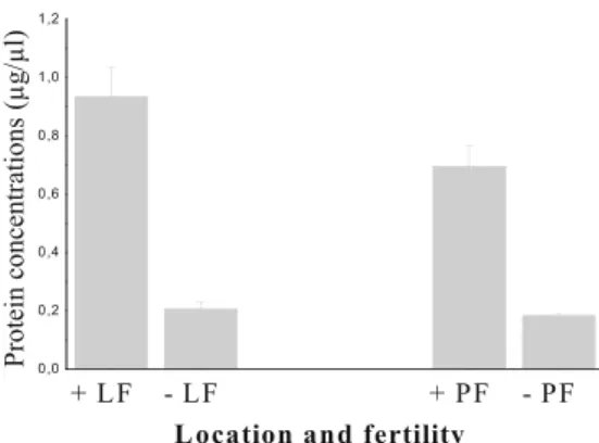
Protein levels detected in fertile liver fluids were
significant-ly higher (0.93231 ± 0.10435μg/μl) than those in infertile
fluids (0.21 ± 0.026μg/μl; P 0 0.0005) (Fig.2). This result
was also observed in pulmonary hydatic fluids (0.695 ±
0.07166 versus 0.18167±0.01078μg/μl, P00.00001) (Fig.2).
In addition to the data shown in Table 2, our results
indicated that the concentration of proteins was not exclu-sively dependent only on the cyst fertility but also on the cyst location, the environment of exchange between the host
and parasite, and host tissue vascularity (Table3).
Discussion
Our comparative study showed nitrite concentration varia-tions in all hydatic patients tested. These differences could
be related to several parameters. The presence of
signifi-cantly high levels of (NO2−+ NO3−) in the serum of hydatic
patients before surgery supports the possible involvement of NO in antihydatic action. NO level increase seems to be associated with the stimulation of the cell-mediated immune system during a long-term parasitic infection. In previous studies, our team has reported that during human hydatido-sis, IFN-γ led to the elevated production of nitrite through
the NOs2 pathway (Touil-Boukoffa et al.1998; Ait Aissa et
al.2006; Amri et al.2007). A correlation between (NO2−+
NO3−) and IFN-γ levels indicates that NO production by the
host is induced in large part by IFN-γ, knowing that this Th1 cytokine is highly required in the upregulation of nitric oxide production by the monocyte/macrophage system
(Paul-Eugène et al.1994; Touil-Boukoffa et al.1998).
The formation of NO and its products has been reported in a majority of parasitic infections. Significant increase of
(NO2− + NO3−) was detected in the serum of patients
infected with Entamoeba coli (Karaman et al. 2009). In
addition, human schistosomiasis results in an increase in
the production of serum (NO2− + NO3−) (Abost-Shousha
Fig. 1 Comparison of (NO2−+ NO3−) levels in pulmonary hydatic
fluids according to the degree of fertility. Data are means ± standard
errors of the means for each point. P00.00322 for (NO2− + NO3−)
levels detected in fertile hydatic fluids (26.55±4.25μM; n010) versus
infertile fluids (4.53±0.62μM; n04). Values for P<0.05 were
consid-ered statistically significant
Fig. 2 Protein levels in hydatic liver and pulmonary fluids according to the fertility. Data are means ± standard errors of the means for each
point. P00.0005 for protein levels detected in fertile liver hydatic
fluids (0.93231±0.10435μg/μl; n013) versus infertile fluids (0.21±
0.026μg/μl; n06). P00.00001 for pulmonary fertile fluids (0.695±
0.07166μg/μl; n08) versus infertile fluids (0.18167±0.01078 μg/μl;
n06). + Fertile, − infertile, LF liver fluid, PF pulmonary fluid. Values
for P<0.05 were considered statistically significant
Table 3 (NO2−+ NO3−) and protein levels according to the fertility of
hydatic fluid in uncommon cyst locations Infrequent cyst location Degree of fertility n (NO2−+ NO3−) concentration Protein levels (μM) (μg/μl) Muscle of left leg − 1 4.10 0.31 Spleen + 1 31.11 1.29 Pancreas − 1 4.76 1.80
Symbols (−) and (+) indicate infertile and fertile fluids, respectively
n number of hydatic cysts
et al.1999). An increase nitrite concentration in the serum was also reported in experimental toxocariasis. Mice infected with Echinococcus multilocularis demonstrated an increase in NOs2 expression in peritoneal macrophages (Dai
and Gottstein1999).
However, in our study, concentrations of (NO2−+ NO3−)
declined after surgery in the absence of postoperative com-plication such as an infection. This decline may have a role in hydatidosis pathophysiology returning to normal levels
(Ait Aissa et al.2006; Parsak et al.2007).
Interestingly, we observed that patients with intact and
viable cysts showed elevated (NO2− + NO3−) levels
com-pared to those with calcified cysts. This result was likely related to the inhibition of the immunogenicity. The calcifi-cation of the cyst indicated the loss of viability, while the detection of high levels of nitrite in sera of broken cysts is due to a probable increase of cyst antigen stimulation and, in turn, more activation of immune cells. This hypothesis is supported by marked hypereosinophilia after breakdown of cysts associated to allergic manifestations and pro-duction of IgE interacting with CD23 inducing human
NOs2 (Paul-Eugène et al. 1994; Bell 1996).
In the present study, a significant increase of (NO2− +
NO3−) concentrations was observed in the serum of patients
with liver cysts in comparison with those bearing cysts in the lung. This difference is probably related to several factors. Hepatic cysts were expected to provide the highest amount of antigen, probably because the high vascularity of this major filtering organ functionally promotes hydatid
development. In addition, Ait Aissa et al. (2006) reported
the presence of NOs2 in hepatocytes and Kupffer cells from liver biopsies of hydatic patients. It has been reported that human PBMCs and leukocytes constitute the cellular source of NO during parasitic infection (Touil-Boukoffa et al.
1998; Ait Aissa et al.2006). The production of high levels
of nitrite in hepatic infections could be correlated with the stimulation of the cell-mediated immune system by E. granulosus antigens.
There is evidence that liver cysts grow at a lower rate
than lung cysts (Larrieu and Frider 2001). In fact, the
presence of hydatic cysts in the lung may cause compression of surrounding structures and lead to the cough reflex, resulting in a release of antigenic material and hemoptysis
(Gottstein and Reichen2002; Blanton2007). Our results are
in-line with these data and support the reasoning that differ-ences in the host immune response are intimately related to the cyst location, suggesting a dependent relationship be-tween cyst location and NO serum levels in hydatic patients. During the lung infection, molecular interactions be-tween inflammatory cells and larvae were investigated
(Muro and Pérez-Arellano 2010). Human epithelial cells
and alveolar macrophages may constitute a cellular source of production of NO. However, NO produced by surface
epithelial cells may therefore act as an important barrier to invasion of the respiratory tract by inhaled organisms (Liew
and Cox1991; Taylor Robinson et al.1994).
The reduction of nitrite production in relapsing patients correlates with the lack of IFN-γ production, which is related to a decrease in the CD4+ T cell count and is of predictive value in E. granulosus follow-up (Touil-Boukoffa
et al.1998).
The patients with double location showed a significant
increase in (NO2−+ NO3−) concentration. This observation
could mean an enhanced immunological response when cysts are multiple. This observation was reported also by
Refik et al. (2005).
The presence of NO products in the cyst fluid was
pre-viously reported by our team (Ait Aissa et al. 2006). This
result is probably a consequence of antigenic burden and IFN-γ induction. The free radical can probably diffuse
through the cystic walls (Ait aissa et al. 2006). In this
context, we suggest the possible involvement of activated macrophages present in the adventitial layer in local innate immune response, knowing that these cells are a major source of inducible NO synthase, and they are highly acti-vated by IFN-γ. This local response could provoke
infertil-ity and death of the hydatic cysts (Shepherd et al. 1991;
Rigano et al.1995,2007; Vuitton2003; Amri et al.2007).
Our hypothesis correlates with the structure of hydatic cysts. In fact, immune cells that lodged in the adventitial layer are unable to penetrate to the germinal layer due to the physical barrier imposed by the laminar layer. Our data suggest a possible local NO production by the larvae. Currently, more investigations are undertaken by our team to clarify this hypothesis. However, these findings do not exclude the possibility of presence of NOs in the scolex, although larvae NOs expression has not been identified yet (Amanvermez
and Celik2002). In the current study, our results show high
levels of (NO2− + NO3−) in fertile liver and lung fluids
compared to infertile fluids. This observation was also
reported by Amanvermez and Celik (2002) in cattle hydatic
fluids. We have noted with interest that the production of
(NO2−+ NO3−) in splenic hydatic fluid was higher than that
in the liver or lung. This increase was probably related to the high vascularity of this organ.
Similarities of human hydatic cyst fluid components and the host serum have been reported, but in reduced amounts as compared to serum, suggesting that the host proteins can penetrate the membranes of the hydatic cyst (Goodchild and
Kagan 1961; Khorsandi and Tabibi 1978). However, our
results indicate that the concentration of proteins is related to the cyst location and organ vascularity. The presence of con-sistent protein concentrations in hydatic fluids underlines the tight relationship between the host and parasite. Compared to
the protein levels, the (NO2−+ NO3−) production appears to
Collectively, our findings show that the NO production during human hydatidosis depends upon the cyst location, the status and viability of the cyst, the number of cysts, and the clinical stage of hydatic patients. The presence of NO products in hydatic fluids underlines the strong relationship between the immune systems of host and may be associated with the location and fertility of the cyst. Assessment of NO production may be a useful tool in the evaluation of the effector mechanisms and clinical manifestations of hydati-dosis and represents a useful marker of the clinical aggres-siveness of this parasitic infection. In future studies, we intend to evaluate the clinical usefulness of NO in the treatment of human echinococcosis.
Acknowledgments The authors wish to thank the technical and
surgical staff of the Mustapha Bacha Hospital of Algiers for providing serum and cyst samples. A special thanks goes to Professor Hamrioui. They thank all the voluntary participants in this study. They are grateful to Dr. Wietzerbin for helpful discussions. This work was supported by a grant from the ANDRS (National Agency for Development of Scientific Research).
References
Abo-Shousha S, Khalil SS, Rashwan EA (1999) Oxygen free radical and nitric oxide production in single or combined human
schis-tosomiasis and fascioliasis. J Egypt Soc Parasitol 29:149–156
Ait Aissa S, Amri M, Bouteldja R, Wietzerbin J, Touil-Boukoffa C (2006) Alterations in interferon-gamma and nitric oxide levels in
human echinococcosis. Cell Mol Biol 52:65–70
Amanvermez R, Celik C (2002) Effectiveness of free radicals in
hydatid cysts. J Egypt Soc Parasitol 32:259–269
Amri M, Ait Aissa S, Belguendouz H, Mezioug D, Touil-Boukoffa C
(2007) In vitro antihydatic action of IFN-γ is dependent on the
nitric oxide pathway. J Interferon Cyt Res 27:781–787
Ascenzi P, Bocediand A, Gradoni L (2003) The anti-parasitic effect of nitric oxide. IUBMB Life 55:573–578
Bell RG (1996) IgE, allergies and helminth parasites: a new perspec-tive on an old conundrum. Immunol Cell Biol 74:337–345 Blanton R (2007) Echinococcosis. In: Behrman RE, Kliegmann RM,
Jenson HB (eds) Nelson textbook of pediatric, 18th edn. Saunders,
Philadelphia, pp 1516–1518
Bradford MM (1976) A rapid and sensitive method for the quantitation of the microgram quantities of protein utilizing the principle of
protein-dye binding. Anal Biochem 72:248–254
Dai WJ, Gottstein B (1999) Nitric oxide-mediated immunosuppression following murine Echinococcus multilocularis infection. Immunol
97:107–116
Eckert J, Deplazes P (2004) Biological, epidemiological, and clinical aspects of echinococcosis, a zoonosis of increasing concern. Clin
Microbiol Rev 17107–135
Getting started manual for Origin version 7.5 (2003) OriginLab Corporation
Goodchild CG, Kagan IG (1961) Comparison of proteins in hydatid fluid and serum by means of electrophoresis. J Parasitol 47:175– 180
Gottstein B, Reichen J (2002) Hydatid lung disease. Clin Chest Med 23:397–408
Karaman U, Kiran TR, Colak C, Iraz M, Celik T, Karabulut AB (2009) Serum malondialdehyde, glutathione and nitric oxide levels in patients infected with Entamoeba coli. Int J Med Medical Sci
1:235–237
Khorsandi HO, Tabibi V (1978) Similarities of human hydatid cyst fluid components and the host serum. Acta Med Iran 21:161–172 Larrieu EJ, Frider B (2001) Human cystic echinococcosis: contribu-tions to the natural history of the disease. Ann Trop Med Parasitol 95:679–687
Liew FY (1992) Regulation of nitric oxide synthase in macrophages. In: Moncada S, Stamler J, Gross S, Higg EA (eds) The biology of nitric oxide synthase 2: enzymology, biochemistry and
immunol-ogy. Portland Press, London, pp 223–229
Liew FY, Cox FF (1991) Nonspecific resistance mechanisms: the role
of nitric oxide. Immunol Today 12:17–21
Moncada S, Higgs EA (1991) Endogenous nitric oxide: physiology,
pathology and clinical relevance. Eur J Clin Invest 21:361–374
Muro A, Pérez-Arellano JL (2010) Nitric oxide and respiratory
helminthic diseases. J Biomed Biotech 9581081:1–8
Nussler AK, Di Silvio M, Billiar TR, Hoffman RA, Geller DA, Selby R, Madariaga J, Simmons RL (1992) Stimulation of the nitric oxide synthase pathway in human hepatocytes by cytokines and endotoxin. J Exp Med 176(1):261–4
Parsak CK, Hanta I, Koltas IS, Sakman G, Akcam T, Kuleci S, Alabaz O (2007) The effectiveness of nitric oxide derivates in hydatid disease. Chir Gastroenterol 23:296–299
Paul-Eugène N, Kolb JP, Damais C, Yamaoka K, Dugas B (1994) Regulatory role of nitric oxide in the IL-4-induced IgE production by normal human peripheral blood mononuclear cells. Lymphokine
Cytokine Res 13:287–293
Refik M, Mehmet N, Duemaz R (2005) Postoperative changes in serum cytokines profile and nitric oxide levels in patients with
cystic echinococcosis. Parasite 1:265–269
Rigano R, Profumo E, Ioppolo S, Notargiacomo S, Ortona E, Teggi A, Siracusano A (1995) Immunological markers indicating the effec-tiveness of pharmacological treatment in human hydatid disease.
Clin Exp Immunol 102:281–285
Rigano R, Buttari B, Profumo E, Ortona E, Delunardo F, Margutti P, Mattei V, Teggi A, Sorice M, Siracusano A (2007) Echinococcus granulosus antigen B impairs human dendritic cell differentiation and polarizes immature dendritic cell maturation towards a Th2 cell response. Infect Immun 75:1667–1678
Shepherd JC, Aitken A, McManus DP (1991) A protein secreted in vivo by Echinococcus granulosus inhibits elastase activity and
neutrophil chemotaxis. Mol Biochem Parasitol 44:81–90
Sun J, Zhang X, Mark Broderick M, Harry Fein H (2003) Measurement of nitric oxide production in biological systems by using Griess
reaction assay. Sensors 3:276–248
Taylor Robinson AW, Lie FY, Severn A (1994) Regulation of the immune response by nitric oxide differentially produced by Th1
and Th2 cells. Eu J Immunol 24:980–984
Touil-Boukoffa C, Bauvois B, Sancéau J, Hamrioui B, Wietzerbin J (1998) Production of nitric oxide (NO) in human hydatidosis:
relationship between nitrite production and interferon-γ levels.
Biochem 80:739–744. doi:10.1016/S0300-9084(99)80027-3
Vuitton DA (2003) The ambiguous role of immunity in echinococcosis:
protection of the host or of the parasite? Acta Trop 85:119–
132
Wen H, Yang WG (1997) Public health importance of cystic
echino-coccosis in China. Acta Trop 67:133–145
Zeghir-Bouteldja R, Amri M, Ait Aissa S, Bouaziz S, Mezioug D, Touil-Boukoffa C (2009) In vitro study of nitric oxide metabolites effects on human hydatid of Echinococcus granulosus. J Parasitol
Res 624919:1–7
Zhang W, Li J, McManus DP (2003) Concepts in immunology and diagnosis of hydatid disease. Clin Microbiol Rev 16:18–36
Parasitol Res
View publication stats View publication stats