HAL Id: hal-01459537
https://hal.archives-ouvertes.fr/hal-01459537
Submitted on 20 Sep 2018
HAL is a multi-disciplinary open access
archive for the deposit and dissemination of sci-entific research documents, whether they are pub-lished or not. The documents may come from teaching and research institutions in France or abroad, or from public or private research centers.
L’archive ouverte pluridisciplinaire HAL, est destinée au dépôt et à la diffusion de documents scientifiques de niveau recherche, publiés ou non, émanant des établissements d’enseignement et de recherche français ou étrangers, des laboratoires publics ou privés.
Khalid El Karkouri, Pierre Pontarotti, Didier Raoult, Pierre-Edouard Fournier
To cite this version:
Khalid El Karkouri, Pierre Pontarotti, Didier Raoult, Pierre-Edouard Fournier. Origin and Evo-lution of Rickettsial Plasmids. PLoS ONE, Public Library of Science, 2016, 11 (2), pp.e0147492. �10.1371/journal.pone.0147492�. �hal-01459537�
Origin and Evolution of Rickettsial Plasmids
Khalid El Karkouri1, Pierre Pontarotti2, Didier Raoult1, Pierre-Edouard Fournier1* 1 Unité de Recherche en Maladies Infectieuses et Tropicales Emergentes (URMITE), UM63, CNRS7278, IRD198, INSERMU1095, Institut Hospitalo-Universitaire Méditerranée-Infection, Aix-Marseille Université, Faculté de Médecine, 27 boulevard Jean Moulin, 13385 Marseille cedex 5, France, 2 Aix Marseille Université, CNRS, Centrale Marseille, I2M UMR 7373, Equipe Evolution Biologique et Modélisation, Marseille, France
*pierre-edouard.fournier@univ-amu.fr
Abstract
Background
Rickettsiaspecies are strictly intracellular bacteria that have undergone a reductive geno-mic evolution. Despite their allopatric lifestyle, almost half of the 26 currently validated Rick-ettsiaspecies have plasmids. In order to study the origin, evolutionary history and putative roles of rickettsial plasmids, we investigated the evolutionary processes that have shaped 20 plasmids belonging to 11 species, using comparative genomics and phylogenetic analy-sis between rickettsial, microbial and non-microbial genomes.
Results
Plasmids were differentially present among Rickettsia species. The 11 species had 1 to 4 plasmid (s) with a size ranging from 12 kb to 83 kb. We reconstructed pRICO, the last com-mon ancestor of the current rickettsial plasmids. pRICO was vertically inherited mainly from Rickettsia/Orientiachromosomes and diverged vertically into a single or multiple plasmid(s) in each species. These plasmids also underwent a reductive evolution by progressive gene loss, similar to that observed in rickettsial chromosomes, possibly leading to cryptic plas-mids or complete plasmid loss. Moreover, rickettsial plasplas-mids exhibited ORFans, recent gene duplications and evidence of horizontal gene transfer events with rickettsial and non-rickettsial genomes mainly from theα/γ-proteobacteria lineages. Genes related to mainte-nance and plasticity of plasmids, and to adaptation and resistance to stress mostly evolved under vertical and/or horizontal processes. Those involved in nucleotide/carbohydrate transport and metabolism were under the influence of vertical evolution only, whereas genes involved in cell wall/membrane/envelope biogenesis, cycle control, amino acid/lipid/ coenzyme and secondary metabolites biosynthesis, transport and metabolism underwent mainly horizontal transfer events.
Conclusion
Rickettsial plasmids had a complex evolution, starting with a vertical inheritance followed by a reductive evolution associated with increased complexity via horizontal gene transfer as well as gene duplication and genesis. The plasmids are plastic and mosaic structures that OPEN ACCESS
Citation: El Karkouri K, Pontarotti P, Raoult D, Fournier P-E (2016) Origin and Evolution of Rickettsial Plasmids. PLoS ONE 11(2): e0147492. doi:10.1371/journal.pone.0147492
Editor: Chih-Horng Kuo, Academia Sinica, TAIWAN
Received: November 4, 2015
Accepted: January 5, 2016
Published: February 11, 2016
Copyright: © 2016 El Karkouri et al. This is an open access article distributed under the terms of the
Creative Commons Attribution License, which permits unrestricted use, distribution, and reproduction in any medium, provided the original author and source are credited.
Data Availability Statement: All relevant data are within the paper and its Supporting Information files.
Funding: The study was funded by the Mediterranean-Infection foundation,http://en. mediterranee-infection.com/article.php?larub= 83&titre = the-foundation.
Competing Interests: The authors have declared that no competing interests exist.
may play biological roles similar to or distinct from their co-residing chromosomes in an obli-gate intracellular lifestyle.
Introduction
Rickettsia species (Order Rickettsiales, Family Rickettsiaceae) are obligate intracellular α-pro-teobacteria associated with diverse eukaryotic hosts. The genus Rickettsia emerged approxi-mately 150 million years ago after several transitions from a presumably free-living ancestor of Rickettsiales to an intracellular life and then to primarily infecting arthropod lineages approx. 525–425 million years ago [1,2]. Rickettsiae can infect humans or animals, mostly through arthropod bites, and can cause a range of mild to fatal diseases such as epidemic typhus and Rocky Mountain spotted fever. Twenty-six species currently have standing in the nomencla-ture, but many isolates await taxonomic classification [3]. Rickettsia species diverged into three phylogenetic groups: the spotted fever group (SFG) associated with ticks, fleas and mites and causing spotted fevers, the typhus group (TG) including R. prowazekii and R. typhii associated with body lice and rat fleas, respectively, and causing typhus, and the so-called ancestral group (AG) containing R. bellii and R. canadensis, associated with ticks [4,5]. However, recent studies have showed that other Rickettsia lineages exist, notably associated with protists, leeches or arthropods [1,2].
Deciphering rickettsial genomes revealed unexpected genetic and evolutionary features. These bacteria have small genomes (1.1–1.8 Mbp) that have undergone a reductive evolution, possibly in relation to their strict intracellular lifestyle in which some metabolic pathways are progressively lost as host cells provide the missing metabolites. Consequences of this progres-sive genomic reduction included: i) an enrichment in A+T content, polyA/T homopolymers, conjugative clusters and mechanisms of adhesion to host and motility; ii) a high degree of inter-species genomic synteny; iii) an increased virulence; and iv) a variable distribution of plasmids [2,6–11]. Although initially thought to be devoid of plasmids, such mobile genetic elements (MGEs) have now been identified in at least 11 Rickettsia species. In addition, these plasmids may be diversely distributed among strains of a species, some strains having no, one or several plasmids [12–22]. Finally, the presence of plasmids may also vary according to the passage history or intra-specific variability, as was observed in R. akarii and R. bellii [21,22]. However, the mechanisms underlying the observed differences in plasmid content among spe-cies and strains remain unknown.
Rickettsial plasmids (RPs) were first detected in R. felis in which two isoforms of the putative conjugative pRf plasmid were identified [14]. Based on phylogenomic analysis, the genes in the R. felis pRf plasmid were suggested to have been acquired through a single horizontal gene transfer event in R. felis or in an ancestor before the divergence of the R. felis/ R. akari clade [14,17]. Furthermore, on the basis of the presence of the pRf plasmid and one unpublished sequence of a putative plasmid in R. bellii OSU85-389, it was hypothesized that the primitive rickettsial ancestor harbored a plasmid system that was lost in certain lineages [23]. Moreover, Baldridge et al. [21,22] suggested multiple possible origins of RPs as well as likely horizontal transfers from plasmids to chromosomes and vice versa using phylogenetic analyses of the parA, hsp1 and hsp2 genes.
However, to date, most of the studies on RPs have been based on a limited number of chro-mosomic and/or plasmidic sequences. Thus, the putative origin(s) and evolutionary processes of RPs remain mostly unknown. As recent studies have demonstrated that plasmids, not virus,
are key vectors of genetic exchanges between bacterial chromosomes, even between distant phylogenetic groups [24,25], we accepted the opportunity provided by the expanded number of publicly available genomes from arthropod-associated Rickettsia species to study the RPs by comparative genomic analysis and to reconstruct the evolutionary scenario of forces that shaped their structures and predicted functions. We inferred the origin, evolution and putative roles of RPs.
Material and Methods
Genomic sequences and annotation
Genomic sequences of plasmids and chromosomes (100% and 94% of which are both complete and circular, respectively) belonging to 26 species and 35 strains of Rickettsia and Orientia gen-era were downloaded from the NCBI ftp server (ftp://ftp.ncbi.nih.gov/Genome/). These species were collected and identified in diverse arthropod hosts (ticks, insects and fleas) or clinical patients (humans) from Africa, Europe, America, Azia and Australia from 1941 to 2002, and they are members of either spotted fever group (SFG), typhus group (TG) and ancestral groups (AG) rickettsiaes. To avoid potential biases across the originally published and unpublished data, that were generated by different gene identification and annotations, all genomes were subjected to CDSs (CoDing Sequences) predictions with the same AMIgene software [26] and automatic functional re-annotation against the RickBase [17] and non-redundant NR data-bases using pipRick (an in-house annotation pipeline written in Perl language) including the BLASTp algorithm [27]. The re-annotated plasmids were then manually curated and either complete (coverage> = 80% of the longest homolog), split (gene with at least two CDSs), frag-ment (coverage<80% of the longest homolog) or chimeric genes were distinguished [14,17]. Functional classification of gene families (COG ID and Letters) was searched using COGnitor and COGsoft against COG database [28,29]. Thus, a standardized database named Rickett-siaDB was constructed for further investigations.
Comparative genomics and phylogenetics
In order to examine evolutionary relationships between Rickettsia plasmids, proteins were sub-jected to a reciprocical best BLAST hit (BBH) algorithm with all-against-all search (coverage of the query length> = 60% and E-value < 10-5) using COGsoft software [28,29]. Each putative orthologous groups of plasmidic rickettsial genes was named pRIGs, and then a manual cura-tion was performed to detect false negatives. A gene content microarray data matrix (pRIGs x plasmids) based on the presence and absence of genes including the degradation levels (com-plete: red, split: yellow, fragment: blue, chimeric: green, and absent or remnant: violet) was then constructed using TMeV software (http://sigenae.org/index.php?id=88). Sequence data-sets of each pRIG and COGID were extracted and packaged into single fasta files for further analysis.
Evolutionary relationships between rickettsial plasmids, Rickettsia and Orientia chromo-somes as well as microbial and non-microbial genomes were, first, examined using BLASTp and tBLASTn algorithms (Cutoffs: aa sequence identity> = 25%, coverage query> = 60%, E-value<10-5), search against RickettsiaDB (excluding plasmid sequences), non-redundant NR
and RefSeq_genomic databases to identify their corresponding best homologs. The genes which did not exhibit, any significant hit neither with rickettsial chromosomes nor with other genomes from NCBI databases and having an E-value>0.01, were considered as ORFans. New visually inspected datasets that gathered sequences of rickettsial plasmids (each pRIG and COGID) and their corresponding homologs were downloaded and packaged into single FASTA files to perform phylogenetic analysis. Split genes were concatenated while highly
degraded sequences (either very short fragments or remnants) were removed. Moreover, redundant sequences of the same gene from different BLAST analysis were removed to retain some representatives. Multiple sequence alignments were carried out using CLUSTALX and/or MUSCLE applications [30,31]. Phylogenetic trees were computed with MEGA version 6 [32]. Both Neighbor-Joining (NJ) and Maximum Likelihood (ML) methods, respectively, under the JTT amino acid substitution matrix and the WAG model plus the Nearest-Neighbor-Inter-change (NNI) were examined. Each tree was constructed by examining the following parame-ters: uniform sites, gamma (Γ) distribution of parameter α to account for substitution rate heterogeneity among sites and deletion either complete or partial (> = 90%). The robustness of the tree nodes was estimated by Bootstrap Percentage (BP) using 1000 and 100 replicates for NJ and ML, respectively. For each pRIG, either single or multiple phylogeny (ies) were exam-ined to retain a consistent dataset for both evolutionary methods.
The evolutionary events including vertical and horizontal gene transfers (VGT and HGT) were identified using distinct cutoffs from best BLAST hits and/or their BP confidence in each phylogenetic trees as follows: VGT (proteins with 28> = aa identity < = 100%), evident HGT (aa identity> = 50% and BP> = 60) and probable HGT (25> = aa identity < = 80%). The recent duplications (i.e., inparalogs after the divergence of each species) were identified in phy-logenetic trees or in case of ORFans by BLASTp with an identity cutoff> = 80%. To perform plasmid grouping and their putative evolutionary origins, the supertree algorithm based on the subtree prune-and-regraft distance [33] was also used from NJ trees of 10 selected genes pres-ent in most plasmids. The phylogenetic results were summarized into evolutionary networks between the rickettsial plasmids and microbial as well as non-microbial genomes using CYTOSCAPE software [34]. In the networks, the nodes represent the genes while the edges represent the links connecting them. Last, complete sequences of all plasmids were started from the dnaA-like gene and ended with the parA and some unknown genes, and then multi-ple pairwise alignments (cutoff: Evalue<10-5) were performed according to plasmid grouping
using BLASTn and viewed under genoPlotR tool [35].
Statistical analysis
The correlation between Rickettsia plasmids and chromosomes sizes was performed using the correlation coefficient R of Pearson and the coefficient of determination R². Statistical analyses were performed using the R Commander software package (http://r-forge.r-project.org).
Results/Discussion
RPs are in a process of reductive evolution
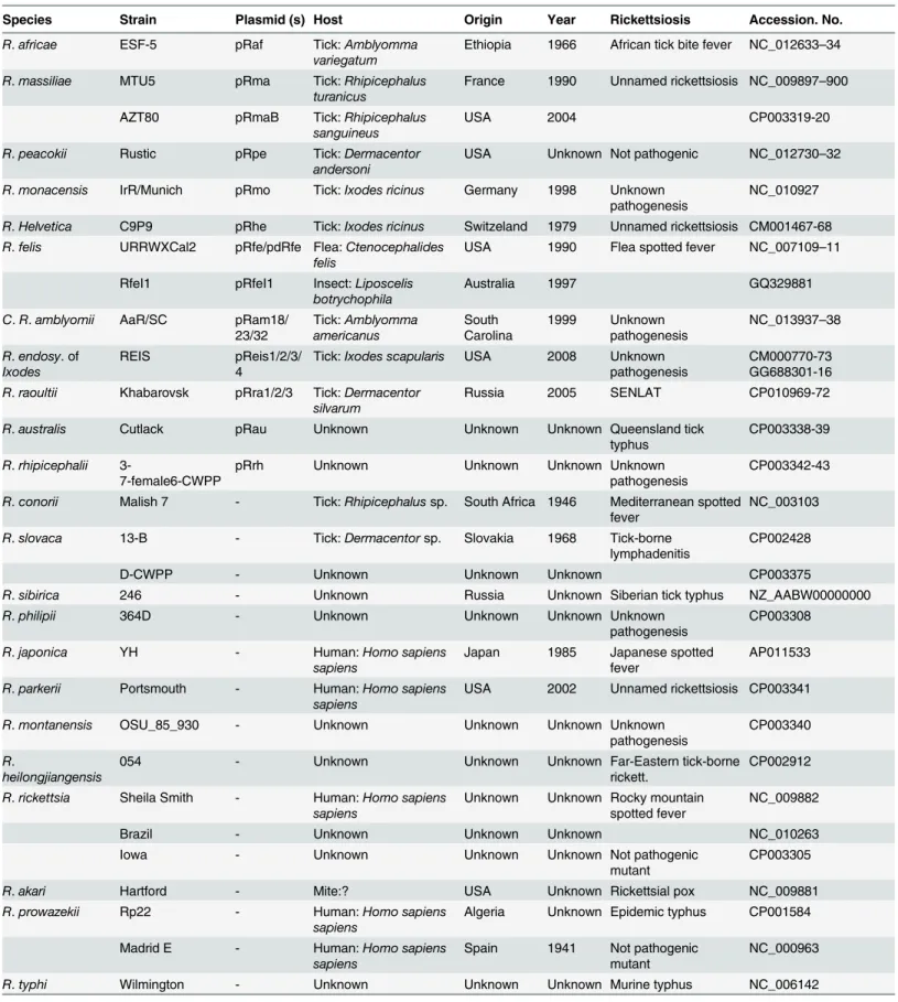
A total of 20 plasmids occurred in 11 SFG Rickettsia species, represented by 13 strains collected from various geographical locations worldwide (Table 1). In contrast, 9 SFG and TG species, represented by 15 strains, had no plasmids (Table 1). These included R. conorii, R. montanen-sis, R. parkeri, R. rickettsii, R sibirica, R. slovaca, R. prowazekii, R. typhi, and R. canadensis in which the absence of plasmid was confirmed in at least two strains by whole-genome sequenc-ing (sequences available in GenBank) and/or pulsed-field gel electrophoresis (PFGE) [21,22]. In another three species (R. japonica, R. philipii and R. heilongjiangensis) the absence of plas-mid needs to be confirmed in more than one strain. In R. akari and R. bellii (two SFG and AG species, respectively), Baldridge et al. [21,22] detected plasmids in some strains by PFGE, but the currently available genomes (Rak strain Hartford and Rbe strains RML369-C and OSU-89-389) had none. This may be explained by strain differences (i. e., plasmid loss in nature), as observed in R. africae [9] or their passage history in cell culture, as observed in R. felis [36] or other bacteria such as Borrelia burgdorferi [37]. Among the other 5 genera within the order
Table 1. Rickettsia and Orientia genomes extracted from the National Center for Biotechnology Information (NCBI) database.
Species Strain Plasmid (s) Host Origin Year Rickettsiosis Accession. No.
R. africae ESF-5 pRaf Tick: Amblyomma
variegatum
Ethiopia 1966 African tick bite fever NC_012633–34 R. massiliae MTU5 pRma Tick: Rhipicephalus
turanicus
France 1990 Unnamed rickettsiosis NC_009897–900 AZT80 pRmaB Tick: Rhipicephalus
sanguineus
USA 2004 CP003319-20
R. peacokii Rustic pRpe Tick: Dermacentor andersoni
USA Unknown Not pathogenic NC_012730–32 R. monacensis IrR/Munich pRmo Tick: Ixodes ricinus Germany 1998 Unknown
pathogenesis
NC_010927
R. Helvetica C9P9 pRhe Tick: Ixodes ricinus Switzeland 1979 Unnamed rickettsiosis CM001467-68 R. felis URRWXCal2 pRfe/pdRfe Flea: Ctenocephalides
felis
USA 1990 Flea spotted fever NC_007109–11 RfeI1 pRfeI1 Insect: Liposcelis
botrychophila
Australia 1997 GQ329881
C. R. amblyomii AaR/SC pRam18/ 23/32 Tick: Amblyomma americanus South Carolina 1999 Unknown pathogenesis NC_013937–38 R. endosy. of Ixodes REIS pReis1/2/3/ 4
Tick: Ixodes scapularis USA 2008 Unknown pathogenesis
CM000770-73 GG688301-16 R. raoultii Khabarovsk pRra1/2/3 Tick: Dermacentor
silvarum
Russia 2005 SENLAT CP010969-72
R. australis Cutlack pRau Unknown Unknown Unknown Queensland tick
typhus
CP003338-39
R. rhipicephalii
3-7-female6-CWPP
pRrh Unknown Unknown Unknown Unknown
pathogenesis
CP003342-43
R. conorii Malish 7 - Tick: Rhipicephalus sp. South Africa 1946 Mediterranean spotted fever
NC_003103
R. slovaca 13-B - Tick: Dermacentor sp. Slovakia 1968 Tick-borne
lymphadenitis
CP002428
D-CWPP - Unknown Unknown Unknown CP003375
R. sibirica 246 - Unknown Russia Unknown Siberian tick typhus NZ_AABW00000000
R. philipii 364D - Unknown Unknown Unknown Unknown
pathogenesis
CP003308
R. japonica YH - Human: Homo sapiens
sapiens
Japan 1985 Japanese spotted fever
AP011533
R. parkerii Portsmouth - Human: Homo sapiens sapiens
USA 2002 Unnamed rickettsiosis CP003341
R. montanensis OSU_85_930 - Unknown Unknown Unknown Unknown
pathogenesis
CP003340
R.
heilongjiangensis
054 - Unknown Unknown Unknown Far-Eastern tick-borne
rickett.
CP002912
R. rickettsia Sheila Smith - Human: Homo sapiens sapiens
Unknown Unknown Rocky mountain spotted fever
NC_009882
Brazil - Unknown Unknown Unknown NC_010263
Iowa - Unknown Unknown Unknown Not pathogenic
mutant
CP003305
R. akari Hartford - Mite:? USA Unknown Rickettsial pox NC_009881
R. prowazekii Rp22 - Human: Homo sapiens
sapiens
Algeria Unknown Epidemic typhus CP001584
Madrid E - Human: Homo sapiens
sapiens
Spain 1941 Not pathogenic mutant
NC_000963
R. typhi Wilmington - Unknown Unknown Unknown Murine typhus NC_006142
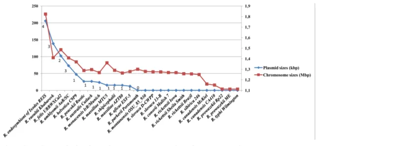
Rickettsiales (Anaplasma, Ehrlichia, Neorickettsia, Orientia, Wolbachia), all available genome sequences were devoid of plasmids [23]. RPs ranged in number and size per species from 1 to 4 plasmids and from 12 kb to 83 kb, respectively, and contained 15 to 85 genes (Table A inS1 File). A decrease in number and pooled plasmid size correlated with the decrease in chromo-some size in rickettsiae (Fig 1, Figure A inS2 File). Such results had previously been observed in prokaryotes [38]. We did not find any evident correlation between presence, number and/or size of plasmids, and rickettsial host range (Table 1).
The plasmidic gene content (plasmidome) counted 260 pRIGs (i.e., putative orthologous groups of plasmidic rickettsial genes) constructed from 747 protein-coding genes (Fig 2). Of these genes, 65% were full-length genes, whereas 35% were partially degraded (i.e., either split, fragment or chimerical). These genes can also be either highly degraded remnants, absent or lost in several plasmids. Overall, the degradation levels varied among plasmids, ranging from 16 to 40% in larger plasmids (size>47 kbp) to 44 to 59% in smaller plasmids (<40 kbp), sug-gesting that RPs are in a progressive degradation process and size reduction (Fig 3). This may
Table 1. (Continued)
Species Strain Plasmid (s) Host Origin Year Rickettsiosis Accession. No.
R. canadensis McKiel - Tick: Haemaph.
leporispalustris
Canada Unknown Unknown pathogenesis
NC_009879
CA410 - Unknown Unknown Unknown CP003304
R. bellii RML369-C - Tick: Dermacentor
varabilis
USA 1966 Unknown
pathogenesis
NC_007940
OSU-89-389 u. s. Unknown Unknown Unknown NC_009883
O. tsutsugamushi Boryong - Human: Homo sapiens sapiens
Japan 1995 Scrub typhus NC_009488.1
Ikeda - Human: Homo sapiens
sapiens
Japan 1979 NC_010793.1
Data were reported from [2,3] as well as PATRIC (http://patricbrc.org) and GENBANK (http://www.ncbi.nih.gov) databases. -, species without any plasmid. u.s., unavailabale sequences.
doi:10.1371/journal.pone.0147492.t001
Fig 1. Rickettsial plasmid sizes (pooled) and numbers plotted with their chromosome sizes. doi:10.1371/journal.pone.0147492.g001
also contribute to variations in the coding capacity of the plasmids which ranged from 75 to 92% (Table A inS1 File).
Overall, these data suggest that modern RPs are in a process of reductive evolution similar to that observed in rickettsial chromosomes [17]. Such a phenomenon is typical of symbiotic/ parasitic lifestyles [7,12,13,17,39]. However, this finding is in contrast with the megaplasmids of the free-living Bacillus cereus group which are a fusion of smaller plasmids [40]. RPs also exhibited G+C contents ranging from 32 to 35% (Table A inS1 File). Such values are similar to those of the rickettsial chromosomes (Table A inS1 File), suggesting that similar evolutionary forces such as vertical and horizontal gene transfers (VGT and HGT), duplications and selec-tion pressure, impacted both genomic components.
Fig 2. Gene content array ofRickettsia plasmidome obtained from BBH analysis. The right three columns showed the pRIG references of putative ortholog gene groups, the total number of genes for each pRIG and its corresponding annotation including its COGID form COG database. Each member of the pRIG was annotated as either complete (red), split (yellow), fragment (blue), chimeric (green) and absent (or remnant, violet).
doi:10.1371/journal.pone.0147492.g002
Fig 3. Frequencies of complete and degraded (split, fragment or chimeric) genes identified in the plasmidome ofRickettsia.
Origin and divergence of RPs
We performed phylogenetic reconstructions of all 260 pRIGs to quantify the evolutionary pro-cesses that may have shaped the 20 current RPs, and to reconstruct the gene content of their last common ancestor and their evolutionary scenarios. The phylogenetic analysis revealed that some pRIGs contained sub-clusters of orthologous genes from multiple origins (see below e.g., parA gene); These sub-clusters were subsequently considered as distinct pRIGs, thus increasing the plasmidome pRIG content from 260 to 275. A total of 136 phylogenetic trees of 184 (67%) pRIGs were retained, for which both neighbor-joining (NJ) and maximum likeli-hood (ML) methods displayed similar topologies. For the remaining 91 (33%) pRIGs no phy-logeny could be inferred.
Vertical evolution. Phylogenetic trees of 12 pRIGs (4.4%), conserved in most plasmids, exhibited two phylogenetically-related clades, one including 5 to 20 RPs and the other made of 1 to 26 chromosomes from Rickettsia and/or Orientia (Figures A1 to A9 inS3 File). Moreover, the supertree constructed from 10 of the 12 pRIGs confirmed these two phylogenetically-related clades, one containing all the 20 RPs and the other all Rickettsia/Orientia chromosomes (Fig 4).
This supertree was used as a reference for all following analyses. Phylogenetic analysis of a second set of 41 (15%) pRIGs showed the same two related clusters in each tree, one containing 1 to 11 RPs and the other 1 to 30 Rickettsia/Orientia chromosomes (Figures A10 to A49 inS3 File). Moreover, the analysis of another four (1.4%) pRIGs (mostly present as fragments), did not result in any reliable phylogeny, but they best matched with homologs in rickettsial chro-mosomes (data not shown). In all, our results suggest that the current RPs have a vertical origin (VGT) from a last common plasmidic ancestor that we named“pRICO” (Fig 5).
Both“pRICO” and “cRICO” (i.e., the last common ancestor of the current Rickettsia/Orien-tia chromosomes) may have been vertically inherited from a last common chromosomal ances-tor,“RICO” (Figs4and5). However, we observed that the internal phylogenetic clades inferred from plasmids and chromosomes were not often identical. This may be due to the facts that i) each species can harbor 1 to 4 plasmid(s) with similar or distinct evolutionary his-tory, ii) both plasmids and chromosomes are in a process of gene degradation and loss, and iii) the selection pressure shaping plasmids and chromosomes may be similar or different in each species and/or between species. Moreover, 8 (3%) pRIGs were also vertically transmitted to RPs but matched non-Rickettsia/Orientia species, suggesting that they might have been hori-zontally acquired by pRICO from unknown species (Figures A50 to A53 inS3 File). Thus, our data strongly support that pRICO harbored around 65 (23%) genes, suggesting that it might have had an estimated size around 60 to 70 kbp (Fig 5). The vertical inheritance is also corrobo-rated by the presence of plasmids in two distinct rickettsial groups (SFG and AG). In Salmo-nella enterica serovars, virulence plasmids were also described to be vertically transmitted [41], although no gene phylogeny and evolutionary relationships were carried out between Salmo-nella plasmids and chromosomes as well as microbial and non-microbial sequenced genomes to infer their origin and evolution.
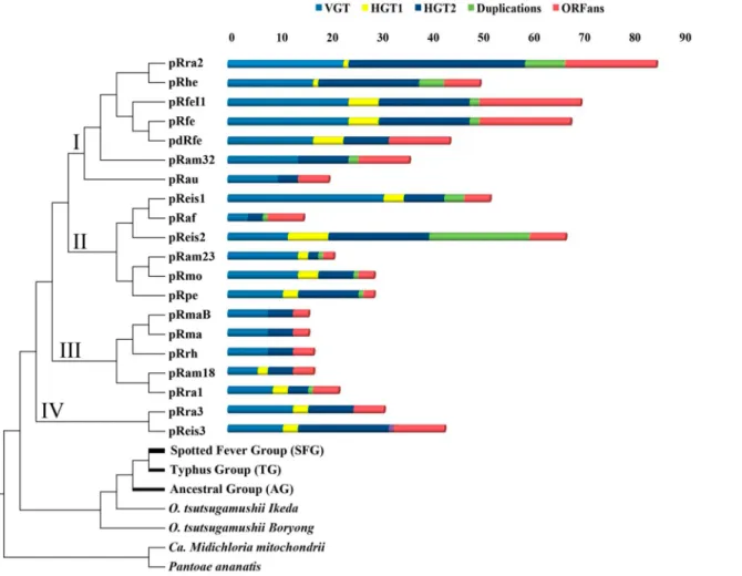
In the supertree, the pRICO ancestor diverged into a single or multiple plasmid(s) in rickett-sial species, and that these plasmids clustered into four putative groups (I to IV) (Fig 4): group I included 4 large and 3 small plasmids of 5 species (pRra2/pRhe/pRfe/pRfeI1/pdRfe/pRam32/ pRau); group II clustered 2 large and 4 small plasmids belonging to 5 species (pReis1/pRaf/ pReis2/pRam23/pRmo/pRpe); group III contained 5 small plasmids of 4 species (pRam18/ pRrh/pRra1/pRma/pRmaB); and group IV gathered one large and one small plasmids from two species (pReis3/pRra3). At the inter-species level, plasmids of the same group showed vari-able sequence conservations (Fig 4and Figures BA to BD inS2 File). At the intra-species level,
plasmids either belonged to the same group with high sequence conservation, as was the case for R. felis (pRfe, pdRfe and pRfeI1) and R. massiliae (pRma and pRmaB) strains (Fig 4and e.g., Figure BA inS2 File). In contrast, the plasmids from either R. raoultii (pRra1/2/3), Rickett-sia endosymbiont of I. scapularis (pReis1/3, except pReis1/2) or R. amblyomii (pRam18/23/32) strains were resolved into distinct phylogenetic groups with variable sequence conservation, although they lived in the same bacterial strain as well as the same host-species (Fig 4and Fig-ures BE to BG inS2 File).
The current RPs conserved highly variable numbers of genes acquired by VGT across the branches of the tree, ranging from 4 genes (6% out of the 65 pRICO genes) in the small pRaf plasmid to 31 (47% out of 65) in the large pReis1 plasmid (Fig 4). Therefore, the loss of ances-tral genes in RPs ranged from 53% in pReis1 to 94% in pRaf. The latter was the compacted RP in which reduction was the greatest driving force.
Horizontal evolution. The plasmidome exhibited 23 (8%) pRIGs resulting from evident horizontal gene exchanges (HGT1, plasmid-chromosome and/or plasmid-plasmid) within a given Rickettsia species, and/or between distinct Rickettsia species (Fig 5, Figures A37 and A54 to A74 inS3 File). RPs contained variable numbers of pRIGs resulting from HGT1 across the
Fig 4. Supertree obtained from 10 genes ofRickettsia plasmids as well as Rickettsia and Orientia chromosomes using subtree prune-and-regraft distance (D = 64). Candidatus Midichloria mitochondrii (YP_004679220.1) and Pantoea ananatis (YP_005193440.1) were used as outgroups. The list of genes used are two transposases, patatin-like phospholipase, thymidylate kinase, heat shock protein, dnaA-like replication initiator proteins, helix-turn-helix DNA-binding domain, cell surface antigen Sca12, conjugative transfer protein containing traD domain, and leucine rich-repeat containing protein. In the right, summary of evolutionary events that shaped rickettsial plasmids.
tree ranging from 0 in the small pRaf plasmid to 7 in the large pReis2 plasmid (Fig 4). Unex-pectedly, 4 pRIGs (1.4%, originally present in pRICO) of pRfe, pdRfe, pRfeI1, pRam32 and pRau exhibited close phylogenetic relationships with a gene cluster in R. prowazekii chromo-somes (Rpr22_687 to Rpr22_698 and RprME_862 to RprME_879, Figures A1, A5, A57 and A100 inS3 File). Similarly, 6 pRIGs (out of the 34 pRIGs acquired by HGT2) in pRpe showed close phylogenetic relationships with a gene cluster in the R. raoultii chromosome (Rra_909 to Rra_916, Figure C inS2 File, Figures A87 to A92 inS3 File). These data suggest that both Rick-ettsia species may have had one (for R. prowazekii) or an additional (for R. raoultii) plasmid that was disrupted in the course of the reductive genomic evolution in the genus Rickettsia, with subsequent HGT1 integration of plasmidic genes in their chromosomes. Alternatively, HGT1 events may have occurred between the pRfe, pRam or pRau plasmids and the R. prowa-zekii chromosome, and between pRpe and the R. raoultii chromosome, respectively.
Thirty-four (12%) pRIGs exhibited evident horizontal gene transfers (HGT2, plasmid-genome) with known non-Rickettsia/Orientia species, mostly belonging toα/γ-proteobacteria lineages (Fig 5, Figure D inS2 File, Figures A75 to A111 inS3 File). Moreover, 55 (20%) pRIGs displayed putative horizontal gene exchanges (HGT2) of unknown origin, but matching homo-logs of non-Rickettsia/Orientia species, also mostly belonging toα/γ-proteobacteria lineages
Fig 5. Evolutionary scenario of rickettsial plasmids. doi:10.1371/journal.pone.0147492.g005
(Fig 5, Figure E inS2 File, Figures A39 to A44, A46, A47, A100 and A112 to A136 inS3 File). RPs harbored highly variable numbers of genes exchanged by HGT2 events ranging from 0 in the small pRam23 plasmid to 24 in the large pRra2 plasmid (Fig 4).
Finally, five pRIGs (1.8%, originally present in 65 pRIGs inherited by VGT) and one pRIG (0,4%, originally present in the 34 pRIGs exchanged by HGT2) displayed evidence of addi-tional gene transfers (HGT3, plasmid-genome) between Rickettsia and distant lineages includ-ing Cardinium endosymbiont of Encarsia pergandiella, Candidatus Amoebophilus asiaticus (Bacteroidetes) as well as bacterial endosymbionts of either Trichoplax adhaerens or Hydra magnipapillata (Bacteria) (Fig 5, Figure D inS2 File, Figures A10, A11, A55, A57, A58 and A101 inS3 File) [42–44].
ORFans. The plasmidome contained a set of 64 pRIGs (23%, one pRIG in 1 to 8 plasmid (s)) classified as ORFans, including 40 that are specific for one given plasmid (Fig 5). RPs har-bored highly variable numbers of ORFans ranging from 2 in the small pRaf plasmid to 18 in the large pRra2 plasmid (Fig 4). These ORFans may have been horizontally acquired from microbes or eukaryotes not yet identified or that have disappeared, or have the de novo origin (gene genesis) either from non-coding regions, through an overprinting phenomenon of genes and/or underwent gene degradation processes [45–47].
Duplications. Recent duplication events drove 34 pRIGs (12%, one inparalog can be pres-ent 1 to 5 times in 1 to 4 plasmid(s)) after the divergence of pRICO (Fig 5). Of these, 21, 9 and 4 pRIGs were duplicated from pRIGs acquired by VGT and HGT2 (Figures A1, A4, A6 to A8, A10, A38, A50, A55 to A58, A60, A61, A81 to A86, A97, A100 and A129 inS3 File) as well as from ORFan pRIGs, respectively. The plasmids contained highly variable numbers of inpara-logs across the tree ranging from 0 in the small plasmid pRaf to 20 in the large plasmid pReis2 (Fig 4). Overall, the gene proliferation by recent duplications in plasmids was found to be simi-lar to duplication phenomenon in the corresponding chromosomes (see phylogenetic trees).
In summary, the current RPs were vertically transmitted from their last common ancestor pRICO. They evolved dynamically under an obligate intracellular lifestyle in two main phases: i) reductive evolution including pseudogenisation as well as gene degradation and loss, and ii) gene gain and innovation via HGT and proliferation-duplication as well as gene genesis. This mode of evolution is described in parasitic and symbiotic organisms as a recurrent biphasic model dominated by longer phases of genome reduction and simplification, punctuated by shorter phases of episodic complexification [39]. Although the expansion via HGT and gene duplications occurred in RPs, the contraction via gene degradation and loss severely contrib-uted to the reduction in gene content and plasmid size, and thus probably led to plasmid degra-dation and/or loss in several rickettsial species. This evolution of RPs seems to be in
accordance with a recent study which revealed that the prevailing mode of evolution in bacteria is genome reduction, which is partially compensated by the gain of new gene families via HGT [48]. The HGT process conferred on RPs a potential to exchange and disseminate their gene pools with their co-residing chromosomes and genomes of closely-related and distant phyloge-netic lineages, while duplication events enabled an internal gephyloge-netic amplification. This indi-cates that RPs were able to overcome the genetic barriers to HGT between distant phylogenetic lineages such asα-, γ-proteobacteria and/or Bacteroidetes. Similar horizontal transfer events between phylogenetically-distant clades were previously described (e.g., between plasmids from α-proteobacteria and Deinococci), underlying the key role of plasmids in genetic exchange throughout the microbial world [25]. Furthermore, all the identified evolutionary processes variably occurred across RPs suggesting that rickettsiaes may have been adapted to distinct biotic and/or abiotic factors for the invasion, survival and/or defense in eukaryotic host cells. Snel et al. [49] suggested that gene loss is under negative selection, while the process that adds genes is under positive selection.
Impact of evolution on functional diversity of RPs
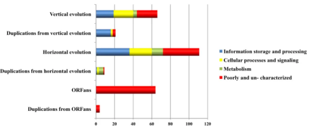
We examined the impact of the identified evolutionary forces (i. e., VGT, HGT, gene genesis, duplications and genome reduction) on the functional diversity of the RPs by a comparative analysis of the COG functional categories of the plasmidome (Fig 6). Overall, VGT had a weaker influence than HGT on pRIGs involved in information storage and processing (6% vs 14%, resp.), metabolism (1.4% vs 4%, resp.) and poorly- or un-characterized genes (8% vs 14%, resp.). In contrast, both forces had a similar impact on pRIGs related to cellular processes and signaling (7.6% vs 8.7%, resp.).
Information storage and processing categories
In information storage and processing categories, both VGT and HGT forces drove high and distinct proportions of pRIGs associated with replication, recombination and repair (5 vs 13%, resp.), but less and similar proportions on those involved in transcription (0.7 vs 0.3%, resp.) and translation (0 vs 0.3%, resp.) (see details in Table B inS1 File). Moreover, duplication forces acted on pRIGs resulting more from VGT than HGT events, and involved in replication, recom-bination and repair (6 vs 0.3%, resp.) (see details in Table B inS1 File). The abundance of HGT pRIGs from rickettsial and non-rickettsial chromosomes and duplications from VGT pRIGs in the replication, recombination and repair category were mainly due to mobile transposable ele-ments (i.e., transposases and integrases, 9 out of 13% and 5.1 out of 6%, resp.). These data from rickettsial plasmids corroborate a recent large-scale evolutionary study that showed that micro-bial plasmids share mainly genes encoding proteins involved in DNA transposition and recom-bination, some functions strongly linked to HGT process [25]. The single pRIG associated to translation was also exchanged with rickettsial chromosomes. Thus, VGT, HGT and duplication forces have driven pRIGs (e.g., dnaA-like, transposases, integrases, resolvases and relaxases) that may contribute to maintenance, rearrangements and plasticity of plasmids, making them self-replicative and simultaneously dynamic to gene exchange with various evolutionary lineages and consequent diversification of rickettsial biological activities and phenotypic traits.
Cellular processes and signaling categories
In cellular processes and signaling categories, both VGT and HGT forces acted in nearly equal proportions on pRIGs related to intracellular trafficking and secretion (tra operons) (4.3 vs 4%, resp.) (see details in Table B inS1 File). Based on the classification of plasmids by their mobility [38], our data predict that three (15%) large plasmids (pRra2/pReis1/pReis3) are
self-Fig 6. COG functional categories ofRickettsia plasmidome originated from vertical and horizontal evolutions as well as duplications.
transmissible or conjugative (i.e., having MOB or relaxases and TRA modules), whereas 8 (40%) small or large plasmids (pRam32/pRra3/pReis2/pRau/pdRfe/pRfe/pRfeI1/pRhe) are mobilisable (i.e., having MOB and/or degraded or no TRA modules). Some of these mobilisable replicons (e.g., pRra3) could be transmissible if using conjugative machineries of co-residing self-transmissible replicons (e.g., pRra2) [38,50,51]. The 9 (45%) remaining small plasmids (e.g., pRaf/pRam12/pRam23/pRrh/pRpe/pRmo/pRra1/pRma/pRmaB), lacked MOB and TRA modules and may not be transmissible. The three mobility types in RPs are common in various bacterial phyla includingα-proteobacteria [38]. Overall, the presence of conjugative system in RPs may have strong implications in their evolutionary dynamics and history as well as in the acquisition and spread of ecologically important traits (novel and/or ancestral among various bacteria) that may be associated to rearrangement, adaptation and/or pathogenesis of rickett-siae. This is consistent with the analysis of gene sharing networks among 329 proteobacteria that suggested that gene transfer in this phylum is frequently mediated by conjugation [52].
Moreover, both VGT and HGT forces drove in a similar way pRIGs related to posttransla-tional modification and chaperones (0.7 vs 0.3%, resp.) and defense mechanisms (0.3 vs 0.7%, resp.), but not pRIGs associated to transduction mechanisms (e.g., 2 vs 0.3%, resp.) (see details in Table B inS1 File). Among these processes we can cite pRIGs associated to adaptation (e.g., hsp), defense (type I restriction-modification system methyltransferase subunit) and stress (e.g., spoT) that may help the survival of host bacteria in an obligate intracellular life style. However, HGT, but not VGT forces, drove 3.3% of pRIGs related to cell wall/membrane/enve-lope biogenesis and cycle control, including genes associated to plasmid segregation (e.g., parA/B) and regulator of cell division and wall (e.g., mobile mystery protein B, putative lytic transglycosylase, glycosyltransferase, group 1 family protein). In addition, duplication forces drove equal proportions of pRIGs resulting from both VGT and HGT events, and associated with intracellular trafficking and secretion, posttranslational modification and chaperones and cycle control (0.3 vs 0.3%, resp.).
Metabolism categories
In metabolism categories, both VGT and HGT forces drove equal proportions of pRIGs involved in inorganic ion transport and metabolism (0.3 vs 0.3%, resp.) (see details in Table B inS1 File). However, only VGT forces acted on 1.1% of pRIGs related to nucleotide/carbohy-drate transport and metabolism (thymidylate kinase, major facilitator superfamily MFS-type transporter). In contrast, only HGT forces drove 4.4% of pRIGs associated with amino acid/ lipid/coenzyme and secondary metabolites biosynthesis, transport and metabolism (e.g., L-allo-threonine aldolases, 3-oxoacyl-(acyl-carrier-protein) synthase III, bioA, B, C, D and F). Furthermore, duplication forces acted on 1.8% of pRIGs including bioA, B, C, D and F. Overall, VGT and duplication forces drove pRIGs common to RPs and Rickettsia/Orientia chromo-somes which may increase the biological activities of some rickettsiae, whereas HGT forces enabled some RPs to acquire novel metabolic functions absent in Rickettsia/Orientia chromo-somes that may complete host-metabolic gaps and confer host-adaptive phenotypes to obligate intracellular lifestyle.
Poorly and uncharacterized categories
In poorly and uncharacterized categories, VGT, HGT and de novo gene genesis drove distinct proportions of pRIGs (7.6, 13.8 and 23.3%, resp.) which have known or unknown functions and/or poorly or uncharacterized COG assignments and that remain to be elucidated for the biology of Rickettsia species (see details in Table B inS1 File). Moreover, duplication forces acted on similar proportions of pRIGs resulting from VGT, HGT and de novo genes genesis
(1.1, 0.3 and 1.4%, resp.). Among the poorly and uncharacterized categories, VGT forces drove some pRIGs (2.5%) related to adhesion and/or infection such as those encoding the cell surface antigen proteins Sca and patatin-like phospholipase Pat [10,53], whereas HGT forces acted on some pRIGs (5.8%) coding ankyrin containing proteins Ank, tetratricopeptide repeat-containing proteins Tpr known to play a role in protein-protein interactions in eukaryotes, and suspected to be associated to adaptation to hosts and/or to virulence [14].
In several human bacterial pathogens, such as Salmonella spp., Escherichia coli and Borrelia spp., virulence plasmids have been identified [37,41,54]. However, among the most pathogenic rickettsiae, R. prowazekii and R. conorii do not harbor any plasmid whereas R. australis contains one plasmid. These data suggest that the role of RPs in rickettsial virulence, remains uncertain and requires further investigations. In addition, the functional diversity of RPs observed in the present study suggests that some of them may be used as transformation vectors for rickettsiae, as was recently described using a shuttle vector system derived from R. amblyomii plasmids that enabled stable transformations of diverse rickettsiae [55,56]. Thus, the genetic manipulation of rickettsiae using RPs may provide new insights in the pathogenesis of these obligate intracellular bacteria as well as the dissemination of their genotypic and phenotypic traits.
Conclusions
This study offers an overview of the evolution and functional diversity of rickettsial plasmids. The current RPs have been shaped by multiple evolutionary forces in a bottleneck ecological niche. Initially, they were vertically inherited and then may have evolved under a biphasic model including a strong reductive evolution as well as innovations by horizontal gene transfer and gene duplication and genesis. This study demonstrates for the first time that plasmids of obligate intracellular bacteria can evolve under both vertical and reductive forces similar to those shaping their co-residing chromosomes. The evolutionary processes that have shaped RPs suggest that they might be influenced by molecular interactions between rickettsiae and eukaroytic host cells including biological needs for adaptation and survival, as suggested for Buchnera aphidicola in which metabolic requirements could determine when genome reduc-tion occurs [39]. RPs may play diverse functional roles that are either novel or similar to those of rickettsial chromosomes via various evolutionary mechanisms. Overall, this study provides insights into rickettsial plasmids and their biology and evolution.
Supporting Information
S1 File. (PDF) S2 File. (PDF) S3 File. (PDF)Acknowledgments
The study was funded by the Mediterranée-Infection foundation.
Author Contributions
Conceived and designed the experiments: KEK PP DR PEF. Performed the experiments: KEK PEF. Analyzed the data: KEK PP DR PEF. Wrote the paper: KEK PP DR PEF.
References
1. Weinert LA, Werren JH, Aebi A, Stone GN, Jiggins FM. Evolution and diversity of Rickettsia bacteria. BMC Biol. 2009; 7: 6. doi:10.1186/1741-7007-7-6PMID:19187530
2. Merhej V, Raoult D. Rickettsial evolution in the light of comparative genomics. Biol Rev Camb Philos Soc. 2011; 86: 379–405. doi:10.1111/j.1469-185X.2010.00151.xPMID:20716256
3. Parola P, Paddock CD, Socolovschi C, Labruna MB, Mediannikov O, Kernif T, et al. Update on tick-borne rickettsioses around the world: a geographic approach. Clin Microbiol Rev. 2013; 26: 657–702. doi:10.1128/CMR.00032-13PMID:24092850
4. Stothard DR, Clark JB, Fuerst PA. Ancestral divergence of Rickettsia bellii from the spotted fever and typhus groups of Rickettsia and antiquity of the genus Rickettsia. Int J Syst Bacteriol. 1994; 44: 798– 804. PMID:7981106
5. Raoult D, Roux V. Rickettsioses as paradigms of new or emerging infectious diseases. Clin Microbiol Rev. 1997; 10: 694–719. PMID:9336669
6. Andersson JO, Andersson SGE. Genome degradation is an ongoing process in Rickettsia. Mol Biol Evol. 1999; 16: 1178–1191. PMID:10486973
7. Andersson SGE, Zomorodipour A, Andersson JO, Sicheritz-Ponten T, Alsmark UCM, Podowski RM, et al. The genome sequence of Rickettsia prowazekii and the origin of mitochondria. Nature. 1998; 396: 133–140. PMID:9823893
8. Darby AC, Cho NH, Fuxelius HH, Westberg J, Andersson SG. Intracellular pathogens go extreme: genome evolution in the Rickettsiales. Trends Genet. 2007; 23: 511–520. PMID:17822801
9. Fournier PE, El Karkouri K, Leroy Q, Robert C, Giumelli B, Renesto P, et al. Analysis of the Rickettsia africaegenome reveals that virulence acquisition in Rickettsia species may be explained by genome reduction. BMC Genomics. 2009; 10: 166. doi:10.1186/1471-2164-10-166PMID:19379498
10. Sahni SK, Narra HP, Sahni A, Walker DH. Recent molecular insights into rickettsial pathogenesis and immunity. Future Microbiol. 2013; 8: 1265–1288. doi:10.2217/fmb.13.102PMID:24059918
11. Bechah Y, El Karkouri K, Mediannikov O, Leroy Q, Pelletier N, Robert C, et al. Genomic, proteomic, and transcriptomic analysis of virulent and avirulent Rickettsia prowazekii reveals its adaptive mutation capabilities. Genome Res. 2010; 20: 655–663. doi:10.1101/gr.103564.109PMID:20368341
12. McLeod MP, Qin X, Karpathy SE, Gioia J, Highlander SK, Fox GE, et al. Weinstock, Complete genome sequence of Rickettsia typhi and comparison with sequences of other rickettsiae. J Bacteriol. 2004; 186: 5842–5855.
13. Ogata H, Audic S, Renesto-Audiffren P, Fournier PE, Barbe V, Samson D, et al. Mechanisms of evolu-tion in Rickettsia conorii and R. prowazekii. Science. 2001; 293: 2093–2098. PMID:11557893
14. Ogata H, La SB, Audic S, Renesto P, Blanc G, Robert C, et al. Genome sequence of Rickettsia bellii illuminates the role of amoebae in gene exchanges between intracellular pathogens. PLoS Genet. 2006; 2: e76. PMID:16703114
15. Ogata H, Renesto P, Audic S, Robert C, Blanc G, Fournier PE, et al. The genome sequence of Rickett-sia felisidentifies the first putative conjugative plasmid in an obligate intracellular parasite. PLoS Biol. 2005; 3: e248. PMID:15984913
16. Blanc G, Ogata H, Robert C, Audic S, Claverie JM, Raoult D. Lateral gene transfer between obligate intracellular bacteria: evidence from the Rickettsia massiliae genome. Genome Res. 2007; 17: 1657– 1664. PMID:17916642
17. Blanc G, Ogata H, Robert C, Audic S, Suhre K, Vestris G, et al. Reductive genome evolution from the mother of Rickettsia. PLoS.Genet. 2007; 3: e14. PMID:17238289
18. Ellison DW, Clark TR, Sturdevant DE, Virtaneva K, Porcella SF, Hackstadt T. Genomic comparison of virulent Rickettsia rickettsii Sheila Smith and avirulent Rickettsia rickettsii Iowa. Infect Immun. 2008; 76: 542–550. PMID:18025092
19. Fournier PE, Raoult D. Intraspecies diversity of Rickettsia conorii. J Infect Dis. 2009; 199: 1097–1098. doi:10.1086/597211PMID:19275477
20. Felsheim RF, Kurtti TJ, Munderloh UG. Genome sequence of the endosymbiont Rickettsia peacockii and comparison with virulent Rickettsia rickettsii: identification of virulence factors. PLoS ONE. 2009; 4: e8361. doi:10.1371/journal.pone.0008361PMID:20027221
21. Baldridge GD, Burkhardt NY, Felsheim RF, Kurtti TJ, Munderloh UG. Plasmids of the pRM/pRF family occur in diverse Rickettsia species. Appl Environ Microbiol. 2008; 74: 645–652. PMID:18065613
22. Baldridge GD, Burkhardt NY, Labruna MB, Pacheco RC, Paddock CD, Williamson PC, et al. Wide dis-persal and possible multiple origins of low-copy-number plasmids in rickettsia species associated with blood-feeding arthropods. Appl Environ Microbiol, 2010; 76: 1718–1731. doi:10.1128/AEM.02988-09
23. Gillespie JJ, Beier MS, Rahman MS, Ammerman NC, Shallom JM, Purkayastha A, et al. Plasmids and rickettsial evolution: insight from Rickettsia felis. PLoS ONE. 2007; 2: e266. PMID:17342200
24. Halary S, Leigh JW, Cheaib B, Lopez P, Bapteste E. Network analyses structure genetic diversity in independent genetic worlds. Proc Natl Acad Sci U S A. 2010; 107: 127–132. doi:10.1073/pnas. 0908978107PMID:20007769
25. Tamminen M, Virta M, Fani R, Fondi M. Large-scale analysis of plasmid relationships through gene-sharing networks. Mol Biol Evol. 2012; 29: 1225–1240. doi:10.1093/molbev/msr292PMID:22130968
26. Bocs S, Cruveiller S, Vallenet D, Nuel G, Medigue C. AMIGene: Annotation of MIcrobial Genes. Nucleic Acids Res. 2003; 31: 3723–3726. PMID:12824403
27. Altschul SF, Madden TL, Schaffer AA, Zhang J, Zhang Z, Miller W, et al. Gapped BLAST and PSI-BLAST: a new generation of protein database search programs. Nucleic Acids Res. 1997; 25: 3389– 3402. PMID:9254694
28. Tatusov RL, Koonin EV, Lipman DJ. A genomic perspective on protein families. Science. 1997; 278: 631–637.
29. Kristensen DM, Kannan L, Coleman MK, Wolf YI, Sorokin A, Koonin EV, et al. A low-polynomial algo-rithm for assembling clusters of orthologous groups from intergenomic symmetric best matches. Bioin-formatics. 2010; 26: 1481–1487. doi:10.1093/bioinformatics/btq229PMID:20439257
30. Larkin MA, Blackshields G, Brown NP, Chenna R, McGettigan PA, McWilliam H, et al. Clustal W and Clustal X version 2.0. Bioinformatics. 2007; 23: 2947–2948. PMID:17846036
31. Edgar RC. MUSCLE: multiple sequence alignment with high accuracy and high throughput. Nucleic Acids Res. 2004; 32: 1792–1797. PMID:15034147
32. Tamura K, Stecher G, Peterson D, Filipski A, Kumar S. MEGA6: Molecular Evolutionary Genetics Anal-ysis version 6.0. Mol Biol Evol. 2013; 30: 2725–2729. doi:10.1093/molbev/mst197PMID:24132122
33. Whidden C, Zeh N, Beiko RG. Supertrees Based on the Subtree Prune-and-Regraft Distance. Syst Biol. 2014; 63: 566–581. doi:10.1093/sysbio/syu023PMID:24695589
34. Shannon P, Markiel A, Ozier O, Baliga NS, Wang JT, Ramage D, et al. Cytoscape: a software environ-ment for integrated models of biomolecular interaction networks. Genome Res. 2003; 13: 2498–2504. PMID:14597658
35. Guy L, Kultima JR, Andersson SG. genoPlotR: comparative gene and genome visualization in R. Bioin-formatics. 2010; 26: 2334–2335. doi:10.1093/bioinformatics/btq413PMID:20624783
36. Fournier PE, Belghazi L, Robert C, El Karkouri K, Richards A, Greub G, et al. Variations of Plasmid Content in Rickettsia felis. PLoS ONE. 2008; 3: e2289. doi:10.1371/journal.pone.0002289PMID:
18509454
37. Chan K, Casjens S, Parveen N. Detection of established virulence genes and plasmids to differentiate Borrelia burgdorferistrains. Infect Immun. 2012; 80: 1519–1529. doi:10.1128/IAI.06326-11PMID:
22290150
38. Smillie C, Garcillan-Barcia MP, Francia MV, Rocha EP, de la Cruz F. Mobility of plasmids. Microbiol Mol Biol Rev. 2010; 74: 434–452. doi:10.1128/MMBR.00020-10PMID:20805406
39. Wolf YI, Koonin EV. Genome reduction as the dominant mode of evolution. Bioessays. 2013; 35: 829– 837. doi:10.1002/bies.201300037PMID:23801028
40. Zheng J, Peng D, Ruan L, Sun M. Evolution and dynamics of megaplasmids with genome sizes larger than 100 kb in the Bacillus cereus group. BMC Evol Biol. 2013; 13: 262. doi: 10.1186/1471-2148-13-262PMID:24295128
41. Feng Y, Liu J, Li YG, Cao FL, Johnston RN, Zhou J, et al. Inheritance of the Salmonella virulence plas-mids: mostly vertical and rarely horizontal. Infect Genet Evol. 2012; 12: 1058–1063.
42. Chapman JA, Kirkness E.F., Simakov O., Hampson S.E., Mitros T., Weinmaier T, et al. The dynamic genome of Hydra. Nature. 2010; 464: 592–596. doi:10.1038/nature08830PMID:20228792
43. Driscoll T, Gillespie JJ, Nordberg EK, Azad AF, Sobral BW. Bacterial DNA sifted from the Trichoplax adhaerens(Animalia: Placozoa) genome project reveals a putative rickettsial endosymbiont. Genome Biol Evol. 2013; 5: 621–645. doi:10.1093/gbe/evt036PMID:23475938
44. Duron O. Lateral transfers of insertion sequences between Wolbachia, Cardinium and Rickettsia bacte-rial endosymbionts. Heredity. 2013; 111: 330–337. doi:10.1038/hdy.2013.56PMID:23759724
45. Yin Y, Fischer D. On the origin of microbial ORFans: quantifying the strength of the evidence for viral lat-eral transfer. BMC Evol Biol. 2006; 6: 63. PMID:16914045
46. Yomtovian I, Teerakulkittipong N, Lee B, Moult J, Unger R. Composition bias and the origin of ORFan genes. Bioinformatics. 2010; 26: 996–999. doi:10.1093/bioinformatics/btq093PMID:20231229
47. Tautz D, Domazet-Loso T. The evolutionary origin of orphan genes. Nat Rev Genet. 2011; 12: 692– 702.
48. Puigbò P, Lobkovsky AE, Kristensen DM, Wolf YI, Koonin EV. Genomes in turmoil: quantification of genome dynamics in prokaryote supergenomes. BMC Biol. 2014; 21: 12:66. doi: 10.1186/s12915-014-0066-4PMID:25141959
49. Snel B, Bork P. Genomes in flux: the evolution of archaeal and proteobacterial gene content. Genome Res. 2002; 12:17–25. PMID:11779827
50. Garcillan-Barcia MP, Francia MV, de la Cruz F. The diversity of conjugative relaxases and its applica-tion in plasmid classificaapplica-tion. FEMS Microbiol Rev. 2009; 33: 657–687. PMID:19396961
51. Torres TG, Pistorio M, Althabegoiti MJ, Cervantes L, Wibberg D, Wibberg D, et al. Rhizobial plasmid pLPU83a is able to switch between different transfer machineries depending on its genomic back-ground. FEMS Microbiol Ecol. 2014. 88: 565–578. doi:10.1111/1574-6941.12325PMID:24646299
52. Kloesges T, Popa TO, Martin W, Dagan T. Networks of gene sharing among 329 proteobacterial genomes reveal differences in lateral gene transfer frequency at different phylogenetic depths. Mol Biol Evol. 2011. 28: 1057–1074. doi:10.1093/molbev/msq297PMID:21059789
53. Rahman MS, Gillespie JJ, Kaur SJ, Sears KT, Ceraul SM, Beier-Sexton M, et al. Rickettsia typhi Pos-sesses Phospholipase A(2) Enzymes that Are Involved in Infection of Host Cells. Plos Pathog. 2013; 9: e1003399. doi:10.1371/journal.ppat.1003399PMID:23818842
54. Johnson TJ, Nolan LK. Pathogenomics of the virulence plasmids of Escherichia coli. Microbiol Mol Biol Rev. 2009; 73: 750–774. doi:10.1128/MMBR.00015-09PMID:19946140
55. Burkhardt NY, Baldridge GD, Williamson PC, Billingsley PM, Heu CC, Felsheim RF, et al. Development of shuttle vectors for transformation of diverse Rickettsia species. PloS ONE. 2011; 6: e29511. doi:10. 1371/journal.pone.0029511PMID:22216299
56. Wood DO, Hines A, Tucker AM, Woodard A, Driskell LO, Burkhardt NY, et al. Establishment of a repli-cating plasmid in Rickettsia prowazekii. PloS ONE. 2012; 7: e34715. doi:10.1371/journal.pone. 0034715PMID:22529927