HAL Id: inserm-01071442
https://www.hal.inserm.fr/inserm-01071442
Submitted on 14 Oct 2014
HAL is a multi-disciplinary open access
archive for the deposit and dissemination of
sci-entific research documents, whether they are
pub-lished or not. The documents may come from
teaching and research institutions in France or
abroad, or from public or private research centers.
L’archive ouverte pluridisciplinaire HAL, est
destinée au dépôt et à la diffusion de documents
scientifiques de niveau recherche, publiés ou non,
émanant des établissements d’enseignement et de
recherche français ou étrangers, des laboratoires
publics ou privés.
Investigating the real role of HIF-1 and HIF-2 in iron
recycling by macrophages
Carole Peyssonnaux
To cite this version:
Carole Peyssonnaux. Investigating the real role of HIF-1 and HIF-2 in iron recycling by macrophages.
Haematologica, Ferrata Storti Foundation, 2014, pp.e112-4. �10.3324/haematol.2013.102319�.
�inserm-01071442�
Investigating the real role of HIF-1 and HIF-2 in iron
recycling by macrophages
Most of the iron required for erythropoiesis is acquired by heme iron recycling of senescent erythrocytes by tissue macrophages (mainly spleen, bone marrow and liver macrophages). This process, called erythrophagocytosis (EP), is essential for mammalian iron homeostasis. Perturbations in EP occur in several diseases, including ane-mia of chronic disorders, hemochromatosis and tha-lassemia.1
The senescent red blood cell is internalized by phagocy-tosis and degraded within acidified early phagosomes. Heme is transported into the cytosol by the recently iden-tified heme transporter HRG1 (Heme-responsive gene 1).2
Heme is then catabolized by heme oxygenase 1 (HO-1) leading to the release of iron. DMT-1 (Divalent Metal Transporter-1) and natural resistance-associated macrophage protein 1 (Nramp1), play essential roles in heme iron recycling3,4but their functional roles remain to
be determined. Iron is released from macrophages by the iron exporter ferroportin (FPN) or stored within ferritin. During EP, these different genes, such as HO-1 and FPN, are regulated at post-transcriptional level (through the
haematologica 2014; 99:e112
L
ETTERS TO THE
E
DITOR
Figure 1. Iron home-ostasis parameters in
WT, HIF-1α∆M and
HIF-2α∆M mice treated with PBS or PHZ. 12 week-old WT, HIF-1α∆
M or HIF-2∆
Mmale mice (n ≥ 4) were injected for two consecutive days with 50 mg/kg of PHZ or PBS and were killed 48 h after the last injec-tion. The mice were maintained on an iron deficient diet immedi-ately after the first injection to avoid a compensatory increase in dietary iron absorp-tion. (A) HIF-1, HIF-2 and F4/80 immunoflu-orescence on PBS or PHZ-treated WT mice. (B) Red blood cells
(RBC), hemoglobin,
splenic index (defined as the spleen/body weight ratio), plasma iron, ferritin levels and (C) liver Iron, liver FPN and HO-1 mRNA levels (by Q-PCR normalized to cyclophilin A) in WT and HIF-1α∆
Mor HIF-2∆M mice treated with PBS or PHZ.
A
B
Iron Regulatory Proteins), and/or at a transcriptional level by heme through a pathway involving Bach1 (BTB and CNC homology 1)/Nrf2 (nuclear factor erythroid 2-related factor 2).5
Hypoxia and its cellular mediators (hypoxia inducible factors: HIF-1 and HIF-2) have been shown to transcrip-tionally regulate essential genes involved in EP (e.g. HO-1, DMT-1, FPN)6-8in a number of cell lines or tissues and have,
therefore, been suggested to have a role in erythrophagocy-tosis,9-11a concept that can be read in a number of recent
reviews, with no associated references. Nevertheless, the physiological role of HIF-1 and HIF-2 in iron recycling of erythrocytes has never been investigated. We, therefore, felt it was important to establish the ‘real’ role of HIF in iron recycling based on scientific data and not extrapolation from other reviews.
haematologica 2014; 99:e113
L
ETTERS TO THE
E
DITOR
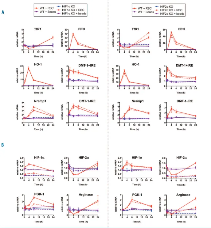
Figure 2. mRNA expression patterns of iron recycling genes after EP in HIF-1 or HIF-2 deficient BMDMs. Q-PCR time course analysis of (A) TfR1, FPN, HO-1, DMT-1+IRE, DMT-1-IRE, Nramp1, (B) HIF-1α, HIF-2 α, PGK-1, Arginase mRNA levels in WT, HIF-1 (HIF-1α KO) or HIF-2 deficient (HIF-2α KO) BMDMs incubated or not (-) with either latex beads (purple line) or RBCs (pink line). Red blood cell aging was achieved by incubating RBCs with a calcium ionophore solution. BMDMs were incubated with latex beads or RBCs (ratio 100 RBCs/BMDM) for 1 hour, then incubated 5 min in a hypotonic solution to lyse non-ingested RBCs and washed twice with PBS. Values were normalized to WT. RNA extraction, reverse transcription and quantitative PCR have been previously described.8All samples were normalized to the threshold cycle value for cyclophilin A. Sequences of the primers used are available upon request. n=3 per time point. Statistical values are repre-sented in Online Supplemental Figure S2.
A
We found that HIF-1 and HIF-2 were stabilized in the macrophages of the spleen in conditions of increased ery-thropoiesis due to acute hemolytic anemia induced by phenylhydrazine (PHZ) (Figure 1A). To determine the impact of the deletion of macrophage HIF-1 and HIF-2 in iron recycling in vivo, we evaluated several parameters of iron homeostasis in HIF-1lox/lox-LysM Cre (HIF-1∆M),12
HIF-2lox/lox-LysM Cre (HIF-2∆M)13
mice and control littermates in basal or PHZ conditions. We first confirmed the decrease in HIF-1 and HIF-2 expression in the respective conditional knockout mice and validated the lack of compensatory mechanism (Online Supplementary Figure S1A). At steady state, the red blood cell, hematocrit, hemoglobin, plasma iron and ferritin levels, as well as the splenic indices were similar between WT and mutant mice (Figure 1B). Two days after PHZ treatment, the expected significant decrease of hematologic parameters, as well as the increased splenic index, ferritin (Figure 1B) and liver iron (Figure 1C) levels were observed similarly in WT and KO mice. The increased expression of liver HO-1 and FPN, two key genes involved in iron recycling, was identical between WT and mutant mice (Figure 1C). Importantly, the increased plasma iron, underlying increased iron recycling was intact in the KO mice (Figure 1B). The recovery from anemia was identical in WT and mutant mice (Online Supplementary Figure S1B). We next used a characterized cellular model of iron recy-cling by macrophages,14which tends to mimic the natural
clearance of senescent RBCs, to investigate more thorough-ly whether this lack of effect was due to the inability of HIF-1 and HIF-2 in macrophages to regulate key genes involved in EP. Primary cultures of murine bone-marrow derived macrophages (BMDMs) were incubated with latex beads or artificially aged murine RBCs. Latex beads had no effects on any tested genes by quantitative PCR, showing that phagocytosis itself was not able to elicit gene expres-sion responses (Figure 2). Four hours after the incubation of senescent RBCs, TfR1 mRNA levels were significantly decreased in BMDMs, concomitant to an increase in FPN and HO-1, indicating an increased iron content, as previ-ously described14,15(Figure 2A). DMT-1+IRE mRNA levels
were maximally induced as early as one hour after EP, while Nramp-1 accumulation was delayed, peaking at 8 h. Unexpectedly, after EP, we found an increase in HIF-1 expression, reaching maximal levels at 4 h and returning to basal levels at 24 h. Conversely, HIF-2 mRNA levels decreased maximally at 4 h (Figure 2B). To determine the role of HIFs in the transcriptional regulation of these iron recycling genes, we derived macrophages from HIF-1∆M and
HIF-2∆M mice. The levels of HIF-1α and HIF-2 α mRNAs
were markedly decreased in the macrophages deficient for HIF-1α and HIF-2α, respectively (Figure 2B). Neither the deletion of HIF-1 nor HIF-2 modified the expression of any tested genes involved in iron recycling (Figure 2A). Importantly, the response of prototypic HIF-1 and HIF-2 target genes, such as PGK-1 and Arginase, was significantly decreased in HIF-1 and HIF-2 deficient macrophages, respectively. Finally, deletion of one isoform did not affect the expression of the other, ruling out a putative compen-satory mechanism.
In conclusion, in contrast to other cells or tissues, macrophage HIF-1 and HIF-2 are not required in the regu-lation of iron recycling genes. While HIF-1 has been shown to regulate TfR1 and HO-1 in other contexts, and has been anticipated to have a role in erythrophagocytosis,9-11 we
show here that macrophage-specific deletion of HIF-1 does not affect iron recycling. Besides, while HIF-2 is essential for iron absorption by regulating DMT-1 and FPN in the enterocytes,6,8it is not essential for the regulation of these
genes during erythrophagocytosis. Our findings that
nei-ther HIF-1 nor HIF-2 are necessary to regulate key iron recycling genes are important to avoid erroneous conclu-sions drawn from reviews.
Jacques R.R. Mathieu,1,2,3,4Mylène Heinis,1,2,3,4
Sara Zumerle,1,2,3,4Stéphanie Delga,1,2,3,4Agnès Le Bon,1,2,3
and Carole Peyssonnaux1,2,3,4*
1INSERM, U1016, Institut Cochin, Paris; 2CNRS, UMR8104,
Paris; 3Université Paris Descartes, Sorbonne Paris Cité, Paris;
and 4Laboratory of Excellence GR-Ex, Paris, France
Correspondence: carole.peyssonnaux@inserm.fr doi:10.3324/haematol.2013.102319
Key words: iron recycling, macrophage, HIF-1. HIF-2.
Acknowledgments: we would like to thank Sophie Vaulont and Olivier Hermine for critical reading of the manuscript. We also thank the Immunobiology platform of the Cochin Institute for help with the FACS analysis. This study was supported by a funding from the European Research Council under the European Community’s Seventh Framework Program (FP7/2011-2015 Grant agreement n. 261296).
The online version of this article has a Supplementary Appendix. Information on authorship, contributions, and financial & other disclo-sures was provided by the authors and is available with the online version of this article at www.haematologica.org.
References
1. Beaumont C, Delaby C. Recycling iron in normal and pathological states. Semin Hematol. 2009;46(4):328-38.
2. White C, Yuan X, Schmidt PJ, Bresciani E, Samuel TK, Campagna D, et al. HRG1 is essential for heme transport from the phagolysosome of macrophages during erythrophagocytosis. Cell Metab. 2013;17(2):261-70.
3. Soe-Lin S, Apte SS, Andriopoulos B Jr, Andrews MC, Schranzhofer M, Kahawita T, et al. Nramp1 promotes efficient macrophage recy-cling of iron following erythrophagocytosis in vivo. Proc Natl Acad Sci USA. 2009;106(14):5960-5.
4. Soe-Lin S, Apte SS, Mikhael MR, Kayembe LK, Nie G, Ponka P. Both Nramp1 and DMT1 are necessary for efficient macrophage iron recy-cling. Exp Hematol. 2010;38(8):609-17.
5. Beaumont C. Multiple regulatory mechanisms act in concert to con-trol ferroportin expression and heme iron recycling by macrophages. Haematologica. 2010;95(8):1233-6.
6. Anderson ER, Xue X, Shah YM. Intestinal hypoxia-inducible factor-2alpha (HIF-factor-2alpha) is critical for efficient erythropoiesis. J Biol Chem. 2011;286(22):19533-40.
7. Carraway MS, Ghio AJ, Carter JD, Piantadosi CA. Expression of heme oxygenase-1 in the lung in chronic hypoxia. Am J Physiol Lung Cell Mol Physiol. 2000;278(4):L806-12.
8. Mastrogiannaki M, Matak P, Keith B, Simon MC, Vaulont S, Peyssonnaux C. HIF-2alpha, but not HIF-1alpha, promotes iron absorption in mice. J Clin Invest. 2009;119(5):1159-66.
9. Chepelev NL, Willmore WG. Regulation of iron pathways in response to hypoxia. Free Radic Biol Med. 2011;50(6):645-66. 10. Evstatiev R, Gasche C. Iron sensing and signalling. Gut. 2012;61(6):
933-52.
11. Torti SV, Torti FM. Iron and cancer: more ore to be mined. Nat Rev Cancer. 2013;13(5):342-55.
12. Peyssonnaux C, Datta V, Cramer T, Doedens A, Theodorakis EA, Gallo RL, et al. HIF-1alpha expression regulates the bactericidal capacity of phagocytes. J Clin Invest. 2005;115(7):1806-15. 13. Imtiyaz HZ, Williams EP, Hickey MM, Patel SA, Durham AC, Yuan
LJ, et al. Hypoxia-inducible factor 2alpha regulates macrophage func-tion in mouse models of acute and tumor inflammafunc-tion. J Clin Invest. 2010;120(8):2699-714.
14. Delaby C, Pilard N, Hetet G, Driss F, Grandchamp B, Beaumont C, et al. A physiological model to study iron recycling in macrophages. Exp Cell Res. 2005;310(1):43-53.
15. Knutson MD, Vafa MR, Haile DJ, Wessling-Resnick M. Iron loading and erythrophagocytosis increase ferroportin 1 (FPN1) expression in J774 macrophages. Blood. 2003;102(12):4191-7.
haematologica 2014; 99:e114