THlE CRYSTAL STRUCTURE OF NARSARSUITE, Na2T10S14O1 0
by
DONALD RALPH PEACOR
S.B., TUFTS UNIVERSITY
lV'
T
(1958)SUBMITTED IN PARTIAL FULFILLMENT OF THE REQUIREMENTS FOR THE
DEGREE OF MASTER OF SCIENCE at the MASSACHUSETTS INSTITUTE OF TECHNOLOGY June, 1960 Signature of Author .nrr...-. .. .;..Ff-.
Departaezt, of Geotogy, May 15, 1960
Certified by ...
,Thesis Supervisor Accepted by
Chairman, Departmental Committee on Graduate Students
The Crystal Structure of '1arsarsukite, Na2Ti03i4010
Donald R. Pacor
Submitted to the Department of Geology on May 21, 1960
in partial fulfillment of the requirements for the degree of Master of Science.
Abstract
The space group of nareareukite has been confirmed
to be 14/. The unit cell dimensions are a = 10.T'60 t .Q002 A and c o 7,948 ±
.001 1,
and the unit cell contains4 agTi0Si40 0 * Three-dimensional intensity data were
obtained witf the single-crystal Geiger-counter diffracto-meter. After correcting for Lorentz and polarization
factors and absorption, the intensity data were used to compute a full three-dimensional Patterson synthesis. Restrictions placed on the location of titanium atoms
by equipoint ranks enabled a complete set of
minimum-function maps based on titanium images to be constructed. Despite ambiguities, these maps, in conjunction with
interpretations of the implication diagra 14(xy0) and the Harker line LOOz3, provided the locations of the silicon and titanium atoms, The silicon inversion peak on the Patterson maps was then located, and a complete set of minimum-function maps based on the silicon
inversion peak was constructed. All atoms were located on these maps with no ambiguities. The atom coordinates derived from these minimum-function maps were sent
through 12 cycles of least-squares refinement.
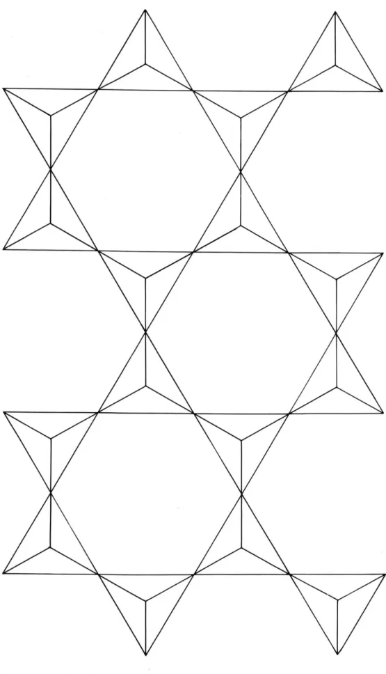
The structure is based on a new type of silica tetrahedra arrangement which has a silicon to oxygen ratio of 4:10, and which may best be described as a tube of tetrahedra. It consists of four-rings of tetrahedra which are arranged around the 4 axes, with
alternating tetrahedra having vertices pointing up and down. The oxygen atoms at these vertices are bonded to
similar fourerings above and below, thus forming an endless tube of four rings of tetrahedra in the c
direction. Titanium octahedra are bonded together In an infinitely long chain which extends along the four-fold axes. These octahedra are bonded to the tube of
tetrahedra through the only oxygen atom of the silica tetrahedra whose bond strength is not completely
satisfied by silicon. Irregularly coordinated sodium atoms occupy large voids between the chains and tubes.
The discrepancy factor, R, based on structure
factors calOulat*d from th* refined atom coordinates, is 14.2 when data with F0 = 0 are includee in the computation and 11.5 when they are not. All
interatomic distanctes are close to previcously.reported values and Pauling's rules are satisfied,
Thests Supervisor: Martin Jo Buerger
Table of contents
Abstract
List -of figures
List of tables y
Acknowledgements 4
Chapter I Introduction 2
Chapter I1 Previous work on narsarsukite 4
Chaptet III Unit cell and space group 6
Space group 6
Unit-cell dimensions 7
Unit-cell contant 9
Chapter IV Measurement of intensities 14 Selection and preparation of material 14
Intensity measurements 14
Chapter V Structure analysis 17
The Patterson synthesis 17
Preliminary considerations 18
Interpretation of the Patterson synthesis 22 , Interpretation of the minimum-function
maps 34
Chapter VI Refinement 40
Chapter VII Final structure 46
Conformity of narsarsukite to iauling's
rules 54
'u11MMsiNm 011-.16 1V List of figures page figure 1. firure 2, figure figure 3, 4, figure 5. figure 64 figure 7. figure 8. figure 9. figure 10.
Diagram of space group 14/m, with
equipoint locations
Graphs of unit cell dimensions vs,
cos 2
Harker line £ooz31
Minimum-function map based on
images of the structure in titanium, with SirTi peaks
Implication map 14(xyO)
trojection on (xyO) of the peaks
of the asymmetric unit of the
threeod imensional minimum-function
which is based on the silicon inversion peak,
Patterson synthesis P(xy)
Frojection of the structure frot
z C to z 0 a
I
on (ryc)Tube of tetrahedra found in narsarsukite and a simplified
version of the same type of
arrangement
Band of silica tetrahedra derived from a mica sheet
8 10 23 30 31 36 39 47 48 50 id wll
page table 1 * table 2,. table 3. table 4. table 5. table 6. table 7. table 8. table 9.
Analysis of Halfbreed Creek narsarsukite by Ellestad with total iron corrected
for PO and Fe20 3 by Schaller
Cell dimensions of all investigators Derivation of unit.cell contents
Weights of peaks to be expected in Patterson synthesis of narearsukite
assuming half-ionization
Limitations placed on atom positions
by equipoint rank
List of equipoints by rank
Patterson levels to be compared to yield the minimum function based on the
silicon inversion peak
Coordinates of atoms derived from the
minimum-function maps and coordinates
and isotropic temperature factors obtained from the refinement
Interatomic distances 4 11 13 19 20 21 35 41 52 List of. tables
Acknowledgements
The author is especially indebted to Professor M. J. Buerger who originally suggested the thesis
topic and who supervised the entire structure
determination. The suggestions offered by all the graduate students in crystallography, including
Tibor Zoltai, Charles Prewitt, Charles Burnham and
Roberto Poljak, are gratefully acknowledged. Their
help in the use of the facilities of the M. I. T. Computation Center was especially appreciated.
Specimens were kindly provided by Dr. David B. Stewart of the United States Geological Survey.
Introduction
Before this investigation no work had been
done on the crystal structure of narsarnukite.
However, certain properties of narsarsukite seemed to indicate possible uniqueness in its crystal structure, It is tetragonal, with two perfect
prismatic cleavages ((100)) and ((110))4 The silicon to oxygen ratio of its formula (4:11) corresponds to that of the amphibole group of minerals. This combination of symmetry, cleavage and formula is unknown in previously solved crystal structures.
In addition, previous work indicated that a structure determination based on the minimum function method developed by Ms J. Buerger would be a good test of this method for two reasons.
First, narsarsukite was known to contain 72 atoms
in its body-centered cell, which indicates a structure analysis of moderate Complexity. Secondly, the
presence of the moderately heavy atom titanium makes identification of Patterson peaks much easier than otherwise, thus almost guaranteeing the ultimate
solution of the problem.
All material used in this study came from Sage
ChapterII
Previous work on narsarsukite
Narsarsukite has been described from only a limited number of localities, which are all in
Narsarsuk, Greenland, the original type locality,
or the Sweetgrass Hills of Montana. Gossner and
Strunz were the first to attempt x-ray studies on the ty -e material. Their results include:
Space groupt h 14/
Cell dimensions:
aelO,78 kX units c. 7.99 kX units Unit-cell contents:
4(NapTii14031) with Fe, Mn, and Mg
substituting for Ti, and F and OH
substituting for 0.
Warren and Amberg, working on Greenland material,
obtained the following results using single-crystal methods.2
Space group: s~mI4, C4.I4, or C54 14/m Cell dimensions:
a*10.74 kX units c 7,90 kX units
Unit-cell contents:
4(Na2TSi401 1 ) with Ti replaced
by minor Fe, Mg, Mn, and A1.
Graham made the original discovery of narsarsukite from Montana.3 An analysis of this material is shown in Table 1.
Analysis of Halfbreed Creek narsarsukite by Ellestad with total iron corrected for FeC and
Fe203 by Schaller (after Graham3).
SIC2 62.30 1 A1203 0.32 FeO 0.47 Fe2 03 3*13 Mgo o.46 CaO 0.18 Na2 *1501 K20 0*41 Ti02 16.80 99.38 % total
A second Montrna narsarsukite locality was discovered
near Sage Creek by Stewart in 1950 near Graham's original locality.9 His crystals showed many forms, and
morphological studies resulted in demonstrating that narsarsukite has the point group 4/m The center of
symmetry (which cannot be determined using x-ray techniques) was confirmed by a negative test for piezoelectricity,
etch-pit symmetry, and symmetrical zoning as exhibited by minor color differences. An x-ray powder study gave the
following unit cell and space group.
Space groupt 14/m
Cell dimensions: a= 1072A a= 7.94,gJAk
Computations using these cell dimensions, the analysis
by Graham on material from his locality, and a specific
gravity determination (fz
?.783±.014)
resulted in the following unit-cell contents.4(Nag 89K.0 3Ca,0 1) (Ti 8 0Fe"' 05Ms 4 Fe" )Ol
Unit cell and space group
A. Space group
Selection of material was especially easy because of the two perfect cleavages of narsarsukite. A small cleavage prism was readily obtained and oriented with the optical goniometer. Precession photographs were taken of several levels in different orientations* The intensity distribution of these photographs corresponded with the diffraction symmetry 4/mI-/. All reflections
could be indexed on the basis of a tetragonal cell with
a10.77t05A and c7.97't.05A, Reflections with
h+k+lmPn+l were absent, showing that the cell is body centered. It could not be determined if the true space group of narsarsukite was I4, 14/m, or 14 because of the inversion center introduced by x-ray diffraction. To resolve this ambiguity, a single crystal with several
forms was chosen and examined under a binocular microscope. Two lines of evidence indicate that the point group of narsarsukite is 4/m and not 4 or 4. First, there are eight faces which have indices of the general form, (hkl), and which are about equally developed on the crystal
examined. If these faces are symmetrically equivalent, and their equal development suggests this, then they
constitute the ditetragonal dipyramid form of point group 4/m. The possibility still exists that these faces represent the
7 Second, there is a small pit on (010) which appears to be an etch pit. The only symmetry of this figure is a mirror plane which is normal to the four-"fold axis of the crystal. This mirror plane is prohibited by point groups
4 and 4, but required by point group 4/m. There is the
possibility, however, that this pit is a growth imperfection.
If so, its symmetry might be controlled by the symmetry of
the two nearby faces of general indices and it was shown above that these might represent equal development of two forms of symmetry lower than 4/m. Thus the symmetry
of the pit might be false.
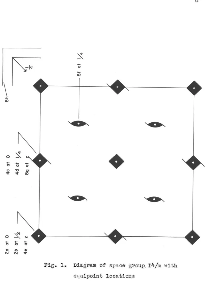
None of the above evidence is conclusive* In the absence of contrary evidence, however, the space group was taken to be 14/m. This is in agreement with the results obtained by Stewart discussed in the previous chapter. A diagram of this space group projected on
(xyO) is shown in Fig. 1. B. Unit-cell dimensions
It was desireable to have more exact unit-cell
dimensions, particularly for the computation of reflection directions to be used during the collection of reflection intensities. A crystal was oriented with the optical goniometer and precession camera in preparation for its use in the precision-Weissenberg method of determining unit-cell dimensions.
0
0
Fig. 1. Diagram of space group. 14/m with equipoint locations
.ACW..,
Weissenberg photographs of all levels normal to a which
were within recording range (levels 0-6) were first
taken, since these would be needed as reference in later intensity determinations. The crystal was then transferred to the preeision-Weissenberg camera and two photographs
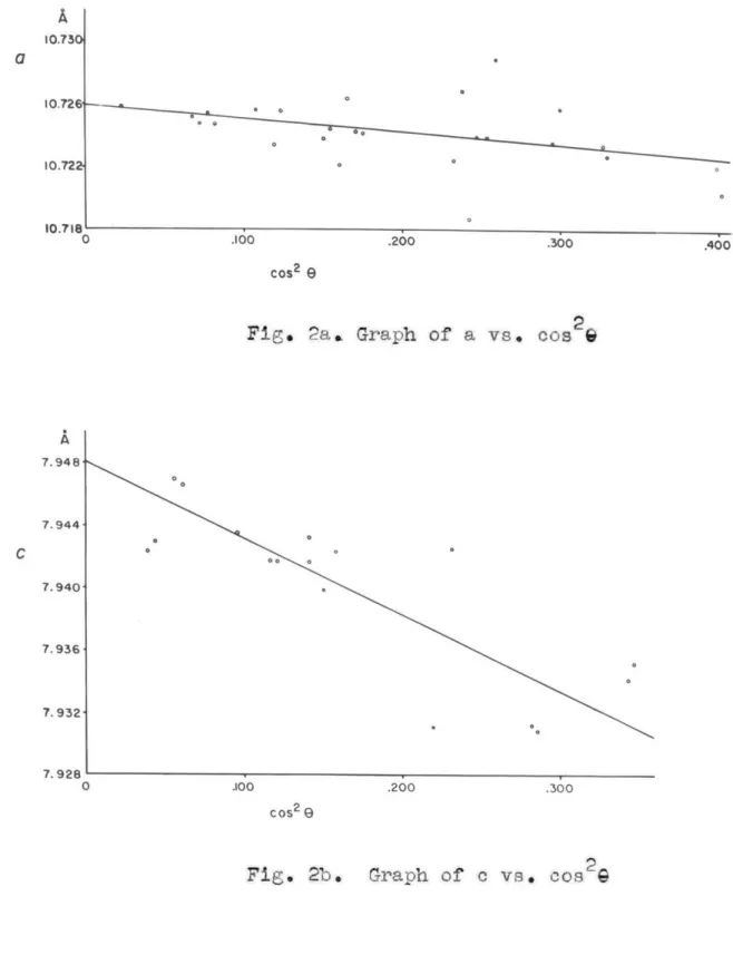
(each with the crystal in a different orientation) were taken which allowed very accurate determination of the unit-cell dimensions. Plots of a and c versus cos2%
were prepared and are shown in Fig. 2. As cos2 approaches 0, the spacing approaches the true value. The precision value of a was needed to compute values of e for the different values of cos 2, Thus the original inaccuracy in a was introduced into the value of co This dimension could therefore not be determined with as great accuracy as the former. The results of this determination along with those of other investigators are tabulated in Table 2.
All results are consistent and within experimental error. C. Unit-cel? content
The unit cell content was determined using the standard relation nM
f
VN, wheresn a number of formula weights per cell
m . weight in grams of one formula weight
density, g./cc.
V s volume of unit cell in A3
N a Avogadro's number, 6.023 X 10²³
Data used included:
1. a specific gravity of 2.783 t.014 g./cc. as
0 0
.100 .200 .300
cos2 e
Fig. 2a. Graph of a vs. cos 2
.100 .200 .300
COS2 (
Fig. 2b. Graph of c vs. cos2G
A 10.750i 10.726 10.722 10.718' 0 .400 A 7.948- 7.944- 7.940- 7.936- 7.932-7.928
11 Table 2
Cell dimensions of all investigators
a i
Gosaner and Strunz 10.80 A* 8.01 A*
Warren and Amberg 10.76* 7.92*
Stewart lO.72 7.94
Peacor 10.7260
7*948
26 the analysis on Halfbreed Creek, Montana material (Table 1).
3. the unit-cell dimensions of this investigation.
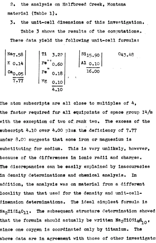
Table 3 shows the results of the computations, These data yield the following unit-cell formulat
Na7. 58 Ti 3.22 311 5.90 043.48
K 0.14 Fe 0.60 Al 0.10 Ca0.05 Fe 0.18 16.00
7.77 Mg 0.10
4.10
The atom subscripts are all close to multiples of 4,
the factor required for all equipoints of space group 14/m
with the exception of two of rank two. The excess of the
subscript 4.10 over 4,00 plus, the ,deficiency of 7.77
under 8.00 suggests that some iron or magnesium is
substituting for sodium. This is very unlikely, however, because of the differences in ionic radii and charges, The discrepancies can be easily explained by inaccuracies in density determinations and chemical analysis. In addition, the analysis was on material from a different locality than that used for the density and unit-cell-dimension determinations. The ideal simplest formula is Na2TiSi4011. The subsequent structure determination showed
that the formula should actually be written Na2TiOSi4 0 .O*
since one oxygen is coordinated only by titanium. The
Table 3
Calculation of unit-cell contents
V x6.023 X102 3 = nM =1533
Oxide Weight Weight "Molecular Number of Number of Number of
per cent per cent x weight" of "molecules" metal atoms oxygen atoms
cell mass oxide of oxide per cell per cell
8102 62.30% 955.1 60.06 15.90 15.90 31.80 A1203 0.32 4.9 101.96 0.05 0.10 0.15 FeO 0.47 7.2 71.85 0.10 0.10 0.10 Fe203 3.13 48.0 159.70 0.30 0.60 0.90 MgO 0.46 7.1 40.32 0.18 0.18 0.18 Oao 0.18 2.8 56.08 0.05 0.05 0.05 Na20 15.31 234.7 62.00 3.79 7.58 3.79 K20 0*41 6.3 94.20 0.07 0.14 0.07 TiO2 16.80 257.5 79.90 3.22 3.22 6.44 99.38 43.48
Chapter IV
Measurement of intensities
A. Selection nAd preparation of material
In order to assure maxium accuracy in intensity determinations the selection of a crystal was made with great care, An ideal sample must meet two requirements. First, the crystal must be as small as possible to bring absorption down to a minimum. Secondly, it should be perfectly cylindrical so that absorption corrections may
be eAsily and accurately made. The final crystal chosen
was a cleavage fragment. Because of the development of the ((100)) and ((110)) cleavages this was a nearly
cylindrical prism 4031 mm. in diameter and .250 mm. long.
This was so small that orientation with the optical
goniometer was only approximate because reflections from the cleavages were nearly undetectable. Final perfect orientation was achieved with the procession camera. The crystal was then transferred to the Geiger-counter
apparatus.
B. Intensity measurements
All intensities were measured with the
single-crystal Geiger-counter diffractometer. The single-crystal setting and the Geiger setting for each reflection were
obtained using a computer program prepared by Mr. Charles Moore of these laboratories. Computations were made on
15 This program also gave corrections for the Lorentz and
polarization factors, as well ns values of sin G , for each reflection.
Special care was taken to confine the intensities to the linearity range of the Geiger counter, Aluminum foils were used to decrease the reflection intensities when they were greater than about 120 counts per second. These foils were carefully calibrated for their
absorptions. In order to keep almost all of the
reflections below a counting rate of 120 counts per second
a very low voltage wis used. This unfortunately
resulted in such low intensities that approximately 15 % of the reflections were below the recording range of the Geiger counter. However, since a very low voltage was used, the most intense reflections had intensities
which fell well within the the absolute linearity range of the Geiger counter* It was hoped that this gain in accuracy in measurement of high intensity reflections would make up for the loss of data on reflections of
very low intensity.
There are about 480 reflections in the asymmetric unit of the copper reciprocal sphere of narsarsukite, but only 469 were within the recording range of the
apparatus. These reflections were measured and corrected for the necessary factors, The Lorentz and polarization corrections were provided as noted above by an I. B. M.
16
704 computer program. The linear absorption coefficient was based on the analysis of Graham's Montana narsarsukite. Absorption corrections were made according to the theory developed for a cylindrical specimen by Buerger and
Niizeki.5 The corrected intensities were then reaiy for
17
Chapter V Structure analysis
It was decided that a two-dimensional Patterson projection would contnin too many coalescing peaks
since narsarsukite is a mineral of moderate complexity (72 atoms in the body-icentered cell). Therefore, it
was planned to solve the structure by applying the minimum function to the three-dimensional Patterson
synthesis, which was sure to contain very few coalescing peaks.
A. The Patterson synthesis
The three-dimensional Patterson synthesis was computed on the I. B. M. 704 computer at the M. I. T.
Computation Center using MIFRI,8a standard program for
computing Fourier series. Since the Patterson synthesis has symmetry 4/m and the cell is body centered, it was only necessary to obtain the synthesis for one sixteenth of the volume of the unit cell. This asymmetric unit
was obtained in sections normal to c, with Patterson values computed at intervals of 1/60 along all three
axes. The section was computed with z varying from
0/60 to 15/60, and x and y varying from 0/60 to 30/60. Using the height of the origin peak obtained in the synthesis as a guide, the heights of peaks on the Patterson maps were computed. These are shown in
Table 4. In order to determine the height of the peaks it is first necessary to choose an absolute-zero level on the Patterson maps. Since this choice may be subject to a fairly large error, this same error will be
inherent in the computation of the expected peak heights shown in Table 4, The correlation between expected
peak heights and peak heights actually found may be
somewhat inaccurate because of this,
B. Preliminary considerations
Comparison of the ranks of the various equipoints of space group 14/m with the number of each kind of atom in the unit cell places initial restrictions on some atom positions, The possible positions are listed in Tables 5 and 6. All equipoints have one of the ranks 2, 4, 8, or 16. Since no combination of 16 and 8 will
yield 44, the total number of oxygen atoms in the unit cell, at least four of the oxygen atoms must be located
on an equipoint of rank four or on two equipoints of rank two. Therefore at least four oxygen atoms must occupy one of the equipoints 4e, 4d, 4c, or Pa and 2b. Since there are only four titanium atoms in the unit
cell, titanium must also occupy one of these equipoints. Equipoints 4c and 4d are on the 4 axes while 4c, 2a, and 2b are on the 4-fold axes. It is possible that both oxygen and titanium occupy the same set of axes. For example titanium might occupy position 4c and
19 Table 4
Weights of peaks to be expected in Patterson
synthesis of narsarsukite astuming half ionization.
Z 2 v 4175
Peak due to ZX Z2 Approximate height of
atom pair peak expected with an
origin peak height of
2150
Single peak Double peak
Ti~i 20 x 20=400 206 412 SisSi 12 x 12.144 74 148 Na:Na 10.5 x 10.5.110.25 57 114 0:0 9 x 9*81 42 84 TitSi 20 x 12V240 124 248 TINa 20 x 10,5.210 108 216 TiSO 20 x 9.180 93 186 SiaNa 12 x 1O.5*l26 65 130 sito 12 x 9.108 56 112 Nato 10.5 X 9e94.5 49 98
Table 5
Limitations placed on atom positions by
equipoint rank
Atom Number Fossible equipoint rank
in cell distributions Ti 4 4 2 2 Na 8 8 44 4 2 2 16 8 8 8 4 8 4 4 4 16 16 16 16 16 16 16 16 16 16 16 16 4 2 2 4*2 2 8 4 8 2 4 4 4 4 84 4 4 4 4 4 4 4 4 4 4 4 4 2 4 2 4 2 2 4 4 4 2 4 4 4 4 8 8 4 4 4 4 4 88 4 4 4 4 4 84 4 4 4 4 4 24 4 A 4 4 4 4 4 4 4 4 11 4 4 2 2 4 4 4 4 4 4 4 2I3 4 2 2 4 4 4 P 2 4 4 4 4 4 P 2 4 4 4 4 4 4 4 2 2 16 16 16 16 84 A 4 4
* Where 3 or more positions of rank but two must be equipoint 4.
4 appear all 16
21 Table 6
List of equipoints by rank
rank equipoints
2 Pa, 2h
4 4c, 4d, 4e
8
Se,
8g, 8hoxygen position 4d. Reference to the space group
diagram (Fig. 1) shows that this distribution consists of strings of alternating titanium and oxygen atoms. Titanium atoms would be located along the
Z
axes atz 0/60, 30/60, and 60/60 while oxygen atoms would be at z a 15/60 and 45/60. Thus the interval between
titanium and oxygen atoms would be 15/60 a c/4. The observed e/4 distance (1.99
A)
compares favorably with known TI-O distances (e.g. 1.96A)t strongly suggestingthat this type of titanium-oxygen distribution is correct. There are four possible arrangements of the strings of alternating titanium and oxygen atoms, however, each corresponding to a combination of the equipoints given above. Each arrangement yields a Ti-*O distance of c/4.
C. Interpretation of the Patterson synthesis
A plot of the Harker line [Ooz] from z a 0 to
Z a 30/60 is shown in Fig. 3. Peaks can be distinguished at a : 23/60, 14/60, and 30/60. Reference to the space
group diagram (Fig. 1) showed that the titanium and oxygen atoms which may alternate along the 4 or Z axes should be separated by an interval of z a 15/60. Since
there are four oxygen and four titanium atoms per unit
cell, there should be a peak of height 4(Tit0) at z a 15/60 of the Harker line. Table 4 shows that this height is 4x93 a 372, which agrees well with the peak
(D 25/60 CD 260 15/60 10/60 560 0 2 4 6 8 10 12 14 16 18 20 Peak height
X
ooIn addition, titanium atoms should be separated by
an interval of z = 30/60, as should oxygen atoms. There should therefore be a peak at z = 30/60 of the
Harker line with height 2(Ti:Ti)* 2(0:0). Table 4 shows this height to be 412484 a 496, which is close to the height of 520 actually found. This is consistent with a titanium-oxygen chain distribution along the 4 or 4 axes.
Since the minimum function was to be used in
determining the crystal structure, it was necessary to locate an inversion peak in order to begin the image-seeking procean. Buerger has shown how to locate
inversion peaks through the use of correlation minimum-function maps.6 Since this case involves a four-fold axis with a perpendicular mirror plane, an inversion peak can be located on any level z by exactly
super-imposing level z on level 0 and contouring the minimum function. This was done for all levels from z * 0 to
z : 30/60. If silicon occupies the equipoint with general coordinates, the symmetry 4/m should produce a peak of weight 4(si:Si) on the Harker line on the same level as the silicon inversion peak* Examination of the Harker line had shown one peak which was still unaccounted for at z a 23/60. The correlation map of
level z a 23/60 was therefore thought to offer the best
25
This level does in fact show a strong peak believed to
be a possible inversion peak, as well as its expected
rotation-equivalent peak. The heights of these two
peaks are several times greater than the computed
expected values of the silicon peaks, showing that they are multiple peaks. A minimum-function solution based
on a multisle peak yields a multiple solution, so these
peaks could not be used in the minimum-function image seeking process. It was decided to attempt to find an inversion peak by another process, since no other
candidate inversion peaks could be located by the
correlation minimum-function method.
Another procedure was suggested by the restrictions
placed on the titanium locations by the equipoint distribution, The possible titanium locations are
listed in Table 5. All coordinates of equipoints Pa, Pb, 4c, and 4d are fixed, but the z coordinate of
equipoint 4e is variable. Reference to the space group diagram shows that the probable titanium-oxygen chain
distribution suggested above requires this coordinate to be 15/60, since the titanium atoms must be separated
by an interval of z a 30/60. With this qualification, it can easily be seen from the space-group diagram that all four possible titanium distributions differ
from each other only in the location of the origin. This requires that the Patterson-peak distribution
26
resulting only from TitTi vectors be the same for all four cases. Therefore it is not possible to ascertain which distribution is the correct one merely by noting the locations of the high TisTi peaks on the Patterson maps, Since, in all four possible distributions,
titanium atoms lie on the 4 or 4 axes separated from one another by an interval of z a 30/60, a method is available
for readily solving the structure, Consider, first, only two of the titanium atoms of one of the possible equipoint distributions, separated only by an Interval of z a 30/60. The Patterson maps contain an image of the crystal structure in both of these atoms, If these two images can be brought together and the minimum function mapped, the result will be an approximation to the
crystal structure. In particular, if all Patterson maps differing by an interval z : 30/60 are exactly
super-imposed and the minimum function mapped, the correct solution results. Notice that it makes no difference where the two titanium atoms are located in the actual
structure, just as long as they lie one above the other separated by the given interval. There are, however, ambiguities in the minimum-function solution described above. First, oxygen atoms are distributed like the titanium atoms (on the 4 or Z axes separated by an
interval of z a 30/60). There is also an image of the structure in the oxygen atoms. The above mapping
27 procedure will therefore yield a solution based on
these images, superimposed on the solution based on the images of the structure in the titanium atoms6
This should cause little difficulty however, since the Patterson peaks based upon titanium as an image point far out-weigh peaks based upon oxygen. The minimum-function solution will thus contain high peaks
representing the structure* Superimposed on these will
be a ghost due to the structure seen from the oxygen atoms. A second ambiguity in, the minimum-function maps is
caused by the presence of a false inversion center with
coordinates x g
t,
y a I in each level of the minimum function. An ambiguity of two thus results in thelocation of each atom. This ambiguity is caused by the centering translation of the unit cell. Consider the two titanium atoms upon which the minimum-function solution is to be based. These are located one above
the other separated by an interval of z : 30/60. Now refer to the space group diagram. In all possible
titanium distributions, it can be seen that these first two atoms are related to a similar pair by the component of the unit-cell centering translation which is normal to c. These second two atoms are also located one
above the other, separated by an interval of £ a 30/60.
The fatterson maps also contain images of the structure in the pair of atoms. The images seen by each pair
of atoms are therefore related by this translation,
which is equivalent to an inversion center at x a 1,
y a of each level. The result produced by comparing an two Patterson levels separated by an interval of
z 30/60 is a minimum-function map with a double
solution to the structure, One solution is related to the other by the component of the centering translation normal to o, which is equivalent to an inversion center at x w 4, y
4
for each level of the minimum function.A third problem is the lack of knowledge of the absolute origin in the minimum-function maps. The
titanium atom will be represented by a high peak at the origin of one of the minimum-function maps. But there are four possible equipoints where the titanium may be located and only one of these includes the real origin of the crystal, The set of minimum-function maps will
contain the true solution to the structure, but the absolute unit-cell coordinates of the atoms will depend on the placement of this set of maps in crystal space.
If the titanium atom can be given its true location, then the set of minimum-function maps may be given their true placement and the absolute coordinates of
the atoms will be known,
The minimum-function mapping procedure outlined above was carried out for the asymmetric unit of the unit cell, by usingtwo titanium atoms as image points.
29
Levels of the Patterson function separated by an interval of z v 30/60 were compared. Levels 0/60 to 15/60 were successively compared with levels 30/60 to 45/60 to yield a set of 15 minimum-function maps. These maps contain, of course, the ambiguities discussed above,
A high peak of the expected weight representing the
titanium atom is at the origin. It was only necessary then to determine which position of the four possibilities that this titanium occupied, to give the entire set of minimum-function maps their correct placement in crystal
space, Only one large peak and its inversion equivalent was found to have general coordinates (Fig. 4), Since
silicon is the only relatively heavy atom which may have general coordinates (Table 5), this peak height
(320) was compared with the computed expected height of
the silicon peak. This height 2(SitTi): 248 is close to the height actually found. The location of the
silicon atom was ths determined relative to the titanium atom, with the exception of the ambiguity due to the
inversion center.
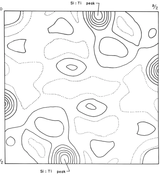
An implication map 14(xyO) was next prepared
(Fig. 5). This is a projection of an approximation to the crystal structure, with ambiguities, on (xyO). There are high peaks on this map corresponding to all possible titanium positions. Since this map is a
SI Ti peak 0
a/2
Si:
Ti
peak
Fig. 5.
Implication map r4 (xyo)
b12
one of these two equipoints. The set of minimummfuCtion
maps was placed so that the high titanium peak of these maps was superimposed on the high peak at 0,1,0 of the
implication map. Since each type of map is an
approximation to the crystal structure, there should be good correlation of high peaks, if the titanium atom is
located at equipoint 4c or 4d, There is, however, no
peak on the implication map oorresponding to either of
the possible silicon peaks on the minimum-funotion maps,
The possible titanium positions 4a and 4d are thus
eliminated, The other positions which the titanium may occupy (4e, fa, 2b) are loeated at 0,0,0 of the implication map. Minimum-function maps were placed over the implication maps so that the high titanium peak was superimposed on the high peak at 0,0,0 of the
implication map, There is good correlation, not only of the silicon peaks, but of all minimum-ftnction peaks,
with implication peaks, The titanium atoms are thus
located either on oquipoint 4e (with a e 15/60) or Pa and 25. In addition, the silicon atom has been determined relative to the titanium atom with the
exception of the ambiguity due to the inversion center, It can easily be seen from the minimum4unction
maps that the silicon and titanium atoms are separated by an interval of z * 4/60. If the titanium is on
WN U0 ilgiiii u w-wlm
33
equipoint 4e(0,0,15/60) the silicon atom is on level
11/60. This yields a high silicon reflection peak on
the Harker line C00zl at z x 22/60. There is, in fact,
a high peak there, the only one on the Harker line unaccounted for. On the other hand, if the titanium atom is located on equipoint 2a(0,0,0), the silicon atom would be on level z a 4/60, A high peak would thus
appear on the Harker line at z * 8/60. The location of
a trough in the Patterson funotion at z = 8/60 eliminates
this possibility, The titanium atom is thus definitely located at 0,0,15/60 (equipoint 4.), Therefore the levels
of the minimum function with the titanium atom at the
origin can be given their proper placement in crystal space* This locates the silicon atom with an ambiguity
due only to the inversion center in the level in which it occurs,
The location of an inversion peak in the Patterson function can easily be predicted if the locations of the atoms causing it are known. Since the silicon
atom is on level 11/60, the inverseon peak should occur on level 22/60, Patterson maps of levels close to
z a 22/60 were inspected to see if the silicon peak could be found in the predicted location# There are, in fact, inversion peaks of the correct weight
corresponding to each of the two possible siliconatom locations, Using one of these inversion peaks, a
complete set of minimum function maps of the asymmetric
unit was prepared. The Patterson superpositions are listed in Table 7. The initial superpositions resulted
in M2 maps but these could be combined with themselves
using the four-fold axis to obtain M8 maps which are a good approximation to the electron density. This same procedure was carried out using the other possible
silicon inversion peak as the starting point for the
image-seeking process. It was noticed, however, that the set of maps obtained in this case was exactly the same as that based on the other inversion peak, differing from it only in the location of the origin.
Do Interpretation of the minimum-function maps.
A projection on (xyO) of the peaks of the
three-dimensional minimum-function maps based on the silicon inversion peak is shown in Fig. 6. Only peaks in the interval z a 0 to { are shown. The large peak at the origin with coordinates 0,0, is, of course, the titanium peak. The smaller peak at the origin with z = 0 is an oxygen peak,(0I)whose location was suggested above by
the titanium-oxygen chain hypothesis* In addition, the peak at JJ,0 represents the other oxygen atom (o0I whose location was suggested. The silicon peak which served as the basis of the image-forming function is marked with a cross. The next highest peak is located on level
35
Table 7
Patterson levels to be compared to yield the minimun function baed on the silicon
inversion peak,
Levels to Levels which are Resulting
be compared equivalent by minimum** Patterson symmetry function
level 27, 26, 25, 24, 23, 22, 21, 20, 19, 18, 17, 16, 15, 14, 13, 12, 11, 4 3 2 1 0 ~m2 4.3 o-4 ~.5 -*6 -*7 49 -*10 -.11 -12
3,
4, 5, 6, 7., 8, 9, 10, 11, 12, 13, 14, 15, 14, 13, 12, 11, 4 3 2 1 0 1 2 3 4 5 6 7 8 9 10 11 12 15.5 14.5 13,5 12.5 11,5 10,5 9,5 8,5 7,5 6.5 5#5 4,5 3.5 2#5 1.5 0.5 -0050 0 at 0 .. at C TI at .25 -Si at ,V OV at .18-/Si at .19 . 0,1, at 0 Na at 0-0,V at .24 Oil CIL Ti Fig. 6.
Projection on (xy0) of the peaks of the
asymmetric unit of the three-dimensional
minimum-function which is based on the
silicon inversion peak
36
37
z aC, a position of rank 8. Since there are 8 sodium atoms in the unit cell this relatively heavy peak must represent a sodium atom. This then locates all cations and four oxygen atoms with no ambiguities.
Forty oxygen atoms still remain to be located. Four peaks, three of rank 16 and one of rank 8, remain
on the minimum-function peak projection. All of these are peaks of low weight and have close to the expected
oxygen-peak height. Two of the peaks of rank 16 and the peak of rank 8, when repeated by symmetry operations, complete tetrahedral coordination for silicon and yield Si"0 distances close to standard values. In addition, titanium has its usual octahedral coordination and all Ti-O distances are close to values found in other
structures* These three peaks were therefore taken to be approximations to the electron density of oxygen atoms,
They are labled 01, Oiv, and Oy in the projection of the minimum-function peaks. This completes the location of all atoms of the unit cell, One false minimum-function peak remains unlabled.
Four checks were made of the correctness of the proposed structure. First, comparison with the
implication diagram shows good correlation of implication peaks with minimum-function peaks, Second, tie original set of minimum-function maps based on titanium shows one-to-one peak correlation with the maps based
3,
on the silicon inversion peak (with the exception of the ambiguity due to centrosymmetry in the former maps)6 Third, a two-dimensional Iiatterson projection 2(xy) was prepared. Since the titanium atom is located at OOj, and since Patterson peaks involving the relatively heavy titanium atom should dominate this map, it should be a rough approximation to a projection of the crystal
structure. This map was prepared using the I. B. Mt 704
computer at the W. Z. T. Computation Center in conjunction with the Fourier series computation program MIFRlP
It is shown in Pig, 7# There is good peak oorrelation again. tourth, all oation-anion distances compare well with distances previously recognized in other crystal structures,
b/2
Fig. 7.
to
Chapter VI Refinement
Refinement of the atom parameters of narsarsukite
was carried out using the Busing and Levy least-squares program on the I. Bo Y, 704 computer at the Y, I. T
Computation Center, This program permits simul*tanious refinement of all atom coordinates, temperature factors
(overall, individual isotropic, or individual anisotropie) and one or more scale factors. It computes structure
factors and the discrepancy factor, R# based on input
parameters, Therefore, in order to compute a discrepancy
factor for the parameters obtained as the output for a given cycle# a separate computation or another cycle
must be run.
The coordinates of the atoms of narsarsukite derived from the minimum-function map based on the
silicon inversion peak are tabulated in the first section
of Table 8. These were used as input data for the first
cycle of refinement along with the following data:
1. An arbitrary seale factor of 1.0 2. All F, including data with F * 0
3. An arbitrary value of 0.7 for all individual temperature factors
4, ?orm factors assuming half-ionization
Six cycles were run in which only the scale factor and the atom coordinates were allowed to refine. The refined
Table 8
Coordinates of atoms derived from function mapa and coordinates and
temperature factors obtained from
41
the minimum-Isotropie
the ref inement
Atom Coordinates derived Coordinates and isotropic from minimum- temperature factors
function maps obtained from refinement
x y x x y z B Ti 0 0 *25O 0 0 .33 1,2 3 Na *186 .141 .186 .138
i
149 Si 4.008 .294 .192 ,012 .308 .191 .2 0 0 0 0 0 0 0 .9 Oil 0 0 0 0 1 ,5 Oxyy -009 *316 0 -4*038 .301 0 .8 Ory *055 .179 .250 .049 .177 .268 .6 0V9 .128 .392 .183 *133 .403 .192 .6atom coordinates and scale factor of each cycle were
used as input data for the next cycle. All other input
data, including the individual temperature factors,
remained unchanged. The largest change in the fractional coordinate of the atoms during the sixth cycle was
?X1Ot # The stage of the refinement during which the temperature factors were held constant was therefore regarded as completed, The individual isotropic temperature factors, along with the atom coordinates and scale factor, were allowed to vary in all subsequent
cycles* The temperature factor of 0 1 obtained from
Cycle 7 was negative, The least-squares program does
not refine if negative temperature factors are used as
input data, so a method was sought to resolve this
difficulty.
The points of a plot of In F0/F0 vs. sin2 0
should lie approximately along a straight line with a slope which is a function of the overall temperature
factor if values of F are based on the correct structure. The intersection of the line with the In F/F0 axis
provides the scale factor, With perfect experimental
intensity data and perfectly refined atom coordinates, all points of the plot fall exactly on a straight line, The distance of a point from the line is a rough
measure of the accuracy of Fg. It was believed that the
caused by inaccurate intensity data, and that the type of plot described above would confirm this, In
preparation for such a plot, values of Fc for all
reflections were computed using a temperature factor of zero and atom parameters of cycle 6. Values of
sIn 0 for each reflection were provided by the I. B. M.
704 program which had also provided Lorentz and
polarization factors, etc.. The resulting plot showed a wide scatter of points, which indicated that some
data were inaccurate, but there was a majority of points which occupied a wide band with a negative slope,
Arbitrary border lines were drawn outlining this band#
with approximately 130 points falling outside of these limits, It was believed that these represented the
most inaccurate data, and might have caused the
temperature factor of 0 obtained from cycle 7 to be negative, Only those reflections which fell within the
band limits were used as input data for cycle 9
(340 reflections). A small positive number (O.?'O) was used for the temperature factor of Ogg and all
parameters were allowed to vary. During this cycle all temperature factors remained positive, confirming the
fact that the inaccurate data had been responsible for the negative temperature factor.
The points of a plot of In FO/F vs. sin2O should lie approximately along a straight line with neglgible
slope after correcting for the temperature factor.
Since the line now had zero slope, a data rejection
test other than the one described above could be used,
Mr. Charles Prewitt prepared a patch for the
least-squares program which allowed only those data with
IFo-Fel/po<0.25
to be included in the refinement# Thisrejection test has essentially the same effect as the
one used in cycle 9, in that it includes data in the
refinement process which occupy a wide band on the plot
of 1n Fo/Fe vs. sin 2a It has the advantage of
allowing all reflections, whether used in the refinement
process or not, to be included in the input data deck
and therefore to be included in the calculation of the
discrepancy factor. This rejection test also excluded
data with F : 0 (approximately 15% of the reflections)
from the refinement process.
Cycles 10, 11 and 12 were run using the new
rejection test, permitting all parameters to vary. After
cycle 11 it was discovered that incorrect weights had
been applied to the reflection data. All reflections
had been weighted equally, when in fact, all zero-level
reflections have a multiplicity one half that of
reflections of general indices. Cycle 12 was accordingly
run with corrected weights for the zero-level reflections,
The largest fractional coordinate change of this cycle was
4;
Final coordinates ant temperature factors are shown inTable 8. The discrepancy factor based on these coordinates is 14,2 when reflections with F0 = 0 ar included in the computation and 11#5 when these are omitted.
46
Chapter VII Final structure
The structure of narsarsukite is illustrated in
Figs. 8 and 9. Narsarsukite has a new type of silica
tetrahedra arrangement whose silicon to oxygen ratio is 4:10. The formula should be written Na2TiOSi4 010
since some oxygen atoms are coordinated only by
titanium and sodium atoms. Fig. 8 is a projection of the structure from z : 0 to z on (xyO). Fig. 9a
is a three-dimensional representation of a section of the new silica tetrahedra arrangement, while Fig. 9b
is a simplified drawing of this same type of arrangement.
The main points involved in an understanding of the structure of narsarsukite may be appreciated with
reference to Fig. 8. A crystal may be built from this unit by repetitions involving translations uvO and
reflections across mirrors at z
:
0 and z : J, which arethe limits of the portion of the crystal projected. There are two basic units in the structure. First, there is a series of titanium octahedra arranged along the four-fold axes. Each titanium atom shares an oxygen atom with the titanium atoms immediately above and below. There is thus an infinite chain of octahedra which
share corners, the four-fold axis being the axis of the chain.
4yf a2 0 01, 01 Ti 0ir C Si 0 0O;' O-Na ol 0 / 0 00 0 0 0 ,0 00 /0 Pig* 8.
Projection of the struoture from z
:
0 to z a on (xy0)Fig. 9a Fig. 9a.
Fig. 9b.
Fig. 9b
Tube of silica tetrahedra found in narsarsukite Simplified version of the tube of silica
tetrahedra found in narsarsukite
49 The second major unit of the structure is the
network of silica tetrahedra. The tetrahedra are arranged around the Z axes in rings of four, each
tetrahedron sharing a corner with two other tetrahedra of the ring. They are oriented so that alternating
tetrahedra of the ring point up and down. Thus two tetrahedra of each four-ring have vertices pointing up and two down. These vertices are shared by similar
four-rings above and below. This arrangement can be appreciated by noting that the oxygen atoms shared by
adjacent four-rings lie in mirror planes arranged
parallel to the plane of the four-rings. The repetition
of these four-rings in the c direction by the mirror planes yields a tube of tetrahedra arranged around the
axis.
The specific nature of the new silica tetrahedra arrangement can be readily grasped with reference to
the simplified illustration of Fig. 9b. The four-rings of tetrahedra with vertices alternating up and down, bonded to four-rings above and below, are easily
distinguished. This structure may be derived from the mica sheet network, which is a planar arrangement of
six-rings. Let a narrow band, infinitely long, and two six-ring units wide, be separated from a mica sheet, as shown in Fig. 10. If this band is bent around parallel to its long axis, so that the long edges are bonded
0
51 together, the silica tetrahedra tube of narsarsukite
is obtained. The tetrahedra of the mica network which are arranged with unshared vertices pointing upward in Fig. 10 have unshared vertices pointing away from the axis of the tube. Thus each tetrahedron of the
narsarsukite arrangement shares three vertices with other tetrahedra, as in the phyllosilicates.
Each tetrahedron has one corner unshared by other tetrahedra, It is the oxygen at this vertex which is bonded to a titanium atom, thus forming a bond between
the chain of titanium octahedra and the tube of silica tetrahedra, both of which extend indefinitely in the c direction. The sodium atoms occupy the large voids between the chain of titanium octahedra and the tube of
silica tetrahedra,
Interatomic distances are tabulated in Table 9.
All four Si-O distances are approximately equal,
suggesting that the oxygen arrangement around silicon approximates that of a tetrahedron. There are two Si--1Si bond angles which are not symmetry equivalent.
These involve adjacent tetrahedra of a four-ring, and
adjacent tetrahedra of different four-rings. The
Si-O.S1 bond angles are 141.0 and 138.9 respectively.
This compares fairly well with angles of 150 found by Nieuwenkamp in low quartz.3a
Interatomic distances
Metal atom Number and type of Interatomic and oxygen neighbors distance coordinates and coordinates
Si x,y,Z 1 0 x,y,z 1.61 A I 0Iv x,y,z 1.58 1 oV x,y,z 1.65 1 0 y 1.62 Ti x,y,z 1 0 x,y,z 1,90 1 oil x,y,z ".08 4 Ogy xOy#z 1 1 .9 Na X,7# 1 Og x,y#z 2,48 2 Oy x,y,z 2,39 2 oy y,EZ 2.72 2
O
-z 2.515,
l a moderate distortion of the octahedral arrangement ofoxygen around titanium. The distortion involves the two oxygen atoms above and below the titanium atom on the four-fold axis. The titanium atom is closer to
O1(Ti-O v 1.90 i) than to 01 (TiwO : 2,08
1).
In addition, the z coordinate of the remaining four oxygen atoms of the titanium octahedron (*268) is greater than that of the titanium atom (239), These oxygen atoms thus lie in a plane (normal to the four-fold axis) whichis displaced away from the titanium atom toward 014 In
an ideal octahedron this plane includes the titanium atom* The titanium atom is thus displaced away from the center of the octahedron, toward one end* This effect is slightly accentuated by displacement of the four oxygen
atoms (not on the four-fold axis) in the opposite
diretion, Exactly this same type of arrangement occurs
in tetragonal Bati%., In this structure, titanium octahedra are bonded in a chain on the four-fold sxes. The titanium atom is displaced 0.13 A toward one end of the octahedron. Ti-0 distances closely correspond to
those of narsarsukite, as shown by the following tablet
narsarsukits BaTi03
f-0 /fo 1.90A 1,869 A
208
2.107
54
The displacement of all titanium atoms toward the
same end of the crystal is associated with ferroelectrioity in BaTiO3, This property may be exhibited by Na T10810
3*2 .4 ll
if an appropriate phase change from narsarsukite occurs, Reference to Table 9 shows that seven oxygen atoms have Naw- distances which compare well with Na-O distances found in other crystal structures. Two of these oxygen atoms (Oy) have bonds which are probably saturate4 by
silicon. If these two atoms are disregarded, the oxygen coordination around sodium is five, Reference to Fig* 8
shows that the polyhedron formed by these five oxygen atoms is very irregulars It does not actually enclose the
sodium atom since all five oxygen atoms lie to one side of the sodium atom* This is also true, but to a lesser
degree, even when the additional two oxygen atoms are included in the polyhedron.
Conformity of narsarsukite to Pauling's rifles, If the possibility of coordination of O with
oxygen is excluded,
Ov
and 0 11 have electrostaticvalency bonds exactly satisfied, since each is coordinated by two silicon atoms. Assuming a coordination of five
oxygen atoms around sodium, the bonds from sodium to
oxygen have electrostatic valency strenths of 1/5#
Titanium and silicon have electrostatic valeney strengths of 4/6 and I respectively. 0 and 0 are each
55
Their charge (o-2) is slightly oversatiefied
[2(4/6) 44(l/5) 32/15. 01 is ocordinated by one titanium atom, one silicon atom, and two sodium atomsi Its charge is also slightly oversatisfied
j1+4/6 -2(1/53: 62/30 . If a coordination of six is assigned to sodium the electrostatic bonds are exactly
satisfied. The extremely irregular coordination of oxygen around sodium, and the possibility that OV should
be included in the oxygen coordination sphere, makes
exact calculation of electrostatic valencies complicated,
The above calculations show that all bonds are satisfied
within limits of interpretation of the sodium coordination. Several points indicate that coordination may be
much more complex than indleated above. It is possible that the two oxygen atoms (Oy) excluded above should be included in the sodium coordination sphere, especially
since the two oxygen atoms (OXw) with considerably
larger Na-CO distances were included. That these two
oxygen atoms (y) are probably making some contribution
to Na is shown by the relatively large SiO distance
(1.65
1)
which indicates that the oxygen atom may not be as closely bonded to silicon as usual. As notedabove, inclusion of these two oxygen atoms in the sodium acordination polyhedron would make the polyhedron more regular.
56
this oxygen atom cannot contribute its total bondstrength to silicon. The resulting excess of silicon
bond strength may be conpensated for by OV* This
oxygen atom exhibits an 21-- distance (1.58
A)
which is smaller than usual, indicating that it may be contributing more than a bond strength of -1 to silicon. In addition,this oxygen atom also has a very high NacO distanoe, indicating that it may contribute little of its total
bond strength to sodium, The *average" effect of 01, and OV may be such as to yield a six-fold oxygen
coordination of sodium and a four-fold coordination of
SYT
Bibliography
la Gosener, 3,, and Struns, H. (1932), Die chemisoho
Zusammersetzung von Narsarsukitt Zeits " Wrist., 8?, 150-151,
2. Warren, B. E., and Amberg, C. 3, (1934), X-ray
study of narsarsukite, Na2 (ir)i4 0 11
Am. Mineral., 19, 546-548,
3, Graham, William A* P* (1935), An occurrenee of narsarsukite in Montanat Am, Mineral,, 20, 598-601. 3a Nieuwenkamp, V. (1935), Die Kristallstruktur des
Tieft.Cristahalits 5102$ Zeit. Krist., 92, 82-48.
4, Evans, R. 04 (1952), Crystei Chemistry, Cambridge,
Cambridge University Press.
4a. Megaw, Helen D. (1957), Ferroelectricity in Crystals, London, Methuen and Co.
5. Buerger, M. J,, and NiIseki, N. (1958), Correction
for absorption for rod-shaped single crystalas Am, Mineral,, 43, 726a731,
6. Buerger, Martin J. (1959), Vector
Space,
New York,John Wiley and Sons,
7, Busing, R. William, and Levy, Henri A. (1959),
A crystallographic least squares refinement program
for the I. B. BM 704, Oak Ridge Tennessee, Oak Ridge National Laboratory.
56 8. Sly, William G., and Jhoemaker, David Po (1959),
MIFRI. two-and three-diraensional crystallographic Fourier summation program for the I. B M. 704,
Mass. Insti. Tooh..
9. Stewart, D. f. (1959), Narearsukite from Sag, Creek, Sweetgrass Hills, Montana: Am. Mineral.,


