HAL Id: inserm-00139590
https://www.hal.inserm.fr/inserm-00139590
Submitted on 2 Oct 2007HAL is a multi-disciplinary open access
archive for the deposit and dissemination of sci-entific research documents, whether they are
pub-L’archive ouverte pluridisciplinaire HAL, est destinée au dépôt et à la diffusion de documents scientifiques de niveau recherche, publiés ou non,
Preferential association of Hepatitis C virus with
apolipoprotein B48-containing lipoproteins.
Olivier Diaz, François Delers, Marianne Maynard, Sylvie Demignot, Fabien
Zoulim, Jean Chambaz, Christian Trépo, Vincent Lotteau, Patrice André
To cite this version:
Olivier Diaz, François Delers, Marianne Maynard, Sylvie Demignot, Fabien Zoulim, et al.. Preferential association of Hepatitis C virus with apolipoprotein B48-containing lipoproteins.. J Gen Virol, 2006, 87 (Pt 10), pp.2983-91. �10.1099/vir.0.82033-0�. �inserm-00139590�
1
2
3
4
5
6
7
8
9
10
11
12
13
14
PREFERENTIAL ASSOCIATION OF HEPATITIS C VIRUS TO APOLIPOPROTEIN B48-CONTAINING LIPOPROTEINS*
Olivier Diaz#, François Delers§, Marianne Maynard¶, Sylvie Demignot§, Fabien Zoulim¶, Jean
Chambaz§, Christian Trépo¶, Vincent Lotteau# and Patrice André#,||.
From the # IFR 128 Biosciences Lyon Gerland, INSERM U503, 21 avenue Tony Garnier, 69007
Lyon, France, § Université Pierre et Marie Curie, UMRS 505, Paris, France, INSERM, UMRS 505,
Paris, France, ¶ Service d’hépato-gastro-entérologie, Hôtel Dieu, Hospices Civils de Lyon, France, ||
Laboratoire de Virologie, Hôpital de la Croix-Rousse, Hospices Civils de Lyon, France.
Running title: HCV association to ApoB48
Address correspondence to : Patrice André, INSERM U503, 21 avenue Tony Garnier, 69007 Lyon, France. Tel : (33) 4 37 28 23 27 ; Fax : (33) 4 37 28 23 41 E-mail : andre@cervi-lyon.inserm.fr
15
16
17
18
19
20
21
summary: 224 words main text: 4782 words figures: 5tables: 2
This is an author manuscript that has been accepted for publication in Journal of General Virology, copyright Society for General Microbiology, but has not been copy-edited, formatted or proofed.
Cite this article as appearing in Journal of General Virology. This version of the manuscript may not be duplicated or reproduced, other than for personal use or within the rule of ‘Fair Use of Copyrighted Materials’ (section 17, Title 17, US Code), without permission from
the copyright owner, Society for General Microbiology.
HAL author manuscript inserm-00139590, version 1
HAL author manuscript
21
22
23
24
25
26
27
28
29
30
31
32
33
34
35
36
37
Whereas hepatitis C virus (HCV) in cell culture has a density compatible with that of the
Flaviviridae family, in vivo infectious particles are partly found in low density fractions, associated
with triacylglycerol (TG)-rich lipoproteins (TRL). In the blood of infected patients, HCV circulates as heterogeneous particles among which are Lipo-Viro-Particles (LVP), globular particles rich in TG and containing viral capsid and RNA. The dual viral and lipoprotein nature of LVP was further addressed with respect to apolipoprotein composition and post-prandial dynamic lipid changes. TRL exchangeable apoE, CII, CIII, but not the HDL apoA-II, were present on LVP as well as the viral envelope proteins. ApoB100 and B48, the two isoforms of the non-exchangeable apoB, were equally represented on LVP, despite the fact that apoB48 was barely detectable in the plasma of these fasting patients. This indicates that a significant fraction of plasma HCV was associated with apoB48-containing LVP. Furthermore, LVP were dramatically and rapidly enriched in triglycerides after a fat meal. As apoB48 is exclusively synthesized by the intestine, our data highlight the preferential association of HCV with chylomicrons, the intestine-derived TRL. These data raise the question of the contribution of the intestine to the viral load, and suggest that the virus could take advantage of TRL assembly and secretion for its own production and of TRL fate to be delivered to the liver.
37
38
39
40
41
42
43
44
45
46
47
48
49
50
51
52
53
54
55
56
57
58
59
60
61
62
63
64
65
66
67
68
69
70
INTRODUCTIONHCV has been classified within the Flaviviridae family according to the structure of its genome (Pringle, 1999). However, in contrast to flaviviruses and closely related viruses, cell culture of HCV remained problematic for fifteen years and this lack of an appropriate in vitro replication system and of a small animal model impeded the understanding of HCV structure and replication cycle. Therefore, most of our knowledge of the virus cell receptors and of the HCV RNA replication relied on pseudotyped viruses and on biscistronic and subgenomic replicons, which do not allow the study of HCV assembly and secretion and the identification of the elusive nature of the virion. Recently, complete replication and production of infectious HCV particles in tissue culture were performed with HCV genotype 2a full length replicons derived from a patient with fulminant hepatitis (Lindenbach et al., 2005, Wakita et al., 2005, Zhong et al., 2005). This major breakthrough identified a viral structure with size, morphology and density (1.15g/ml) appropriate for a member of the Flaviviridae family. The structure of these virions most likely match that of virions found in the plasma of chronically infected patients, with a density of 1.15g/ml and recognized by anti-HCV envelope antibodies (Kaito et al., 1994, Petit et al., 2005, Takahashi et al., 1992).
Several forms of HCV particles coexist in the plasma of infected patients (Carrick et al., 1992, Kanto et al., 1994, Miyamoto et al., 1992) with a wide range of density (from 1.30g/ml to an unusual low density <1.06g/ml). Low density viral particles are of particular interest since they correlate with plasma infectivity in chimpanzees (Bradley et al., 1991, Hijikata et al., 1993). Interestingly, chimpanzee infection with in vitro produced HCV with a density of 1.14 g/ml led to plasma HCV particles whose specific infectivity was recovered in fractions of lower density indicating that a shift to lower buoyant density was correlated with an increased specific infectivity of HCV grown in vitro (Lindenbach et al., 2006). The low density of some HCV particles was attributed to an association of the virus with triacylglycerol (TG)-rich lipoproteins (TRL) (Prince et al., 1996, Thomssen et al., 1992). Proportions of plasma HCV RNA found associated with TRL vary from patient to patient, with a mean value close to 40% but can reach almost 100% for some patients (Andre et al., 2002, Nielsen et al., 2004, Nielsen et al., 2006, Thomssen et al., 1992, Thomssen et al., 1993). Some of these TRL-like structures have been described as lipo-viro-particles (LVP), whose structure and origin remain to be better defined (Andre et al., 2002, Nielsen et al., 2006).
TRL are very low density particles (d≤ 1.006 g/ml) made of a hydrophobic core of neutral lipids, TG and cholesterol esters, surrounded by a monolayer of phospholipids (PL) and free cholesterol, associated with apoB and other apolipoproteins (Fisher & Ginsberg, 2002). TRL are formed by the assembly of one molecule of apo B with TG within the endoplasmic reticulum lumen. ApoB is a non exchangeable apolipoprotein which remains associated to the particle until its capture and internalization by lipoprotein
71
72
73
74
75
76
77
78
79
80
81
82
83
84
85
86
87
88
89
90
91
92
93
94
95
96
97
98
99
100
101
102
103
104
receptors. In humans, hepatocytes secrete very low density lipoproteins (VLDL), which comprise one apoB100 molecule per particle, whereas enterocytes secrete another class of TRL, chylomicrons, which contain one molecule of apoB48, the truncated form of apoB resulting from the enterocyte-specific editing of apoB mRNA (Patterson et al., 2003). In the circulation, TRL are subjected to TG hydrolysis by lipoprotein lipase releasing free fatty acids, the remodelling of surface lipids and of exchangeable apolipoproteins A, C and E. These modifications give rise to particles of smaller size and higher density, i.e. remnants from chylomicrons, and intermediate density lipoprotein (IDL) and low density lipoprotein (LDL) from VLDL.
LVP are low density globular HCV RNA-containing particles covered with natural antibodies allowing their purification from plasma low density fractions (d<1.055 g/ml) (Andre et al., 2002). They are rich in TG and contain internal structures which appeared as capsids recognized by anti-core protein antibodies after delipidation. Binding and entry of purified LVP into cells was competed by native VLDL and by anti-apoB and anti-apoE antibodies and increased by upregulation of the LDL receptor (Agnello et al., 1999, Andre et al., 2002). Therefore, LVP appear to display some features of TRL-like structures. To further characterize the TRL-like nature of LVP, the apolipoprotein composition of LVP was analysed, as well as its lipid composition during the dynamic transition from the pre-prandial to the post-prandial period.
METHODS
Material - Unless indicated, all chemicals were from Sigma (Saint-Quentin-Fallavier, France). Silica gel thin-layer chromatography plates were from Whatman (Maidstone, United Kingdom). Anti-E1 (A4) or E2 (H52 and H47) monoclonal antibodies and 293T cells expressing E1 and E2 were obtained from Dr. J. Dubuisson (Institut de Biologie de Lille-Institut Pasteur de Lille, France). Anti-apoB (clone 1D1) monoclonal antibody was from the Heart Institute (University of Ottawa, Ontario), and peroxidase-conjugated goat anti-apoB antibodies from Biodesign. Anti-apoCII polyclonal antibody was purchased from Merck Calbiochem (Darmstadt, Germany). Anti-apoAII, apoCIII polyclonal antibodies and anti-apoE monoclonal antibody were obtained from Chemicon (Temecula, California).
Blood samples and patients - Plasma from HCV negative and HCV positive blood donors were obtained from the Etablissement de Transfusion Sanguine, Lyon, France. Eight volunteers attending the Service d’hépato-gastro-entérologie at the Hôtel Dieu Hospital, Lyon, France, were selected in accordance with hospital ethics committee statements and enrolled in the study of the transition from the pre- to post-prandial states and of the lipidomic analysis of their plasma viral population (Table 1). These patients were chronically HCV-infected and had not been given antiviral therapy for at least 6 months. HCV
105
106
107
108
109
110
111
112
113
114
115
116
117
118
119
120
121
122
123
124
125
126
127
128
129
130
131
132
133
134
135
136
137
genotypes were determined by sequencing of the 5’ untranslated region and presence of cryoglobulinemia was checked by routine laboratory examination. Patients were given a breakfast of 900 kcal meal containing 30% fat after an overnight fasting. Peripheral blood was drawn just before breakfast and 90min after the first phlebotomy. EDTA was added to 0.1 mM final concentration and samples were immediately processed.
Preparation of low-density fractions - Plasma were adjusted to 1.055 g/ml with NaBr and centrifugated for4 h at 4°C and 543,000 x g with a TL100 (Beckman Instruments S. A., Gagny, France).Upper low density fraction was extensivelydialyzed at 4°C against 150 mM NaCl-0.24 mM EDTA (pH 7.4)buffer, filtered through 0.22-µm-pore-size filters, and stored at 4°C in thedark, in presence of 2% of inhibitor cocktail.
LVP purification - LVP purification was performed as previously described (Andre et al., 2002). Briefly, protein A-coated magnetic beads (MiltenyiBiotec, Paris, France) were incubated at room temperature with2 ml of the low-density fractions in PBS with gentle rocking for 30 min. Samples were then passed through a magnetic column (Miltenyi Biotec), washed with PBSand collected in 500 µl of DMEM-0.2% bovine serum albumin (BSA). Immunocaptured particles (purified LVP) were stored at 4°C in dark in the presence of 2% of inhibitor cocktail.
Protein, ApoB, and lipid quantitation - Protein concentration was determined according to Lowry method and calculated from a calibration curve using BSA as a standard. ApoB concentration in low-density-fraction and sera was determined byusing immunochemical kits (ApoB kit; bioMérieux S. A., Marcy l'Etoile,France or ApoB kit, SFRI Diagnostics, St-Jean d'Illac, France). Total cholesterol,phospholipid, and triacylglycerol concentrations in sera were calculated with Cholesterol RTU, Phospholipides Enzymatique PAP 150, andTriacylglycerols Enzymatic PAP 150 kits (bioMérieux) with the inclusionof standard curves to calculate the concentrations.
ApoB concentrations in purified LVP were determined by ELISA. Ninety-six-well flat-bottom enzyme-linked immunosorbent assay plates (Maxisorb; Nunc) were coated overnight at 4°C with 100 µl of monoclonal anti-human apoB antibody(5 µg/ml; clone 1609) in PBS andthen saturated with 2% BSA for 1 h. Samples were first incubatedfor 30 min at RT in PBS-0.2% BSA supplementedwith 10 µg of human IgG/ml before being distributed at 100 µl/well. After 2 h of incubation at37°C and washing with PBS-0.05% Tween 20, peroxidase-conjugatedgoat anti-human apoB antibody (1.6 µg/ml) 100 µl/wellin PBS-0.2% BSA was added for 90 minat 37°C. The plates were washed and ortho-phenylenediaminesubstrate was added (150 µl/well). The reactionwas revealed for 10 min and read at 490 nm.Standard curves were established with LDL dilutions ranging from 2 to 100 ng of ApoB/ml. Controls included human IgG-saturatedprotein A-coated magnetic beads prepared under the same conditions.
138
139
140
141
142
143
144
145
146
147
148
149
150
151
152
153
154
155
156
157
158
159
160
161
162
163
164
165
166
167
168
169
170
171
Phopholipids and triacylglycerol compositions of LVP and low-density-fractions were determined by gas chromatography quantitation of their fatty acid content. Diheptadecanoyl phosphatidylcholine and triheptadecanoyl glycerol were added to LVP and low-density-fraction before lipid extraction as internal standards. Lipid extracts obtained from 200µl of LVP or 100µl of low-density-fraction were separated on Silica Gel G60 plates(Merck) with the solvent system hexane/diethyl ether/acetic acid(60:40:1, v/v/v). The silica gel areas corresponding to phospholipids and triacylglycerols were scraped off and transmethylated. Briefly, 1 volume of 5%H2SO4 in methanol was added to the scraped silica gel, and
transmethylationwas carried out at 100 °C for 90 min in screw-capped tubes. Thereaction was terminated by the addition of 1.5 volume of ice-cold5% (w/v) K2CO3, and the fatty acid methyl esters were extracted
with isooctane and analyzed using a PerkinElmer Life Sciences chromatograph model 5830, equipped with a capillary column (30m × 0.32 mm, Supelco) and a flame ionization detection. The columnwas two-step programmed from 135 to 160 °C at 2 °C/min and from160 to 205 °C at 1.5 °C/min; the detection temperature was maintainedat 250 °C. The vector gas was helium at a pressure of 0.8 pounds/squareinch (5520 pascals). Peaks were identified using standard fattyacid methylesters and theabsolute amounts of fatty acid methyl esters present in PL and TG were determinedrelative to the known amount of added 17:0.
HCV RNA quantitation - RNA was extracted from 150 µl of serum, 10 µl of low-density fraction or purified LVP with aQIAamp viral RNA kit (Qiagen S. A., Courtaboeuf, France); RNA was eluted in 50 µl of water and stored at -80°C.HCV-RNA quantitation was performed by real-time PCR of the 5'HCV noncoding region as previously described but with minormodifications (Komurian-Pradel et al., 2001). Briefly, RNA (4 µl) was reverse transcribedwith Thermoscript reverse transcriptase kit (Gibco/BRL)with the RC21 primer (Besnard & Andre, 1994). Real-time PCR were carried out with2 µl of cDNA and the RC1 and RC21 primers by using anLC FastStart DNA Master SYBR Green I kit and a LightCycler apparatus (Roche Diagnostics, Meylan, France).Proportion of HCV-RNA inlow-density fractions was defined as previously described (Andre et al., 2002).
Western blotting - 15µl of purified LVP and 15µl of 100 fold diluted low-density fraction were denaturated in Laemmli buffer and separated on a 5% (apoB) or 10% (E1 and E2) acrylamide gel. ApoB100 and apo48 used as control of migration were obtained respectively from LDL and chylomicrons isolated from healthy plasma donors. Briefly, for apoB100, plasma density was adjusted at 1.055g/ml with NaBr and ultracentrifuged as described above. 15µl of 100 fold diluted low-density fraction were denaturated in laemli buffer and load on the gel. For ApoB48, a post-prandial plasma from one healthy volunteer was immediately ultracentrifuged for 4 h at 4°C and 543,000 x g with a TL100 (Beckman Instruments S. A., Gagny, France). 20µl of 20 fold diluted ApoB48 rich very-low-density chylomicrons fraction were denaturated in Laemmli buffer and load on the gel. After migration, proteins were
172
173
174
175
176
177
178
179
180
181
182
183
184
185
186
187
188
189
190
191
192
193
194
195
196
197
198
199
200
201
202
203
204
205
electrotransferedonto an Immobilon P membrane (Millipore, St. Quentin Yvelines,France). Membranes wereincubated in blocking solution (20 mM Tris-HCl pH 7.4, 150mM NaCl, 0.05% Tween 20, 5% skim milk) overnight at4 °C. All following steps were performed in TBS-Tween 0.05% at RT.After washing, blots were incubated for 90 min with 1D1 human apoB monoclonal antibody (1/10000) or with anti-E1 (A4) or E2 (H52) (1/1000). After washing, membranes were incubated for 1 h with HRP-conjugated goat anti-mouse antibody (Perbio Science, Brebières, France, 1/5000). Immunoreactive proteins were visualized using the ECL detection system (Amersham Biosciences) or SuperSignal FEMTO system (Perbio Science, Brebières, France) and Biomax MR-film (Kodak, Chalon-Sur-Saône, France). Bands were quantified with a videodensitometricsoftware analyzer (Imagemaster, Amersham Biosciences). HCV envelope ELISA - 96 well ELISA plates (Nunc MaxiSorp, Roskilde, Denmark) were coated with 10ng/well of protein A (Sigma) in 100µl of PBS over night at 4°C. Plates were washed three times with washing buffer (PBS containing 0.02% v/v Tween 20) and unspecific binding was blocked by addition of 200µl/well of blocking buffer (PBS containing 2% w/v BSA) for 2 h at 37°C. Low density fractions (d<1.055g/ml), prepared as described in the low density fraction preparation section above from infected (patient) or non-infected plasma (control) , were diluted from 10 to 0µg prot./ml in PBS (10, 5, 3.33, 2.5, 1.67, 1.25, 0.63 and 0). 100µl of each dilution were transferred to the protein A-coated plate and incubated during 60min at 37°C. After washing, free protein-A binding site were blocked by 150µl of human IgG (2.5µg/ml) during 2h at 37°C. An additional wash was performed and 100µl of monoclonal anti-E1 A4 (upper panel), anti-E2 H47 (lower panel) or anti-Measles virus H protein (clone 55)(negative control) antiserum (diluted 1/1000 in PBS) were added to each well and incubated at 37°C for 1 h. A further wash step was performed and 100µl of alkaline phosphatase conjugated anti-mouse IgG (Sigma A2429) diluted 1/1500 in PBS was incubated in each well for 1 h at 37°C. After a final wash step, 100µl of a 2mg/ml PNPP (Sigma N2765) substrate solution was added to each well and developed for 45 min. Absorbance was read at 405 nm.
RESULTS
Apolipoprotein distribution in purified LVP and in the whole d<1.055 g/l plasma fraction. ApoB are non exchangeable apolipoproteins and apoB100 and 48 are specific of hepatic VLDL or intestinal chylomicrons, respectively, in humans. We first determined if HCV was randomly associated to TRL by looking at the composition of LVP in apoB isoforms. As expected, in the whole d<1.055 g/l plasma fraction, apoB100 was the major form of apoB while apoB48 was barely detectable, reflecting the massive predominance of liver-derived lipoproteins in the plasma of fasting patients (fig. 1A and B). In contrast, apoB100 and 48 were equally represented in LVP purified from the same whole fraction. Considering that
206
207
208
209
210
211
212
213
214
215
216
217
218
219
220
221
222
223
224
225
226
227
228
229
230
231
232
233
234
235
236
237
238
239
apoB is a non exchangeable apolipoprotein, these data indicated that viral low density particles are preferentially associated with intestine-derived lipoproteins. For the eight patients studied (Table 1), the proportion of plasma HCV RNA found in the <1.055g/ml fraction varied from patient to patient from 10 to 95% (mean 39%), accordingly with previously report (Andre et al., 2002). Therefore, we could estimate that 5 to 45% (average 18%) of the total viral load in the plasma of these patients was in the form of chylomicron-like particles, considering that apoB48 was found in half of purified LVP. The presence of apoE, CII and CIII, but not of apoAII, in purified LVP further supported the TRL-like nature of these particles (fig. 2), whereas their viral nature was confirmed by western blot experiments with anti-envelope antibodies showing the presence of both E1 and E2 glycoproteins in purified particles (fig. 3A). In addition, anti-envelope antibodies recognized native LVP captured by protein A in an ELISA assay, indicating that E1 and E2 viral glycoproteins are localized at the surface of the particle (fig. 3B).
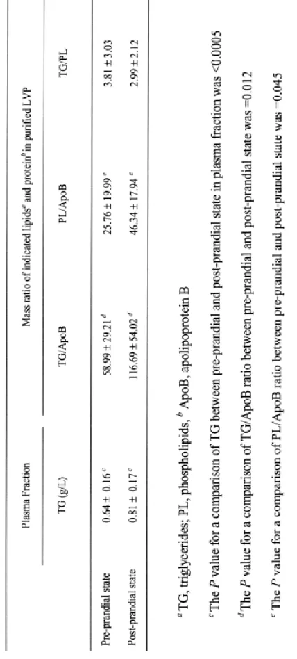
Post-prandial modifications of LVP. In order to further support the hypothesis of a preferential association of HCV to chylomicrons which contributes to LVP production, we studied the dynamic transition between the pre-prandial and the post-prandial periods. For that means, seven HCV-infected volunteers were given a 900 kcal breakfast with 30% fat after an overnight fasting. Peripheral blood was drawn before breakfast and 90 min after the first phlebotomy. The increase observed in plasma TG in all post-prandial samples indicated that fat absorption and chylomicron secretion had occurred during this time period for all patients (table 2). The TG/apoB and PL/apoB ratios of purified LVP significantly increased in 90 min (table 2). By contrast, the TG/PL ratio did not significantly differ in the inter and post-prandial periods indicating that the TG and PL contents of the particle increased in the same proportion. Moreover, the TG/apoB mass ratios in both the pre-prandial and post prandial periods were largely higher in LVP than in the d <1.055g/ml fraction from which LVP were purified (fig. 4A), indicating that LVP are TG-enriched circulating particles in plasma. The fatty acid composition of TG and PL in purified LVP and in the d<1.055 g/ml) fraction was similar in the pre-prandial and in the post-prandial periods (table 3), and very similarly between the two periods (fig.5). These results confirmed the lipoprotein nature of LVP. The rapid and dramatic post-prandial changes observed in the composition of LVP while the composition of the corresponding whole d<1.055 g/l fraction remained steady, further suggested an active contribution of the intestine to LVP production.
DISCUSSION
In the plasma of HCV chronically infected patients, infectious particles are partly found in low density fractions, associated with TRL forming LVP. Some of these LVP are naturally coated with antibody and
240
241
242
243
244
245
246
247
248
249
250
251
252
253
254
255
256
257
258
259
260
261
262
263
264
265
266
267
268
269
270
271
272
273
can be protein A precipitated. Previous analysis of these captured LVP showed that they are globular particles, rich in TG and contain HCV core protein and RNA. In the present study, we show that, in addition to HCV RNA, similarly purified LVP contain at their surface HCV envelope glycoproteins and TRL apolipoproteins, apoB, apoE, apoCII and apoCIII, but not apoAII which is a component of HDL. A major findings is that among TRL apolipoproteins, the two isoforms of apoB, apoB100 and 48, are equally represented, while apo48 is barely detectable in the fasting patient plasma. A direct indication of the association of HCV RNA with apoB48 would require an immunoprecipitation with an anti-apoB48 antibody and the detection of HCV RNA in the captured material. However, ApoB48 results from the edition in enterocytes of a stop codon within the open reading frame of the apoB mRNA (Patterson et al., 2003) leading to a protein lacking the C terminal end of the complete apoB100 molecule. As a result, there is no direct way to capture apoB48 containing lipoproteins and therefore to directly demonstrate the association of HCV RNA with apoB48-containing lipoproteins. Despite this limitation, the strong apoB48 enrichment in protein A captured, HCV RNA positive LVP compare to the plasma lipoproteins strongly suggest a direct association of HCV RNA with apoB48-containing lipoprotein. The rapid and dramatic increase in TG of these purified LVP after lipid ingestion further strengthens the HCV-apoB48 association. These results raise the question of the nature, origin and functions of such particles.
The strong relationship between apoB-containing lipoproteins and viral particles is a specificity of HCV and related virus (Sato et al., 1996). Although ultrastructural analysis of LVP is necessary, these data suggest that LVP are TRL-like particles, in which the two hydrophobic domains of the core protein could be embedded in the neutral lipids of the lipoprotein core (Hope & McLauchlan, 2000, McLauchlan et al., 2002). Glycoprotein E1 and E2, may display an amphipathic helix conformation (Charloteaux et al., 2002) as apolipoproteins, and insert into the surface layer of the particle. With respect to apoE which is born by LVP, it has recently been shown that E2,E3 and E2,E4 genotypes were respectively associated with a significant 3- and 5-fold reduction in the risk of chronic HCV infection compared with E3E4 or E3 and E4 homozygotes (Price et al., 2006). In addition, E2,E2 genotype, was never found in HCV positive patients. The E2 isoform of apoE poorly binds to the LDL receptor (Mahley & Rall, 2000). Since LVP binding to cell can be blocked by anti-apoE antibody (Agnello et al., 1999, Andre et al., 2002), it is likely that the defective binding of apoE2 isoform could result in a poor uptake of LVP. Moreover, these data support a biological role for LVP which, like TRL, may have their fate and their site of clearance directed by their apolipoprotein composition (Field & Mathur, 1995).
Several mechanisms could be involved in the production of LVP. First, LVP could be formed within the blood circulation by the association of mature HCV virions to circulating TRL. However, a recent study reported that HCV RNA quasispecies found in LVP corresponded to a subgroup of the whole plasma viral population (Deforges et al., 2004). This indicates, at least, that LVP are not issued from a random fusion
274
275
276
277
278
279
280
281
282
283
284
285
286
287
288
289
290
291
292
293
294
295
296
297
298
299
300
301
302
303
304
305
306
307
of circulating HCV viruses with plasma lipoproteins. Although natural antibodies against LVP may introduce some bias in selecting a particular LVP subpopulation, the most likely hypothesis is that LVP are formed within the endoplasmic reticulum of lipoprotein-secreting cells, in which apoB and TG are assembled to form TRL. Indeed, immunoprecipitation of TRL with an anti-apoB antibody precipitated 50% of HCV RNA from HCV infected liver macerate, indicating that a substantial amount of HCV RNA was already associated with apoB in hepatocytes (Nielsen et al., 2004). Altogether, these studies suggest that HCV association with apoB-containing lipoproteins likely occurs within lipoprotein-secreting cells rather than results from binding of HCV to TRL in the circulation.
Therefore, one should consider the hypothesis of an intestinal production of LVP, based on the association of HCV-RNA and envelope glycoproteins with apoB48-containing TRL. Indeed, the expression of Apobec1, the editing enzyme of the apoB mRNA leading to apoB48 synthesis, is strictly restricted to enterocytes (Patterson et al., 2003) and HCV infection has not been reported to induce Apobec1 expression in hepatocytes (Jacobs et al., 2005, Smith et al., 2003, Su et al., 2002). This hypothesis is further supported by the variation in the lipid enrichment of circulating LVP between the pre- and the post-prandial period of the patient, as expected for intestinal TRL after food intake (Field & Mathur, 1995). Such an hypothesis is consistent with a previous study reporting that the quasispecies populations of LVP and liver HCV RNA did not completely match suggesting a second reservoir beside the liver and with the presence of HCV proteins in enterocytes of chronically-infected patients (Deforges et al., 2004). Further investigations of chronically infected patients for detection of HCV RNA in intestinal biopsies and comparative quasispecies analysis between gut, LVP and plasma are necessary to precisely quantify the contribution of enterocytes to the circulating viral load.
Besides the fundamental challenge to decipher the mechanisms leading to the production of LVP, considering the intestine as a reservoir and replication site of HCV in the form of LVP have important pathophysiological consequences. Proportion of intestinal LVP might be substantial, mean calculated value 18% of the plasma viral load. Since the final destination of intestinal lipoprotein remnants is the liver (Field & Mathur, 1995), an intriguing possibility could be a permanent inoculation of the liver with LVP from the intestine. Binding and internalization of naturally antibody-coated LVP was shown to be mediated by lipoprotein receptors which recognize apolipoproteins on the viral particles (Andre et al., 2002). Neutralizing antibodies directed to the envelope glycoproteins may therefore not be sufficient to control infection of the liver by LVP. Therefore, classical virions, like those produced in vitro, and LVP, could deliver the virus with the possibility to both acutely and chronically infect the host, a feature not achieved by other flaviviruses.
FOOTNOTES
308
309
310
311
312
* This work was supported by a grant from the Ministère de la jeunesse, de l’éducation nationale et de la recherche, INSERM and the ANRS (Agence Nationale de Recherche sur le SIDA et les Hépatites Virales), by a “Programme de Recherche Clinique des Hospices Civils de Lyon”. Lipid analysis was performed at the “Lipidomics Platform” set up at UMR 585 INSERM / INSA-Lyon (IMBL).
REFERENCES
Agnello, V., Abel, G., Elfahal, M., Knight, G. B. & Zhang, Q. X. (1999). Hepatitis C virus and
other flaviviridae viruses enter cells via low density lipoprotein receptor. Proc Natl Acad
Sci U S A 96, 12766-71.
Andre, P., Komurian-Pradel, F., Deforges, S., Perret, M., Berland, J. L., Sodoyer, M., Pol, S.,
Brechot, C., Paranhos-Baccala, G. & Lotteau, V. (2002). Characterization of low- and
very-low-density hepatitis C virus RNA-containing particles. J Virol 76, 6919-28.
Besnard, N. C. & Andre, P. M. (1994). Automated quantitative determination of hepatitis C virus
viremia by reverse transcription-PCR. J Clin Microbiol 32, 1887-93.
Bradley, D., McCaustland, K., Krawczynski, K., Spelbring, J., Humphrey, C. & Cook, E. H.
(1991). Hepatitis C virus: buoyant density of the factor VIII-derived isolate in sucrose. J
Med Virol 34, 206-8.
Carrick, R. J., Schlauder, G. G., Peterson, D. A. & Mushahwar, I. K. (1992). Examination of the
buoyant density of hepatitis C virus by the polymerase chain reaction. J Virol Methods 39,
279-89.
Charloteaux, B., Lins, L., Moereels, H. & Brasseur, R. (2002). Analysis of the C-terminal
membrane anchor domains of hepatitis C virus glycoproteins E1 and E2: toward a
topological model. J Virol 76, 1944-58.
Deforges, S., Evlashev, A., Perret, M., Sodoyer, M., Pouzol, S., Scoazec, J. Y., Bonnaud, B.,
Diaz, O., Paranhos-Baccala, G., Lotteau, V. & Andre, P. (2004). Expression of hepatitis C
virus proteins in epithelial intestinal cells in vivo. Journal of General Virology 85,
2015-2023.
Field, F. J. & Mathur, S. N. (1995). Intestinal lipoprotein synthesis and secretion. Prog Lipid Res
34, 185-98.
Fisher, E. A. & Ginsberg, H. N. (2002). Complexity in the secretory pathway: the assembly and
secretion of apolipoprotein B-containing lipoproteins. J Biol Chem 277, 17377-80.
Hijikata, M., Shimizu, Y. K., Kato, H., Iwamoto, A., Shih, J. W., Alter, H. J., Purcell, R. H. &
Yoshikura, H. (1993). Equilibrium centrifugation studies of hepatitis C virus: evidence for
circulating immune complexes. J Virol 67, 1953-8.
Hope, R. G. & McLauchlan, J. (2000). Sequence motifs required for lipid droplet association and
protein stability are unique to the hepatitis C virus core protein. J Gen Virol 81, 1913-25.
Jacobs, J. M., Diamond, D. L., Chan, E. Y., Gritsenko, M. A., Qian, W., Stastna, M., Baas, T.,
Camp, D. G., 2nd, Carithers, R. L., Jr., Smith, R. D. & Katze, M. G. (2005). Proteome
analysis of liver cells expressing a full-length hepatitis C virus (HCV) replicon and biopsy
specimens of posttransplantation liver from HCV-infected patients. J Virol 79, 7558-69.
Kaito, M., Watanabe, S., Tsukiyama-Kohara, K., Yamaguchi, K., Kobayashi, Y., Konishi, M.,
Yokoi, M., Ishida, S., Suzuki, S. & Kohara, M. (1994). Hepatitis C virus particle detected
by immunoelectron microscopic study. J Gen Virol 75 ( Pt 7), 1755-60.
Kanto, T., Hayashi, N., Takehara, T., Hagiwara, H., Mita, E., Naito, M., Kasahara, A., Fusamoto,
H. & Kamada, T. (1994). Buoyant density of hepatitis C virus recovered from infected
hosts: two different features in sucrose equilibrium density-gradient centrifugation related
to degree of liver inflammation. Hepatology 19, 296-302.
Komurian-Pradel, F., Paranhos-Baccala, G., Sodoyer, M., Chevallier, P., Mandrand, B., Lotteau,
V. & Andre, P. (2001). Quantitation of HCV RNA using real-time PCR and fluorimetry. J
Virol Methods 95, 111-9.
Lindenbach, B. D., Evans, M. J., Syder, A. J., Wolk, B., Tellinghuisen, T. L., Liu, C. C.,
Maruyama, T., Hynes, R. O., Burton, D. R., McKeating, J. A. & Rice, C. M. (2005).
Complete Replication of Hepatitis C Virus in Cell Culture. Science.
Lindenbach, B. D., Meuleman, P., Ploss, A., Vanwolleghem, T., Syder, A. J., McKeating, J. A.,
Lanford, R. E., Feinstone, S. M., Major, M. E., Leroux-Roels, G. & Rice, C. M. (2006).
Cell culture-grown hepatitis C virus is infectious in vivo and can be recultured in vitro.
Proc Natl Acad Sci U S A.
Mahley, R. W. & Rall, S. C., Jr. (2000). Apolipoprotein E: far more than a lipid transport protein.
Annu Rev Genomics Hum Genet 1, 507-37.
McLauchlan, J., Lemberg, M. K., Hope, G. & Martoglio, B. (2002). Intramembrane proteolysis
promotes trafficking of hepatitis C virus core protein to lipid droplets. Embo J 21, 3980-8.
Miyamoto, H., Okamoto, H., Sato, K., Tanaka, T. & Mishiro, S. (1992). Extraordinarily low
density of hepatitis C virus estimated by sucrose density gradient centrifugation and the
polymerase chain reaction. J Gen Virol 73 ( Pt 3), 715-8.
Nielsen, S. U., Bassendine, M. F., Burt, A. D., Bevitt, D. J. & Toms, G. L. (2004).
Characterization of the genome and structural proteins of hepatitis C virus resolved from
infected human liver. J Gen Virol 85, 1497-507.
Nielsen, S. U., Bassendine, M. F., Burt, A. D., Martin, C., Pumeechockchai, W. & Toms, G. L.
(2006). Association between hepatitis C virus and very-low-density lipoprotein
(VLDL)/LDL analyzed in iodixanol density gradients. J Virol 80, 2418-28.
Patterson, A. P., Chen, Z., Rubin, D. C., Moucadel, V., Iovanna, J. L., Brewer, H. B., Jr. &
Eggerman, T. L. (2003). Developmental regulation of apolipoprotein B mRNA editing is
an autonomous function of small intestine involving homeobox gene Cdx1. J Biol Chem
278, 7600-6.
Petit, M. A., Lievre, M., Peyrol, S., De Sequeira, S., Berthillon, P., Ruigrok, R. W. & Trepo, C.
(2005). Enveloped particles in the serum of chronic hepatitis C patients. Virology 336,
144-53.
Price, D. A., Bassendine, M. F., Norris, S. M., Golding, C., Toms, G. L., Schmid, M. L., Morris,
C. M., Burt, A. D. & Donaldson, P. T. (2006). Apolipoprotein epsilon3 allele is associated
with persistent hepatitis C virus infection. Gut 55, 715-8.
Prince, A. M., Huima-Byron, T., Parker, T. S. & Levine, D. M. (1996). Visualization of hepatitis
C virions and putative defective interfering particles isolated from low-density
lipoproteins. J Viral Hepat 3, 11-7.
Pringle, C. R. (1999). Virus taxonomy--1999. The universal system of virus taxonomy, updated
to include the new proposals ratified by the International Committee on Taxonomy of
Viruses during 1998. Arch Virol 144, 421-9.
Sato, K., Tanaka, T., Okamoto, H., Miyakawa, Y. & Mayumi, M. (1996). Association of
circulating hepatitis G virus with lipoproteins for a lack of binding with antibodies.
Biochem Biophys Res Commun 229, 719-25.
Smith, M. W., Yue, Z. N., Korth, M. J., Do, H. A., Boix, L., Fausto, N., Bruix, J., Carithers, R.
L., Jr. & Katze, M. G. (2003). Hepatitis C virus and liver disease: global transcriptional
profiling and identification of potential markers. Hepatology 38, 1458-67.
Su, A. I., Pezacki, J. P., Wodicka, L., Brideau, A. D., Supekova, L., Thimme, R., Wieland, S.,
Bukh, J., Purcell, R. H., Schultz, P. G. & Chisari, F. V. (2002). Genomic analysis of the
host response to hepatitis C virus infection. Proc Natl Acad Sci U S A 99, 15669-74.
Takahashi, K., Kishimoto, S., Yoshizawa, H., Okamoto, H., Yoshikawa, A. & Mishiro, S. (1992).
p26 protein and 33-nm particle associated with nucleocapsid of hepatitis C virus
recovered from the circulation of infected hosts. Virology 191, 431-4.
Thomssen, R., Bonk, S., Propfe, C., Heermann, K. H., Kochel, H. G. & Uy, A. (1992).
Association of hepatitis C virus in human sera with beta-lipoprotein. Med Microbiol
Immunol (Berl) 181, 293-300.
Thomssen, R., Bonk, S. & Thiele, A. (1993). Density heterogeneities of hepatitis C virus in
human sera due to the binding of beta-lipoproteins and immunoglobulins. Med Microbiol
Immunol (Berl) 182, 329-34.
Wakita, T., Pietschmann, T., Kato, T., Date, T., Miyamoto, M., Zhao, Z., Murthy, K.,
Habermann, A., Krausslich, H. G., Mizokami, M., Bartenschlager, R. & Liang, T. J.
(2005). Production of infectious hepatitis C virus in tissue culture from a cloned viral
genome. Nat Med.
Zhong, J., Gastaminza, P., Cheng, G., Kapadia, S., Kato, T., Burton, D. R., Wieland, S. F.,
Uprichard, S. L., Wakita, T. & Chisari, F. V. (2005). Robust hepatitis C virus infection in
vitro. Proc Natl Acad Sci U S A 102, 9294-9.
LEGENDS
Fig. 1. ApoB100 and apoB48 are present in purified LVP. A, Nature of apoB in purified LVP. Plasma from HCV-infected or healthy donors were adjusted to a 1.055g/ml density and centrifugated for 4h at 4°C and 543,000xg. LVP were immunopurified from the low-density fraction as described in experimental procedures. Samples of LVP and of the whole fraction were analyzed by 5% SDS-PAGE under reducing conditions and immunoblotted with 1D1 anti-apoB monoclonal antibody. Lane 1 and 2, the d<1.055g/ml fraction and chylomicrons isolated from a healthy blood donor; respectively; lane 3, mocked-prepared LVP from a healthy blood donor; lane 4 and 5, purified LVP from patient H; lane 6 and 7, the d<1.055g/ml fraction from which LVP were purified from the same patient and a healthy subject, respectively. B, Relative proportions of apoB48 and apoB100 in purified LVP and in the d<1.055g/ml fraction prepared from eight infected patients. ApoB48 and apoB100 spots were quantified by videodensitometry and expressed as % of total apoB. The proportion of apoB48 was significantly higher in purified LVP than in the whole d<1.055g/ml fraction (Student T test p<0.01).
Fig. 2. Presence of apoE, CII and CIII in LVP. LVP were immunopurified from the d<1.055g/ml fraction, as described in experimental procedures, analysed by 12% (apoE) or 15% (apoAII, apoCII and apoCIII) SDS-PAGE under reducing conditions, and immunoblotted with anti-apoE, anti-apoAII or anti-apoCIII monoclonal antibodies (Chemicon International) or anti-apoCII polyclonal antibody (Merck Calbiochem). Lane 1 and 2, mocked-prepared LVP and the d<1.055g/ml fraction from a non infected blood donor, respectively;lane 3 and 4, LVP and the d<1.055g/ml fraction from a chronically infected patient, respectively; lane 5, control plasma from a blood donor. ApoE, CII and III were present in the d<1.055g/ml fraction where apoB-containing lipoproteins reside and in purified LVP. ApoAII, a component of HDL, was neither detected in the d<1.055g/ml fraction nor in LVP.
Fig. 3. Presence of envelope glycoproteins in LVP. A, LVP were immunopurified from the d<1.055g/ml fraction, as described in the experimental procedures, analyzed by 10% SDS-PAGE under reducing conditions, and immunoblotted with the A4 anti-E1 (lower panel) or the H52anti-E2 (upper panel) monoclonal antibodies. Lane 1 and 2, lysates of 293T cells expressing or not E1 and E2 glycoproteins, respectively; lane 3, mocked-prepared LVP from plasma of a healthy blood donor; lane 4, purified LVP from a chronically infected patient; lane 5 and 6, the d<1.055g/ml fraction from which LVP were purified from a HCV patient or a healthy subject, respectively. Western blots are representative of experiments performed with plasma from 3 infected individuals. Note that both viral glycoproteins were detected in purified LVP. B, Detection of protein A-captured LVP in an ELISA with anti-E1 and anti-E2 antibodies .
The d<1.055g/ml fractions were prepared as described in material and methods from chronically infected patients and from non-infected blood donors. The d<1.055g/ml fraction from infected (closed square) or non infected (open square) patients were revealed by anti-E1 A4 (a) or anti-E2 H47 (b). As a control, the d<1.055g/ml fraction from infected (open triangle) or non infected (open circle) patients, stained by anti-H measles clone 55, are presented. Results are means of duplicates (×). Note that protein A-captured LVP from infected patient were only recognized by HCV E1 and E2 envelope antibodies and not by anti-measles virus H envelope antibodies. No material from non infected control was recognized by any antibody.
Fig. 4. Evolution of TG/apoB mass ratio in LVP between the pre-prandial and the post-prandial periods. (a), Mean (dash) and individual TG/apoB mass ratios (spots) in both fractions from 7 patients.. Lipids from purified LVP and the whole <1.055g/ml fraction were extracted and separated by TLC. TG spots were scraped off, and the fatty acids were transmethylated and quantified by gas chromatography, and the apoB content of purified LVP was determinated by ELISA, as described under “Experimental Procedures”. Note that the TG/apoB was significantly higher in LVP than in their respective whole fraction (Wilcoxon T test, p≤0.05). (b), TG/ApoB mass ratio in LVP increase between the pre-prandial to post-prandial periods. Results are expressed as the ratio between the TG/apoB mass ratio in LVP in the post-prandial period vs that in the pre-prandial period for each patient. Patients are identified as patient A to G and indicated by arrows in (a). Note that the TG/apoB mass ratio was significantly increased in LVP during the post-prandial period (distribution-free Wilcoxon T test, p≤0.05).
Fig. 5. Effect of lipid intake on LVP characteristics. A, Lipid and protein mass ratios in the whole d<1.055g/ml fractions and purified LVP in the pre-prandial and post-prandial periods. Results are means from seven patients. B, Parallel modifications of fatty acid composition of LVP and of the fraction d<1.055g/ml between the pre-prandial to post-prandial periods. n-6 and saturated fatty acids were quantified by gas chromatography as described in material and methods and expressed as mol% in triacylglycerol (TG) and phospholipids (PL) of purified LVP and of the whole d<1.055g/ml fraction. Results are means from 11 patients. Note that the fatty acid composition of TG and PL vary in the same proportion in LVP and the whole d<1.055g/ml density fraction between the pre-prandial and the post-prandial periods.
Figure 4
(a)
(b)
(a)
(b)
Figure 5
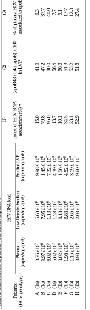
Table 1
ical ch aracteristics o f p atien ts with ch ro n ic h ep atitis C HCV RNA load Patien ts (HC V genot y p e) Pla sma (copies/mg apoB) Low-Density -Fraction (copies/mg apoB) Pruifie d L V P (copies/mg apoB) (1) index of HC V R NA association (%) † (2) (apoB 48 / t ot al apoB ) x 100 in LVP (3) % of plasm a HCV RNA associated to apoB48 ‡ A (1a) 3.76 ×10 7 5.63 ×10 6 9.98 × 10 8 15.0 41.9 6.3 B (1 a) 9.97 ×10 6 7.95 ×10 6 1.45 × 10 8 79.8 47.2 37.7 C (1 a) 6.02 ×10 5 1.13 ×10 6 7.32 × 10 7 95.0 40.0 40.0 D (1a) 5.62 ×10 7 1.28 ×10 7 4.39 × 10 9 13.7 56.4 7.7 E (1b) 8.02 ×10 6 8.13 ×10 5 1.56 × 10 8 10.1 50.3 5.1 F (1b) 1.98 ×10 7 6.83 ×10 6 4.52 × 10 8 34.5 51.3 17.7 G (1b) 1.15 ×10 7 2.65 ×10 6 3.19 × 10 9 23.1 53.2 12.3 H (1b) 3.93 ×10 6 2.08 ×10 6 9.60 × 10 7 52.9 51.8 27.4 † Index of HC V R NA associ at ion wi th LDF: see m et hods‡
colum n 3 cal cul at ed as val u e of (col um n 1 x col u m n 2)/ 100 22Table 2
ffect of li
E
pid intake on LVP characteristics: lipid and protein m
ass ratio
23Ta
ble 3
ty aci d com posi ti on of phosphol ip id s and t ri acy lg ly cerol s from puri fi ed LVP a and whol e l o w-densi ty -fract io n a before ( Pre-prandi al) and after the m
eal ( Post-al ). ation: PL, phosphol ip id s; TG, t ri acy lg ly cerol ; ND, not det ect ed
by weight %. Values represent m
eans ± SEM Pu rif ied LVP (n =7 ) Wh o le f ractio n (n =7 ) Fatty acids PL TG PL TG mol % P re-prandial P o st-p randial P re-prandial P o st-p randial P re-prandial P o st-p randial P re-prandial P o st-p randial 14:0 5. 91 ± 2. 26 5. 75 ± 1. 78 3. 56 ± 0. 90 5. 16 ± 1. 47 0, 61 ± 0. 11 0. 55 ± 0. 11 2. 91 ± 0. 68 4. 84 ± 0. 37 16:0 38. 82 ± 2. 30 35. 61 ± 2. 57 36. 36 ± 2. 14 36. 63 ± 2. 74 38, 32 ± 1. 27 35. 85 ± 2. 63 34. 90 ± 1. 44 35. 84 ± 2. 13 16:1 n -7 0. 49 ± 0. 17 1. 19 ± 0. 25 6. 46 ± 1. 92 5. 30 ± 1. 28 0, 65 ± 0. 20 0. 87 ± 0. 19 3. 16 ± 0. 61 3. 35 ± 0. 63 18:0 15. 51 ± 2. 36 14. 98 ± 2. 42 10. 18 ± 3. 50 9. 09 ± 3. 77 12, 95 ± 1. 39 16. 32 ± 2. 29 6. 20 ± 1. 31 4. 89 ± 0. 31 18:1 n -9 16. 90 ± 4. 07 13. 64 ± 1. 47 32. 42 ± 3. 48 32. 35 ± 2. 78 11, 04 ± 1. 04 10. 25 ± 1. 25 35. 39 ± 3. 68 37. 14 ± 2. 14 18:2 n -6 16. 24 ± 1. 74 16. 78 ± 1. 61 11. 85 ± 1. 77 9. 43 ± 1. 71 20, 05 ± 1. 18 19. 27 ± 1. 45 14. 70 ± 1. 61 10. 67 ± 1. 13 18:3 n -6 ND ND ND ND 0, 18 ± 0. 10 0. 66 ± 0. 37 0. 14 ± 0. 05 0. 13 ± 0. 05 20:3 n -6 3. 38 ± 1. 34 1. 89 ± 0. 41 0. 97 ± 0. 45 0. 14 ± 0. 07 2, 35 ± 0. 27 2. 38 ± 0. 32 0. 54 ± 0. 29 0. 22 ± 0. 07 20:4 n -6 6. 56 ± 0. 95 6. 64 ± 0. 64 1. 82 ± 0. 47 1. 54 ± 0. 44 6, 72 ± 0. 87 6. 58 ± 0. 63 2. 24 ± 0. 64 1. 12 ± 0. 23 20:5 n -3 0. 40 ± 0. 11 1. 27 ± 0. 48 ND 0 .6 4 ± 0. 36 1, 62 ± 0. 90 2. 04 ± 1. 37 ND ND 22:4 n -6 9. 87 ± 2. 19 7. 48 ± 2. 67 2. 46 ± 1. 06 1. 13 ± 0. 29 5, 53 ± 1. 11 1. 70 ± 0. 78 0. 33 ± 0. 13 0. 24 ± 0. 08 22:5 n -3 0. 44 ± 0. 11 2. 92 ± 0. 78 1. 56 ± 0. 69 0. 88 ± 0. 24 0, 64 ± 0. 10 0. 66 ± 0. 09 0. 34 ± 0. 07 0. 23 ± 0. 06 22:5 n -6 1. 01 ± 0. 07 7. 06 ± 1. 40 1. 11 ± 0. 27 1. 75 ± 0. 85 1, 07 ± 0. 48 3. 45 ± 1. 81 0. 67 ± 0. 39 0. 12 ± 0. 03 22:6 n -3 2. 19 ± 0. 49 2. 82 ± 0. 62 0. 68 ± 0. 26 0. 56 ± 0. 24 2, 33 ± 0. 41 3. 23 ± 0. 70 0. 92 ± 0. 26 0. 50 ± 0. 14 ΣsatFA 58. 10 ± 3. 34 54. 78 ± 3. 81 49. 17 ± 4. 73 50. 87 ± 3. 42 61. 82 ± 0. 89 52. 72 ± 1. 76 43. 45 ± 1. 50 45. 57 ± 2. 11 Σn -3 23. 98 ± 3. 15 27. 68 ± 4. 21 12. 93 ± 2. 54 11. 34 ± 2. 02 32. 99 ± 1. 68 30. 60 ± 2. 28 16. 78 ± 2. 94 12. 48 ± 1. 35 Σn -6 1 .1 0 ± 0. 53 3. 21 ± 0. 98 0. 61 ± 0. 39 0. 95 ± 0. 43 3. 56 ± 0. 71 5. 37 ± 1. 00 1. 04 ± 0. 32 0. 78 ± 0. 21 24