HAL Id: inserm-01281366
https://www.hal.inserm.fr/inserm-01281366
Submitted on 2 Mar 2016
HAL is a multi-disciplinary open access
archive for the deposit and dissemination of
sci-entific research documents, whether they are
pub-lished or not. The documents may come from
teaching and research institutions in France or
abroad, or from public or private research centers.
L’archive ouverte pluridisciplinaire HAL, est
destinée au dépôt et à la diffusion de documents
scientifiques de niveau recherche, publiés ou non,
émanant des établissements d’enseignement et de
recherche français ou étrangers, des laboratoires
publics ou privés.
Clinical Features of Spontaneous Partial Healing During
Mycobacterium ulcerans Infection
Estelle Marion, Annick Chauty, Marie Kempf, Yannick Le Corre, Yves
Delneste, Anne Croue, Laurent Marsollier
To cite this version:
Estelle Marion, Annick Chauty, Marie Kempf, Yannick Le Corre, Yves Delneste, et al.. Clinical
Features of Spontaneous Partial Healing During Mycobacterium ulcerans Infection. Open Forum
Infectious Diseases, Oxford University Press, 2016, �10.1093/ofid/ofw013�. �inserm-01281366�
Open Forum Infectious Diseases
M A J O R A R T I C L E
Clinical Features of Spontaneous Partial Healing During
Mycobacterium ulcerans Infection
Estelle Marion,1,2Annick Chauty,1Marie Kempf,3Yannick Le Corre,4Yves Delneste,6Anne Croue,5and Laurent Marsollier2; for the Franco-Beninese Buruli
Research Group
1Centre de Dépistage et de Traitement de l’Ulcère de Buruli de Pobè, Fondation Raoul Follereau, Bénin;2
Atip/Avenir Team, Centre de Recherche en Cancérologie Nantes-Angers (CRCNA), Université et Centre Hospitalier Universitaire (CHU) d’Angers,3
Laboratoire de Bactériologie et d’Hygiène Hospitalière,4
Service de Dermatologie,5
Laboratoire d’Anatomie Pathologique, CHU d’Angers,6
Team“Innate Immunity”, Université d’Angers, Labex IGO, France
Background. Buruli ulcer, caused by Mycobacterium ulcerans, is a necrotizing skin disease leading to extensive cutaneous and subcutaneous destruction and functional limitations. Spontaneous healing in the absence of medical treatment occurs in rare cases, but this has not been well described in the literature.
Methods. In a retrospective case study in an area of Benin where this disease is highly endemic, we selected 26 Buruli ulcer patients presenting features of spontaneous healing from a cohort of 545 Buruli ulcer patients treated between 2010 and 2013.
Results. The 26 patients studied had a median age of 13.5 years and were predominantly male (1.4:1). Three groups of patients were defined on the basis of their spontaneous healing characteristics. The first group (12 patients) consisted of pa-tients with an ulcer of more than 1 year′s duration showing signs of healing. The second (13 patients) group contained patients with an active Buruli ulcer lesion some distance away from afirst lesion that had healed spontaneously. Finally, the third group contained a single patient displaying complete healing of lesions from a nodule, without treatment and with no relapse.
Conclusions. We defined several features of spontaneous healing in Buruli ulcer patients and highlighted the difficulties associated with diagnosis and medical management. Delays in consultation contributed to the high proportion of patients with permanent sequelae and a risk of squamous cell carcinoma. Early detection and antibiotic treatment are the best ways to reduce impairments.
Keywords. Buruli ulcer; M ulcerans; spontaneous healing.
Buruli ulcer is a neglected tropical disease caused by Mycobac-terium ulcerans, an environmental mycobacMycobac-terium. This skin infection, which mostly affects children, has emerged or re-emerged in the last 2 decades, particularly around the Gulf of Guinea in West Africa [1]. The route of transmission has yet to be clearly determined, but inoculation of the derma with the bacterium appears to be necessary [2]. After an estimated incubation period of a few weeks to several months,M ulcerans causes necrotizing hypodermitis, in the form of a nodule, an edema, or a plaque [3]. After destruction of the subcutaneous tissue (caused by a cytotoxic, immunosuppressive and analgesic toxin called mycolactone [4–6]), the skin may break down, lead-ing to the development of largely painless necrotic ulcers with characteristic undermined edges [7].
No specific vaccine against Buruli ulcer is currently available. Bacillus Calmette-Guérin (BCG) vaccination was initially thought to have incomplete but significant short-term protec-tive effects [8,9] or to confer partial protection againstM ulcer-ans osteomyelitis [10]. However, other studies provided no evidence of a protective effect [11,12] or even reported a higher risk of Buruli ulcer in adults and children over the age of 5 years that had been vaccinated with BCG [13].
The severity of Buruli ulcer lesions depends on their form, ex-tent, and localization [14]. Indeed, early lesions (nonulcerative) may differ in clinical appearance (taking the form of a nodule, pla-que, or edema), and these different forms progress differently. For example, a nodule may spontaneously progress to a small circum-scribed ulcer, whereas an edema of the entire limb generally pro-gresses to extensive ulceration with severe functional impairment [14,15]. Other factors may also determine the severity of Buruli ulcer. These factors include human immunodeficiency virus coin-fection [16–18] and time from onset tofirst consultation (which depends partly on local healthcare management) [19,20].
Small Buruli ulcer lesion can be treated with a combination of rifampin and an aminoglycoside for 8 weeks, but larger lesions re-quire additional extensive surgery in dedicated hospitals [21–23]. Even in cases of effective medical treatment, more than 20% of pa-tients present permanent functional sequelae 1 year later [14].
Received 18 January 2016; accepted 22 January 2016.
Correspondence: E. Marion, CRCNA U892, Angers 49100, France (estelle.marion@inserm.fr). Open Forum Infectious Diseases®
© The Author 2016. Published by Oxford University Press on behalf of the Infectious Diseases Society of America. This is an Open Access article distributed under the terms of the Creative Commons Attribution-NonCommercial-NoDerivs licence (http://creativecommons.org/licenses/ by-nc-nd/4.0/), which permits non-commercial reproduction and distribution of the work, in any medium, provided the original work is not altered or transformed in any way, and that the work is properly cited. For commercial re-use, please contact journals.permissions@oup.com. DOI: 10.1093/ofid/ofw013
at Laboratoire Algebre Et Geometrie on March 1, 2016
http://ofid.oxfordjournals.org/
If left untreated, Buruli ulcers progress to chronic ulcerations with a risk of dissemination and bone destruction [14,19,24]. Although many publications mention the occurrence of spon-taneous healing in some patients, the process remains poorly described or studied [7,25–32]. Questions thus remain unre-solved: Are healing cases rare in the Buruli ulcer context? What are the principal clinical features of this healing process? In this context, we performed a retrospective study in a highly specialized Buruli ulcer diagnosis and treatment center in Benin. Moreover, we provide a comprehensive description of the various clinical presentations of the spontaneous partial healing of Buruli ulcer.
METHODS AND RESULTS
Study Design, Data Collection, and Definitions
A retrospective study was conducted at the Centre de Diagnostic et de Traitement de la Lèpre et de l′Ulcère de Buruli (CDTLUB) in Pobè, Benin, which has one of the largest databases for cases of Buruli ulcer confirmed by polymerase chain reaction (PCR) [14]. Wefirst conducted a preliminary analysis of 545 Buruli ulcer patients treated at the CDTLUB in Pobè from 2010 to 2013. For the selection of medical records, we used several key words de-fined on the basis of publications in this field: spontaneous heal-ing, spontaneous clearance, scar, old ulcer, scarring lesion. During this review step, medical records containing at least 1 of the chosen key words were selected. If the clinical records were incomplete (eg, lack of photographic monitoring, patients lost to follow-up), the patient was excluded.
The preselected clinical records were then fully examined and patient information was collected (including age, sex, date of onset, date of diagnosis, clinical features, past medical history, medical and laboratory analysis, lesion examination, imaging data, and clinical monitoring). None of the patients received pharmacological treatment for mycobacterial infection before consulting at the Centre de Dépistage et de Traitement de l ’Ul-cère de Buruli (CDTUB) Pobè.
Cohort Presentation
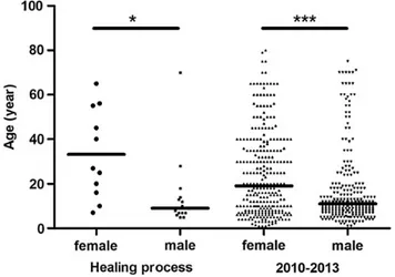
We selected 26 patients (sex ratio M/F =1.4), on the basis of our criteria, from the database of the CDTLUB. Median patient age at diagnosis was 13.5 years (5–70 years), with female patients tend-ing to be older than male patients (Figure1). The age distribution of male and female patients was similar to that for the entire pop-ulation of 545 Buruli ulcer patients from 2010 to 2013 (Figure1). Almost all the patients (25 of 26) had category 3 lesions (World Health Organization nomenclature) presenting as sin-gle lesions of more than 15 cm in diameter or osteomyelitis, or multiple lesions, and/or lesions at critical sites. In 85% of pa-tients (22 of 26), lesions were present on the lower limbs (59% for the total cohort,P < .05, Fisher test). Three patients present-ed multiple active Buruli ulcer lesions (2 patients with 2 lesions, 1 patient with 3 lesions).
We found that 77% (20 of 26) of patients had waited at least 1 year after thefirst signs of an active lesion before consulting at the CDTUB Pobè (13% for the total cohort,P < .001, Fisher test). Complete physical examination of body showed that 13 of the 26 patients had a healed scar with a stellate appearance consistent with an old Buruli ulcer lesion at some distance from the active lesion. An exhaustive study of the clinical re-cords of each patient, including the clinical features of the lesion and the medical history of the patient, led us to define 3 groups on the basis of the healing process observed, as described below.
Group 1: Spontaneous Healing Process in Progress
Patients of this group (n = 12) typically had an ulcer that had been present on a limb for more than 1 year and was healing (Table1). Seven of the 12 patients in this group were male (me-dian age = 9 years) and 5 were female (me(me-dian age = 25 years). The edges of the lesion were defined by skin repair and re-epi-thelialization processes (Figure2). The lesions typically dis-played no edema, plaque, necrosis, or acute inflammation (Figure2). Despite the lack of specific clinical signs of Buruli
ulcer, microbiological analysis based on PCR (73%), acid-fast bacilli (45%), and culture (9%) demonstrated the presence of M ulcerans infection (9%) (Table1). Swabbing was difficult be-cause the edges of the lesions had healed. Biological confirma-tion was obtained principally by biopsy during surgical repair (eg, debridement, skin graft). Most patients (75%) presented physical impairment on arrival at the center and, even with combined drug treatment and surgery, this impairment was ir-reversible in 42% of patients (Table1). The need for surgery and rehabilitation resulted in hospitalization for 2 to 7 months. In 1 patient, the lesion progressed to squamous cell carcinoma 6 months after release from hospital and 3 years after thefirst signs of this lesion. The affected limb had to be amputated.
Figure 1. Age distribution, by sex, of Buruli ulcer patients in the spontaneous
healing cohort and in the general cohort of patients seen between 2010 and 2013. The age distribution is similar for the 2 cohorts, with female patients older
than male patients in both cohorts. Mann–Whitney U test; *P < .05, ***P < .0001.
at Laboratoire Algebre Et Geometrie on March 1, 2016
http://ofid.oxfordjournals.org/
Group 2: Active Buruli Ulcer Lesion After Initial Spontaneous Healing
There were 13 patients in this group (n = 13): 8 were male (me-dian age = 10 years) and 5 were female (me(me-dian age = 40 years) (Table2). Patients presented 1 (77%) or multiple (23%) active Buruli ulcer lesions, with bone involvement in 40% of the pa-tients, mostly on the lower limbs. All of the active lesions
were biologically confirmed, at least by PCR. There was fre-quently a long interval between the onset of symptoms and con-sultation, with 77% of patients waiting more than 1 year after the onset of the active lesion before seeking medical attention (Table2).
During medical examination, 1 or several stellate scars were observed, some distance away from the active lesion (Figure3). The scars were located on the same lower limb as the active le-sion, on the contralateral lower limb, upper limbs, abdomen, or head (Table2). The history of the previous lesions and the ap-pearance of the scars were consistent with the spontaneous healing of an old Buruli ulcer lesion. The interval between total healing of the previous lesion and the occurrence of the active lesion was highly variable: from 4 weeks to more than 1 year. Biological confirmation of M ulcerans infection was not performed on healed tissues at the consultation. However, in 2 cases, the need for surgery to minimize functional impair-ment made it possible to confirm the presence of M ulcerans DNA in the healed tissues.
On arrival, most patients (70%) presented physical impair-ments due to the spontaneously healed old lesion or the active lesion. After drug treatment (100% of patients) and surgery (92% patients), the patients continued to display functional damage. Limb amputation was required in 1 patient, due to the development of a squamous cell carcinoma at the site of the treated lesion after the completion of drug treatment. Hospital stay exceeded 120 days for 46% of the patients, due to the pres-ence of extensive or multifocal lesions, osteomyelitis, or physical impairment.
Group 3: Complete Spontaneous Healing
One patient (n = 1) presentedM ulcerans infection progressing spontaneously towards complete healing without treatment.
Table 1. Characteristics of Patients Presenting a Buruli Ulcer Lesion Undergoing Spontaneous Healing
Patient Age, Years/ Sex Clinical Form Site Patient Delay, Weeks Hospital Stay,
Days Antibiotics Surgery PCR Culture DSE
Physical Impairment, Arrival Physical Impairment, Final Squamous Cell Carcinoma 1 7/F U LL >52 73 C/R No NA NA NA 1 1 0 2 9/M U UL 30 78 S/R Yes + − + 0 0 0 3 7/M U UL >52 105 S/R Yes + − + 1 1 0 4 14/M U UL >52 219 S/R Yes + − − 1 1 0 5 18/M U LL >52 56 S/R No − − − 1 1 0 6 20/F U UL >52 155 S/R Yes − − − 1 1 1 7 10/M U UL >52 99 S/R Yes + − + 1 1 0 8 65/F U UL 52 56 C/R No + − − 0 0 0 9 25/F U UL 18 75 S/R Yes + − + 1 0 0 10 55/F U UL >52 122 C/R Yes + − − 0 0 0 11 5/M U UL >52 127 C/R Yes − − − 1 1 0 12 6/M U UL 52 152 S/R Yes + + + 1 0 0 Mean Age 20 U 83% LL >52 110 100% 75% 73% 9% 36% 75% 58% 8%
Abbreviations: C/R, clarithromycin and rifampicin; DSE, direct smear examination; LL, lower limb; NA, not available; PCR, polymerase chain reaction; S/R, streptomycin and rifampicin; U, ulcer; UL, upper limb.
Figure 2. Typical case of group 1 spontaneous healing in progress (
Mycobacte-rium ulcerans lesions with evidence of healing tissues). Clinical examination showed a large ulcer on the outside of the right knee, measuring approximately 10 × 6.5 cm, with a well demarcated border. The base of the ulcer was clean and there was gran-ulation tissue. Partial healing was observed, and peripheral epithelialization was as-sociated with adhesions restricting the motion of the joint. The black dotted line circumscribes the ulcerative area. Abbreviations: Ad, adhesion; E, peripheral epithe-lialization; Gt, granuloma tissue.
at Laboratoire Algebre Et Geometrie on March 1, 2016
http://ofid.oxfordjournals.org/
The patient, a 27-year-old woman, presented a typical painless nodule (3 cm diameter) on the right arm. Fine-needle aspira-tion was performed on the nodule to confirm M ulcerans infec-tion [33,34].Mycobacterium ulcerans was detected by PCR and Ziehl-Neelsen staining. Nodule excision or antibiotic treatment was refused by the patient. Two months after thefirst consulta-tion, medical staff visited the patient and found that the nodule had disappeared, with a small induration in its place. During interviews, the patient and her family indicated that the patient had received no drug treatment or traditional treatment. Three months later, the patient was seen again. No active lesion was detectable and a small scar was visible (Figure 4). She was seen again 3 years later and had suffered no relapse during this period. This clinical case highlights the ability of some M ulcerans lesions to resolve spontaneously.
DISCUSSION
Spontaneous healing is known to occur in most infectious dis-eases [35–38]. However, there have been few descriptions of spontaneous healing in cases ofM ulcerans infection. There is no medical consensus concerning the relevance of this process in this disease, mostly due to the lack of epidemiological studies. In this context, we conducted thefirst retrospective study of spontaneous healing, in which we reviewed 545 cases of Buruli ulcer.
We included 26 (4.7%) Buruli ulcer patients described as “presenting a spontaneous healing process”, according to our criteria. In most cases, the spontaneous healing was a lengthy process observed in old extensive Buruli ulcer lesions. The delay in seeking medical attention resulted in irreversible phys-ical impairment in most patients. Furthermore, 2 patients sub-sequently displayed squamous cell carcinoma, which is not surprising because the depigmented scar of a healed lesion on black skin is a risk factor of this cancer.
Spontaneous healing may manifest in different ways, as high-lighted by the 3 different clinical groups defined in this study. In group 1, the patients had old ulcerative lesions that were heal-ing. This form is not typical of active Buruli ulcer lesions and its diagnosis is difficult, particularly given the difficulty obtaining samples for biological confirmation (absence of an undermined edge). Moreover, there are several differential diagnoses (vari-cose ulcers, for example), and the history of the disease recorded during the patient interview is critical to facilitate diagnosis. The patients in group 2 had an active lesion occurring at some dis-tance from a typical Buruli ulcer scar [26,27,30]. Again, the his-tory of this lesion is the key to associating the scar with a typical Buruli ulcer that healed spontaneously.
These observations of a spontaneous healing process demon-strate that patients can develop an immune response able to counteract the effects ofM ulcerans and mycolactone and to promote tissue remodeling. The occurrence of a second active Buruli ulcer lesion at some distance from a healed lesion
Ta b le 2 . Char a cte ris tics of P a tients W ith an Activ e Burul i Ulc er Lesion Some Dis tance Fr om a Sponta neousl y Heale d Les ion P a tient Age, Years/ Se x Clinical Form Site Number of Activ e lesions Dis tant Scar P a tient Dela y , W eeks Hospital Sta y Da y s Antibiotics Surgery PCR Cultur e DSE Phy sical Impairment, Arrival Phy sical Impairment, Final Time to Second Lesion, W eeks a Squamous C ell Car cinoma 1 10/F EUOsC 2 LL 2 LL >52 240 S/R Y es + 2 UL −− 1 1 >52 0 2 12/M UOsC LL, UL 2 Ba ck >52 405 S/R Y es + UL, LL + UL + UL, LL 1 1 >52 0 3 56/F UC 2 UL >2 2 UL >52 120 S/R Y es + UL − + UL 1 1 >52 0 4 70/M EUC UL 1 UL >52 55 S/R Y es + − +1 1 N A 0 5 45/F QUC UL 1 UL 24 55 S/R No + + + 0 0 8 0 6 8/M QC UL 1 UL 12 40 S/R Y es + − +0 0 4 0 7 28/M UC UL 1 UL >52 120 S/R Y es + −− 1 1 >52 1 8 7/M OsC UL 1 Abdomen >52 57 S/R Y es + − + 1 1 >52 0 9 16/F UC UL 1 UL 52 68 S/R Y es + − +0 0 4 0 10 7/M OsC 2 LL, 2 UL 1 2 UL, 1LL >52 70 S/R Y es + UL −− 11 8 0 11 5/M UC Buttock 1 Head 36 65 C/R Y es + Buttock + − 0 0 36 0 12 40/F UC UL 1 UL >52 125 S/R Y es + − + 1 1 >52 0 13 13/M OsC 2 UL, 1 LL >2 UL, LL >52 155 S/R Y es + UL, LL − + LL 1 1 >52 0 Mean Age 24 38% Os 92% LL 30% multi >52 121 100% 92% 100% 23% 69% 69% 69% >52 8% Abbr evia tions: C/R, clarithr omycin and rifam picin; DSE, dir ect smear e xamina tion; E, edema; LL, lo w er limb; Os, os teomy elitis; Q, plaque; S/R, s tr ept omycin and rifampicin; U, ulcer; UL, upper limb. aTime to second lesion pr o vided by pa tient his tory .
at Laboratoire Algebre Et Geometrie on March 1, 2016
http://ofid.oxfordjournals.org/
suggests that the immune response involved in healing cannot sterilize the tissue, or even confer protection againstM ulcerans. Therefore, the development of a vaccine againstM ulcerans may be compromised. It seems unlikely that the occurrence of a sec-ond lesion is due to a secsec-ond contamination event involving the inoculation of the skin with the bacillus. Given the high rate of osteomyelitis observed in second lesions, it seems more likely that M ulcerans disseminates systemically, as suggested by other studies [14,16,19,24].
The single patient in group 3 provides a formal demonstra-tion of the possibility of spontaneous healing of Buruli ulcer without the intervention of Western or traditional medicine.
It is reasonable to assume that the number of cases of sponta-neous healing is underestimated, because patients do not seek medical assistance for small, painless cutaneous lesions. Even though we cannot exclude that patients used traditional thera-pies before visiting the doctor, the potential impact of such ther-apies on the course of the disease remains highly speculative.
Finally, this study raises key questions about the contribution of bacterial and host factors to the documented heterogeneity of the clinical presentation of Buruli ulcer. We cannot rule out the possibility that bacterial strain affects virulence, adaptation, and ability to modulate the immune response. However, this effect is not likely to be the key determinant, because molecular [39] ep-idemiological studies have shown thatM ulcerans diversity is poor (within the same endemic area). In this context, our results suggest that host genetic factors are the key determinants of path-ogen control governing the initiation of spontaneous healing.
CONCLUSIONS
It is not currently possible to characterize the pathophysiologi-cal aspects of spontaneous healing in patients with Buruli ulcer, given the small number of patients identified to date, the diffi-culty obtaining access to patients in areas where Buruli ulcer is endemic, and for ethical reasons. Furthermore, there is current-ly no satisfactory experimental model for dissecting the molec-ular pathway underlying the process of spontaneous healing in Buruli ulcer, and such a model would facilitate the development of treatments to induce healing in patients.
Acknowledgments
Members of the Franco-Beninese Buruli Research Group are as follows: Quentin B. Vincent (Laboratory of Human Genetics of Infectious Diseases, Necker Branch, Institut National de la Recherche Médicale [INSERM] U1163 and Université Paris Descartes, Sorbonne Paris Cité, Institut Imag-ine, Paris, France); Laurent Abel (Laboratory of Human Genetics of Infec-tious Diseases, Necker Branch, INSERM U1163 and Université Paris Descartes, Sorbonne Paris Cité, Institut Imagine, Paris, France and
Figure 3. Typical group 2 clinical form (old spontaneously healed Buruli ulcer and an active lesion [n= 13]). The patient presented (A) a stellate scar on the upper part of the
right arm, 8 cm in diameter (B) a typical activeMycobacterium ulcerans ulcerative lesion on the left foot, measuring approximately 10 × 5 cm). The lesion had undermined edges
and was painless.Mycobacterium ulcerans infection was confirmed by polymerase chain reaction and acid-fast bacilli on tissue extracted from this lesion. The yellow dotted
line circumscribes the scar area with a stellate appearance (edema). The white dotted line circumscribes the active lesion (ulceration).
Figure 4. Complete spontaneous healing of aMycobacterium ulcerans lesion.
Scar resulting from the spontaneous complete healing of a nodule in a woman who received no treatment.
at Laboratoire Algebre Et Geometrie on March 1, 2016
http://ofid.oxfordjournals.org/
St. Giles Laboratory of Human Genetics of Infectious Diseases, Rockefeller Branch, Rockefeller University, New York, NY); Christian Johnson (Fonda-tion Raoul Follereau, Paris, France); Alexandre Alcaïs (Laboratory of Human Genetics of Infectious Diseases, Necker Branch, INSERM U1163, Université Paris Descartes, Sorbonne Paris Cité, Institut Imagine, and Unité de Recherche Clinique, Paris Centre Descartes Necker Cochin, Assis-tance Publique-Hôpitaux de Paris, Paris, France, and St. Giles Laboratory of Human Genetics of Infectious Diseases, Rockefeller Branch, Rockefeller University, New York, NY); Estelle Marion and Laurent Marsollier (Atip/ Avenir Team, CRCNA, INSERM U892 et CNRS U6299, Université et CHU d’Angers, Angers, France); Marie Kempf (Laboratoire de Bactériolo-gie, CHU d′Angers, Angers, France); Jean-Paul Saint-André (Laboratoire d′ Anatomie Pathologique, CHU d′Angers, Angers, France); Ambroise Adeye (Centre de Dépistage et de Traitement de l’Ulcère de Buruli, Pobè, Benin); Annick Chauty (Fondation Raoul Follereau, Paris, France and Centre de Dé-pistage et de Traitement de l’Ulcère de Buruli, Pobè, Benin); Didier Agossa-dou (Programme de Lutte Contre la Lèpre et l′Ulcère de Buruli, Ministère de la Santé, Cotonou, Bénin).
Financial support. This work was funded by Institut Nationale de la
Santé et de la Recherche Médicale, Atip Avenir Program, Agence Nationale de Recherche (ANR) sur le Sida et les Hépatites, BU_SPONT_HEAL Project (Infect-ERA, ANR), Région Pays de la Loire, and Fondation Raoul Foller-eau-France.
Potential conflicts of interest. All authors: No reported conflicts. All
authors have submitted the ICMJE Form for Disclosure of Potential Con-flicts of Interest.
References
1. Huang GK, Johnson PD. Epidemiology and management of Buruli ulcer. Expert Rev Anti Infect Ther 2014; 12:855–65.
2. Williamson HR, Mosi L, Donnell R, et al.Mycobacterium ulcerans fails to infect through skin abrasions in a guinea pig infection model: implications for transmis-sion. PLoS Negl Trop Dis 2014; 8:e2770.
3. Trubiano JA, Lavender CJ, Fyfe JA, et al. The incubation period of Buruli ulcer (Mycobacterium ulcerans infection). PLoS Negl Trop Dis 2013; 7:e2463. 4. George KM, Chatterjee D, Gunawardana G, et al. Mycolactone: a polyketide toxin
fromMycobacterium ulcerans required for virulence. Science 1999; 283:854–7. 5. Marion E, Song OR, Christophe T, et al. Mycobacterial toxin induces analgesia in
Buruli ulcer by targeting the angiotensin pathways. Cell 2014; 157:1565–76. 6. Torrado E, Adusumilli S, Fraga AG, et al. Mycolactone-mediated inhibition of
tumor necrosis factor production by macrophages infected withMycobacterium ulcerans has implications for the control of infection. Infect Immun 2007; 75:3979–88.
7. Asiedu K, Sherpbier R, Raviglione MC. Buruli ulcerMycobacterium ulcerans in-fection. W.H.O. Global Buruli Ulcer Initiative. Report 2000. Geneva: World Health Organization; 2000.
8. Uganda Buruli Group. BCG vaccination againstMycobacterium ulcerans infection (Buruli ulcer). First results of a trial in Uganda. Lancet 1969; 1:111–5. 9. Smith PG, Revill WD, Lukwago E, Rykushin YP. The protective effect of BCG
againstMycobacterium ulcerans disease: a controlled trial in an endemic area of Uganda. Trans R Soc Trop Med Hyg 1976; 70:449–57.
10. Portaels F, Aguiar J, Debacker M, et al.Mycobacterium bovis BCG vaccination as prophylaxis againstMycobacterium ulcerans osteomyelitis in Buruli ulcer disease. Infect Immun 2004; 72:62–5.
11. Pouillot R, Matias G, Wondje CM, et al. Risk factors for Buruli ulcer: a case control study in Cameroon. PLoS Negl Trop Dis 2007; 1:e101.
12. Nackers F, Dramaix M, Johnson RC, et al. BCG vaccine effectiveness against Buruli ulcer: a case-control study in Benin. Am J Trop Med Hyg 2006; 75:768–74.
13. Debacker M, Portaels F, Aguiar J, et al. Risk factors for Buruli ulcer, Benin. Emerg Infect Dis 2006; 12:1325–31.
14. Vincent QB, Ardant MF, Adeye A, et al. Clinical epidemiology of laboratory-con-firmed Buruli ulcer in Benin: a cohort study. Lancet Glob Health 2014; 2:e422–30. 15. O’Brien DP, Friedman ND, McDonald A, et al. Clinical features and risk factors of oedematousMycobacterium ulcerans lesions in an Australian population: beware cellulitis in an endemic area. PLoS Negl Trop Dis 2014; 8:e2612.
16. Johnson RC, Ifebe D, Hans-Moevi A, et al. DisseminatedMycobacterium ulcerans disease in an HIV-positive patient: a case study. AIDS 2002; 16:1704–5. 17. Kibadi K, Colebunders R, Muyembe-Tamfum JJ, et al. Buruli ulcer lesions in
HIV-positive patient. Emerg Infect Dis 2010; 16:738–9.
18. Vincent QB, Ardant MF, Marsollier L, et al. HIV infection and Buruli ulcer in Af-rica. Lancet Infect Dis 2014; 14:796–7.
19. Pommelet V, Vincent Q, Ardant MF, et al. Analysis of 81 osteomyelitis cases from a cohort of 1,257 PCR-confirmed Mycobacterium ulcerans infections in Benin. Clin Infect Dis 2014; 59:1256–64.
20. Marion E, Carolan K, Adeye A, et al. Buruli ulcer in South Western Nigeria: a ret-rospective cohort study of patients treated in Benin. PLoS Negl Trop Dis 2015; 9: e3443.
21. Converse PJ, Nuermberger EL, Almeida DV, Grosset JH. TreatingMycobacterium ulcerans disease (Buruli ulcer): from surgery to antibiotics, is the pill mightier than the knife? Future Microbiol 2011; 6:1185–98.
22. World Health Organization. Treatment ofMycobacterium ulcerans (Buruli Ulcer): Guidance for Health Workers. Geneva: World Health Organization; 2012. 23. Chauty A, Ardant MF, Marsollier L, et al. Oral treatment forMycobacterium
ulcer-ans infection: results from a pilot study in Benin. Clin Infect Dis 2011; 52:94–6. 24. Lagarrigue V, Portaels F, Meyers WM, Aguiar J. Buruli ulcer: risk of bone invol-vement! Apropos of 33 cases observed in Benin. Med Trop (Mars) 2000; 60:262–6. 25. Portaels F, Silva MT, Meyers WM. Buruli ulcer. Clin Dermatol 2009; 27:291–305. 26. van der Werf TS, van der Graaf WT, Tappero JW, Asiedu K.Mycobacterium
ulcer-ans infection. Lancet 1999; 354:1013–8.
27. Gordon CL, Buntine JA, Hayman JA, et al. Spontaneous clearance of Mycobacte-rium ulcerans in a case of Buruli ulcer. PLoS Negl Trop Dis 2011; 5:e1290. 28. Dega H, Chosidow O, Barete S, et al.Mycobacterium ulcerans infection. Ann Med
Interne (Paris) 2000; 151:339–44.
29. Herbinger KH, Beissner M, Huber K, et al. Efficiency of fine-needle aspiration compared with other sampling techniques for laboratory diagnosis of Buruli ulcer disease. J Clin Microbiol 2010; 48:3732–4.
30. Muelder K, Nourou A. Buruli ulcer in Benin. Lancet 1990; 336:1109–11. 31. Walsh DS, Portaels F, Meyers WM. Buruli ulcer (Mycobacterium ulcerans
infec-tion). Trans R Soc Trop Med Hyg 2008; 102:969–78.
32. Meyers WM. Mycobacterial infections of the skin. In: Doerr W, Seifert G, eds. Tropical Pathology. Berlin: Springer-Verlag; 1995; 9:291–377.
33. Eddyani M, Fraga AG, Schmitt F, et al. Fine-needle aspiration, an efficient sam-pling technique for bacteriological diagnosis of nonulcerative Buruli ulcer. J Clin Microbiol 2009; 47:1700–4.
34. Phillips RO, Sarfo FS, Osei-Sarpong F, et al. Sensitivity of PCR targeting Mycobac-terium ulcerans by use of fine-needle aspirates for diagnosis of Buruli ulcer. J Clin Microbiol 2009; 47:924–6.
35. Morizot G, Delgiudice P, Caumes E, et al. Healing of Old World cutaneous leish-maniasis in travelers treated withfluconazole: drug effect or spontaneous evolu-tion? Am J Trop Med Hyg 2007; 76:48–52.
36. Costa JM, Saldanha AC, Silva CM, et al. Spontaneous regional healing of extensive skin lesions in diffuse cutaneous Leishmaniasis (DCL). Rev Soc Bras Med Trop 1995; 28:45–7.
37. Jesudasan K, Christian M. Spontaneous healing in paucibacillary leprosy. Indian J Med Res 1985; 81:119–22.
38. Browne SG. Self-healing leprosy: report on 2749 patients. Lepr Rev 1974; 45:104–11.
39. Ablordey AS, Vandelannoote K, Frimpong IA, et al. Whole genome comparisons suggest random distribution ofMycobacterium ulcerans genotypes in a Buruli ulcer endemic region of Ghana. PLoS Negl Trop Dis 2015; 9:e0003681.
at Laboratoire Algebre Et Geometrie on March 1, 2016
http://ofid.oxfordjournals.org/