1692
CONCISE COMMUNICATION
Levels of Matrix Metalloproteinase–9 within Cerebrospinal Fluid in a Rabbit
Model of Coccidioidal Meningitis and Vasculitis
Paul L. Williams,1,2,aStephen L. Leib,5 Perparim Kamberi,2,3David Leppert,6
Raymond A. Sobel,3,4Yoeng-Delphine Bifrare,5 Karl V. Clemons,2,3and David A. Stevens2,3
1Kaweah Delta Health Care District, Visalia,2California Institute for Medical Research and Santa Clara Valley Medical Center, San Jose,3Division of Infectious Diseases and Geographic Medicine, Department of Medicine, Stanford University, Stanford, and4Veterans Affairs Medical Center, Palo Alto, California;5Institute for Infectious Diseases, University of Bern, Bern, and6Department of Research, University Hospital, Basel, Switzerland Matrix metalloproteinase (MMP)–9 is produced by the central nervous system and
inflam-matory cells in a variety of inflaminflam-matory conditions in both animals and humans. MMP-9 promotes inflammation, breakdown of the blood-brain barrier, and vasculitis. Because vas-culitis is seen frequently in patients with coccidioidal meningitis (CM), this study evaluated the presence of MMP-9 within the cerebrospinal fluid (CSF) of rabbits infected intracisternally with Coccidioides immitis arthroconidia. Infected rabbits demonstrated systemic and neuro-logical sequelae to infection, including CSF pleocytosis. Levels of MMP-9 within CSF were assayed by use of zymography and compared with MMP-2 levels, which served as an internal control. Elevated levels of MMP-9 were detectable by day 3, continued to increase through day 10, and declined by day 15 after infection. MMP-9 may contribute to inflammation and vasculitis in this animal model. Future work can focus on evaluation of MMP inhibitors, to gain a better perspective of the role of this MMP in CM.
Matrix metalloproteinases (MMPs) are a family of endopep-tidases produced by a variety of inflammatory cells [1]. In ad-dition, MMPs are made by resident brain and vascular cells during central nervous system (CNS) infection [2]. These enzymes are thought to have a major role in promoting destructive in-flammatory processes associated with infection, including dis-ruption of the blood-brain barrier (BBB), edema formation, and vascular compromise, with resultant ischemic injury to neural tissue [2]. MMP-9 appears to play a pivotal role in the patho-genesis of adverse inflammatory processes, including vasculitis, as reported for human Streptococcus pneumoniae meningitis and corresponding animal models [2–4]. Coccidioidal meningitis is a highly lethal disease in humans, and vasculitis plays an important role in its morbidity and lethality, since, despite appropriate
ther-Received 20 March 2002; revised 16 July 2002; electronically published 1 November 2002.
Presented in part: 41st Interscience Conference on Antimicrobial Agents and Chemotherapy, Chicago, 16 December 2001 (abstract J471).
Financial support: Community Outreach Funds (grant to Kaweah Delta Health Care District); Swiss National Science Foundation (grant 32-61654.00); National Institutes of Health (grant NS-35902); Meningitis Re-search Foundation; Bank of Stockton (Stockton, California).
a Present affiliation: Kaiser Permanente, Fresno, California.
Reprints or correspondence: Dr. Paul L. Williams, Kaiser Permanente, Dept. of Medicine, 7300 N. Fresno St., Fresno, CA 93720 (Pwilliam@ pol.net).
The Journal of Infectious Diseases 2002; 186:1692–5
䉷 2002 by the Infectious Diseases Society of America. All rights reserved. 0022-1899/2002/18611-0023$15.00
apy, up to 40% of patients exhibit this complication, as assessed by computed tomography scanning [5].
We sought to address the pathogenesis of meningeal inflam-mation and vasculitis by assessing the time course of changes in MMP-9 levels in cerebrospinal fluid (CSF) in a rabbit model of Coccidioides immitis–induced meningitis and vasculitis [6]. CSF levels of MMP-9 have not been addressed either in humans with coccidioidal meningitis or in an animal model of C.
im-mitis–induced meningitis. In the present study, over a 2-week
period we compared the CSF levels of MMP-9 in rabbits in-fected intracisternally with C. immitis arthroconidia with levels in uninfected rabbits that received intracisternal injections of sterile saline.
Methods
Preparation of C. immitis arthroconidia. Arthroconidia of C.
im-mitis for intracisternal injection were prepared to a final concentration
of 4arthroconidia/0.25 mL, as described elsewhere [5].
3.2⫻ 10
Animal inoculation and immunosuppression. Five New Zealand white male rabbits (Myrtle’s Rabbitry) weighing∼2.5 kg were anes-thetized as described elsewhere [5] and received, in a class II biologic
safety cabinet, an intracisternal injection of 4
arthroconi-3.2⫻ 10
dia. Five matched control rabbits were injected in a similar fashion with 0.25 mL of sterile saline. All rabbits were given 2.5 mg/kg Solu-Cortef (Steris Laboratories) intramuscularly, beginning the day before infection, the day of infection, and for 3 consecutive days after infection, as described elsewhere [5].
JID 2002;186 (1 December) MMP-9 in Rabbit CSF 1693
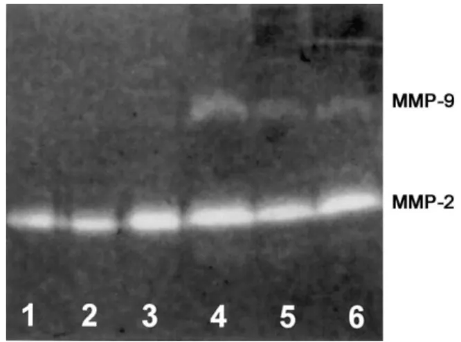
Figure 1. Representative zymography of cerebrospinal fluid (CSF) samples from uninfected control rabbits (lanes 1–3) and rabbits with coccidioidal meningitis (lanes 4–6) 7 days after infection. A gelatinolytic band of 72 kDa for the released matrix metalloproteinase (MMP)–2 was present in all samples, and there were no significant differences between uninfected and infected rabbits. CSF from infected rabbits showed a marked band at 92 kDa, indicating the presence of MMP-9.
and 15 after infection, animals were anesthetized with isoflurane
gas, and∼0.25–0.5 mL of CSF was removed by cisternal puncture
under sterile conditions. After white blood cell count analysis by hemocytometer, the remaining CSF was immediately centrifuged
and frozen at⫺80⬚C until processed for MMP-9 analysis by
elec-trophoresis and zymography. Rabbits were monitored twice daily for neurological complications and clinical signs. On day 15, all rabbits were anesthetized, CSF and blood samples were taken, and rabbits subsequently were killed with sodium pentobarbital (Eu-thasol; Delmarva Laboratories) given intravenously. Brains, spinal cords, and brain basilar arteries of infected and control rabbits were removed and fixed in 10% neutral buffered formalin for sub-sequent histologic examination.
Measurement of MMP-2 and MMP-9 levels in CSF. Samples of CSF (18 mL) were diluted into sample buffer (0.4 M Tris-HCl [pH 6.8], 10% SDS, 34% glycerol, and 1% bromphenol blue) to a loading volume of 24 mL and electrophoresed under nonreducing conditions in 10% polyacrylamide-SDS gels containing type A gel-atin (1 mg/mL) as proteinase substrate [6]. After electrophoresis for 2.5 h at 95 V, MMPs were renatured by removal of SDS by bathing the gel for 1 h in Triton X-100 (2.5% vol/vol). Gels were
then incubated in 10 mM CaCl2, 50 mM Tris, and 50 mM NaCl
(pH 7.65) for 18 h at 37⬚C, to allow proteolysis of the gelatin substrate, were fixed, and were stained with Coomassie blue. The gelatinolytic activity of MMP-9 and MMP-2 was determined by densitometric quantitation of gelatin lysis zones at 92 (MMP-9) and 72 (MMP-2) kDa, using the Image program (version 1.61; National Institutes of Health). Infection did not influence the con-centration of the constitutively produced MMP-2 in CSF of ani-mals (measured at days 0, 3, 7, 10, and 15), compared with un-infected control animals. The amount of induced MMP-9 protein was expressed as a percentage of the amount of MMP-2, which was used as an internal standard. During electrophoresis, MMPs, even if present as proenzyme or in the active form but linked to their natural inhibitors, are activated by partial denaturation in SDS and/or by autoproteolysis with reconstitution of enzymatic activity (renaturation) occurring with Triton-X bathing [7]. Hence, the proteolytic bands for MMP-9 and MMP-2 reflect total levels of enzymes; however, this may not measure the enzymatic activity in vivo at that particular moment. Experiments to show the spec-ificity of the assays for MMP-9 and MMP-2 were done by blocking these gelatinolytic zones by addition of b-aryl-succinic acid hy-droxamate [8] and EDTA, 2 agents that specifically block these MMP enzymes in the incubation buffer (data not shown).
Histologic analysis. Histologic assessment of severity of men-ingitis was undertaken as described elsewhere [9].
Statistical analysis. Normally distributed variables were
pre-sented asmeanⳲ SD, and differences between groups were
com-pared with the Mann-Whitney rank sum test.
Results
Clinical, neurological, and histopathological consequences of
C. immitis infection were documented in all infected rabbits. All
infected rabbits, but no control rabbits, demonstrated impair-ment in mobility, abnormal posture changes (reflecting menin-gismus), fever up to 40.2⬚C, and weight loss. CSF pleocytosis
was demonstrated in all infected rabbits beginning at day 7, whereas none of the control rabbits demonstrated pleocytosis.
C. immitis was recovered from the CSF in all infected rabbits
(⭓20 cfu/mm3) beginning at day 7, and significant growth of C.
immitis was also demonstrated from brain (mean, 3.23 log10cfu/
g) and spinal cord cultures (mean, 2.8 log10cfu/g).
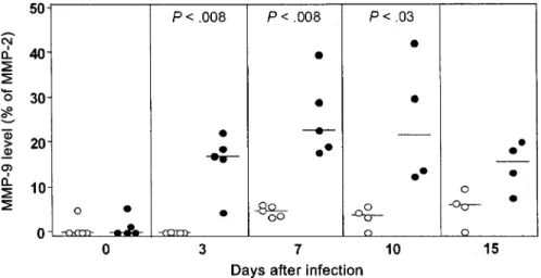
A representative zymogram, comparing day 7 MMP-9 levels in infected rabbits with those in control rabbits, is shown in figure 1. The time course of individual rabbit CSF assessment of the amount of MMP-9 for a 15-day period after infection or sham inoculation is presented in figure 2. One infected rabbit could not be tapped on day 10, and another infected rabbit met criteria for being killed on day 10 after tapping. Thus, one data point is missing for day 10, and another is missing for day 15. In addition, we were unable to tap one control rabbit on both days 10 and 15. Significant amounts of MMP-9 were observed on day 3 in the infected group, peaked at days 7 and 10, and slowly decreased thereafter. Low amounts of MMP-9 in control rabbits were observed on day 7, which persisted until day 15.
Histopathological study of infected rabbits showed evidence of meningitis of moderate or greater severity in 4 rabbits, with 1 rabbit demonstrating mild meningitis. No meningitis was ob-served in any of the control rabbits. Vasculitis involving men-ingeal vessels, brain basilar artery, or both was demonstrated in 4 rabbits. One rabbit did not demonstrate vasculitis but did demonstrate elevation of MMP-9 levels. Severity of meningeal inflammation and the presence of meningeal vasculitis, but not CSF white blood cell count, appeared to be associated with higher maximum levels of MMP-9 (table 1).
1694 Williams et al. JID 2002;186 (1 December)
Figure 2. Matrix metalloproteinase (MMP)–9 concentration (expressed as a percentage of the constitutively released MMP-2) in cerebrospinal fluid of rabbits intracisternally infected with Coccidioides immitis (䢇) and uninfected control rabbits (䡬) over the course of 15 days. A significant increase in MMP-9 level was first detected at 3 days after infection, reached maximum levels at day 7, and persisted at significantly high levels until day 10.P!.05was considered to be significant.
Table 1. Severity of meningitis, presence or absence of meningeal and basilar artery vasculitis, and maximum cerebrospinal fluid (CSF) white blood cell (WBC) count, compared with production of matrix metalloproteinase (MMP)–9, in Coc-cidioides immitis–infected and control rabbits.
Group, rabbit Severity of meningitis Meningeal vasculitis Basilar artery vasculitis Maximum CSF WBC count (day) Maximum MMP-9 concentration (day)a Infected 152b Moderate ⫹ ⫺ 300 (7) 39.58 (7) 153 Severe ⫹ ⫹ 1900 (10) 29.81 (10) 154 Severe ⫹ ⫹ 1400 (10) 42.14 (10) 155c Mild ⫺ ⫺ 1850 (10) 19 (7) 156 Mild-moderate ⫺ ⫹ 1550 (10) 17.8 (7) Control 147d None ⫺ ⫺ 0 6.17 (7) 148 None ⫺ ⫺ 0 5.58 (15) 149 None ⫺ ⫺ 0 9.47 (15) 150 None ⫺ ⫺ 0 5.75 (10) 151 None ⫺ ⫺ 0 6.35 (15)
NOTE. ⫺, Absent; ⫹, present.
a
Expressed as a percentage of the constitutively released MMP-2.
b
Unable to tap at day 10.
c
Killed at day 10 after tap.
d
Unable to tap at days 10 and 15. Discussion
Significant increases in MMP-9 levels were observed over a 2-week period in the 5 C. immitis–infected rabbits that developed clinical meningitis and vasculitis, compared with sham-treated control rabbits. We have previously assayed supernatant of the parasitic-phase, endospore-seeded C. immitis growth medium through time of endosporulation and did not demonstrate MMP-9 production by C. immitis (authors’ unpublished data). This observation is consistent with previous reports implicating in-flammatory cells and resident brain cells, but not the infectious agents, as the source of MMP-9 during CNS infection [2, 10].
MMP-9 has been implicated in the disruption of the BBB during infection, leading to brain edema. This occurs by
disrup-tion of the subendothelial basement membrane via proteolytic activity that degrades type IV and V collagens [3]. In addition, MMP-9 is implicated in stimulating the migration of activated inflammatory cells across the BBB, with resulting further damage to the barrier by cytokine products of these cells [2, 3]. MMP-9 has also been reported to up-regulate production of tumor ne-crosis factor (TNF)–a, a major proinflammatory cytokine [2, 3]. TNF-a can, in turn, positively feed back to inflammatory cells, enhancing their production of MMP-9 and, thus, perpetuating TNF-a production. In an animal model of pneumococcal men-ingitis, these events were closely correlated with the severity of inflammation, including vasculitis, leading to breakdown of vas-cular integrity, decreased blood flow, and eventual thrombosis of
JID 2002;186 (1 December) MMP-9 in Rabbit CSF 1695
vascular lumens. This ultimately leads to focal ischemic and in-farction sequela. Ampel et al. [11] have demonstrated significant levels of TNF-a within the CSF of patients with coccidioidal meningitis.
MMP-9 also appears to play a pivotal role in enhancing inflammation within the meninges and within the brain paren-chyma and vascular tree of humans with tuberculous, candidal, or streptococcal meningitis [3, 12]. In addition, Sorbi et al. [13], have demonstrated that MMP-9 is associated with the pro-duction of vasculitis in humans with temporal arteritis. Thus, MMP-9 activity appears to be a generalized CNS response to a number of inflammatory conditions.
Our protocol for establishing a rabbit model of coccidioidal meningitis and vasculitis calls for immunosuppressing rabbits during the first few days of infection. Immunosuppression could have affected the temporal events influencing MMP-9 levels within the CSF of infected animals (i.e., by slowing production) and may have affected the absolute levels produced. It would likely not have affected the final interpretation of results, how-ever, since control animals were also treated with steroids in exactly the same fashion and had lower levels of MMP-9. The finding of small amounts of MMP-9 in the control group none-theless is of interest and could signify the influence of environ-mental stress factors, anesthesia, and frequent intracisternal punctures in promoting production of MMP-9, possibly by di-rect effects on resident CNS cells.
It is of interest that the amount of MMP-9 demonstrated in this study may promote inflammation and vasculitis in our model. The increase of MMP-9 in the present rabbit model of coccidioidal meningitis is slower and less intense but far more protracted, compared with that in bacterial meningitis [2, 9] and, to a lesser extent, viral meningitis [1]. Hence, the longi-tudinal course of MMP-9 up-regulation is in accordance with the subacute clinical disease.
Our observations here enable the future study of MMP-9 in-hibitors to assess the role of MMP-9 in promoting meningeal inflammation and vasculitis. Recently published data [2, 10] dem-onstrate that 2 inhibitors of MMP-9 (GM6001 and BB1101), when given intraperitoneally or subcutaneously, respectively, to rats with experimental S. pneumoniae meningitis, can block the activity of both MMP-9 and another metalloproteinase, TNF-a–converting enzyme, which correlated with decreasing severity of inflammation, an effect that could be used therapeutically in coccidioidal meningitis. Future studies with our rabbit model will examine the role of endogenous inhibitors of tissue inhibitors of metalloproteinases and whether other MMPs (such as MMP-8 and MMP-13) are up-regulated in coccidioidal meningitis.
Acknowledgments
We thank Katharine Fletcher for excellent secretarial assistance, Da-vid Hewitt (director of the Kaweah Delta District Hospital Laboratory, Visalia, California) for laboratory support, and Leilani Calderon for research assistance.
References
1. Leppert D, Lindberg RL, Kappos L, Leib SL. Matrix metalloproteinases: multifunctional effectors of inflammation in multiple sclerosis and bac-terial meningitis. Brain Res Brain Res Rev 2001; 36:249–57.
2. Leib SL, Leppert D, Clements J, Ta¨uber MG. Matrix metalloproteinases contribute to brain damage in experimental pneumococcal meningitis. Infect Immun 2000; 68:615–20.
3. Leppert D, Leib SL, Grygar C, Miller KM, Schaad UB, Holla¨nder GA. Matrix metalloproteinase (MMP)–8 and MMP-9 in cerebrospinal fluid during bacterial meningitis: association with blood brain-barrier damage and neurological sequelae. Clin Infect Dis 2000; 31:80–4.
4. Azeh I, Ma¨der M, Smirnov A, Beuche W, Nau R, Weber F. Experimental pneumococcal meningitis in rabbits: the increase of matrix metallopro-teinase–9 in cerebrospinal fluid correlates with leukocyte invasion. Neu-rosci Lett 1998; 256:127–30.
5. Williams PL, Johnson R, Pappagianis D, et al. Vasculitic and encephalic complications associated with Coccidioides immitis infection of the central nervous system in humans: report of 10 cases and review. Clin Infect Dis 1992; 14:673–82.
6. Williams PL, Sobel RA, Sorensen KN, et al. A model of coccidioidal me-ningoencephalitis and cerebrospinal vasculitis in the rabbit. J Infect Dis 1998; 178:1217–21.
7. Leppert D, Waubant E, Galardy R, Bunnett NW, Houser SL. T cell gelatinases mediate basement membrane transmigration in vitro. J Immun 1995; 154: 4379–89.
8. Kottirsch G, Koch G, Feifel R, Newmann U. b-aryl-succinic acid hydrox-amates as dual inhibitors of matrix metalloproteinases and tumor necrosis factor alpha converting enzymes. J Med Chem 2002; 45:2289–93. 9. Sorensen KN, Sobel RA, Clemons KV, Pappagianis D, Stevens DA, Williams
PL. Comparison of fluconazole and itraconazole in a rabbit model of coccidioidal meningitis. Antimicrob Agents Chemother 2000; 44:1512–7. 10. Leib SL, Clements JM, Linberg RLP, et al. Inhibition of matrix metallopro-teinases and tumor necrosis factor converting enzyme as adjuvant therapy in pneumococcal meningitis. Brain 2001; 124:1734–42.
11. Ampel NM, Ahmann DR, Delgado, KL, Galgiani JN, Cloud GA. Tumor necrosis factor–a and interleukin-1b in cerebrospinal fluid of patients with coccidioidal meningitis during therapy with fluconazole. The National Institute of Allergy and Infectious Diseases Mycosis Study Group. J Infect Dis 1995; 171:1675–8.
12. Matsuura E, Umehara F, Hashiguchi T, Fugimoto N, Okada Y, Osame M. Marked increase in matrix metalloproteinase 9 in cerebrospinal fluid of patients with fungal or tuberculous meningeal encephalitis. J Neurol Sci 2000; 173:45–52.
13. Sorbi D, French DL, Nuovo GJ, Kew RR, Arbeit LA, Gruber BL. Elevated levels of 92-kD type IV collagenase (matrix metalloproteinase 9) in giant cell arteritis. Arthritis Rheum 1996; 39:1747–53.