Protein Engineering vol.4 no 7 pp 831-835. 1991
A hybrid of bovine pancreatic ribonuclease and human angiogenin:
an external loop as a module controlling substrate specificity?
Rudolf K.Allemann1, Steven A.Benner3
Scott R.Presnell2 and
Laboratory for Organic Chemistry, Swiss Federal Institute of Technology. CH-8092 Zurich, Switzerland
'Present address: Laboratory of Eukaryotic Molecular Genetics, MRC National Institute for Medical Research, Mill Hill, London NW7 IAA, UK ^Present address. Computer Graphics Laboratory, Department of
Pharmaceutical Chemistry, University of California, San Francisco, USA
3To whom correspondence should be addressed
A comparison of the sequences of three homologous ribonucleases (RNase A, angiogenin and bovine seminal RNase) identifies three surface loops that are highly variable between the three proteins. Two hypotheses were contrasted: (i) that this variation might be responsible for the different catalytic activities of the three proteins; and (ii) that this variation is simply an example of surface loops undergoing rapid neutral divergence in sequence. Three hybrids of angiogenin and bovine pancreatic ribonuclease (RNase) A were prepared where regions in these loops taken from angiogenin were inserted into RNase A. Two of the three hybrids had unremarkable catalytic properties. However, the RNase A mutant containing residues 63-74 of angiogenin had greatly diminished catalytic activity against uridylyl-(3' — 5')-adenosine (UpA), and slightly increased catalytic activity as an inhibitor of translation in vitro. Both catalytic behaviors are characteristic of angiogenin. This is one of the first examples of an engineered external loop in a protein. Further, these results are complementary to those recently obtained from the complementary experiment, where residues 5 9 - 7 0 of RNase were inserted into angiogenin [Harper and Vallee (1989) Biochemistry, 28, 1875-1884]. Thus, the external loop in residues 6 3 - 7 4 of RNase A appears to behave, at least in part, as an interchangeable 'module' that influences substrate specificity in an enzyme in a way that is isolated from the influences of other regions in the protein. Key words: angiogenin/evolutionary guidance/folding pathway/
ribonuclease/site-directed mutagenesis
Introduction
Bovine pancreatic ribonuclease A (RNase A) is a monomeric enzyme that hydrolyzes single-stranded RNA and dinucleotides such as uridylyl-(3' — 5')-adenosine (UpA) (Blackburn and Moore, 1982). RNase A appears to play a special role in ruminant digestion (Barnard, 1969), a role that emerged only recently in evolutionary history (Stackhouse et al., 1990). It has little activity as either an inhibitor or a promoter of cell growth.
Angiogenin is a homolog of RNase A first isolated as an angiogenesis factor from tumor-cell-conditioned medium because of its ability to induce the growth of blood vessels in biological assay systems (Fert et al., 1985). Although a physiological role for angiogenin in the growth of tumors is far from certain, angiogenin itself is clearly necessary for this process (Folkman,
1972). The sequences of angiogenin and bovine RNase A are - 6 5 % different (Kurachi et al., 1985; Strydom et al.. 1985). Angiogenin also differs from RNase A in that it shows little enzymatic activity as a ribonuclease against Small substrates; in particular, angiogenin hydrolyzes UpA only ~2% as fast as RNase A (Shapiro et al., 1986). Angiogenin has, however, a characteristic ribonuclease activity against larger RNA molecules. In particular, angiogenin is a somewhat better inhibitor of protein synthesis in the rabbit reticulocyte lysate system than pancreatic RNase A, where its principal target appears to be ribosomal RNA (StClair et al., 1987).
Seminal RNase (D'Alessio and Leone, 1963), a third homologous protein (81% sequence identical with RNase A; Suzuki et al., 1987), has still different catalytic and biological activities (Taniguchi and Libonati, 1974). Unlike the digestive enzyme, seminal RNase is a covalent dimer (Capasso et al., 1983; Mazzarell et al., 1987), catalyzes preferentially the hydrolysis of double-stranded RNA and DNA — RNA hybrids (Taniguchi and Libonati, 1974), and has potent anti-tumor activity both in vitro and in vivo (Matousek, 1973; Vescia et al., 1980; Vescia and Tramontane 1981).
The homology between potential tumor growth promoters, tumor growth inhibitors and digestive RNases is especially interesting in the context of the hypothesis that extracellular RNA molecules serve as intercellular communicator molecules important in mammalian development (Wissler et al., 1986; Benner, 1987, 1988; Benner and Allemann, 1989). Therefore, we examined the sequences of these proteins in an effort to correlate specific sequence differences with the differences in their catalytic and biological activities (Nambiar et al., 1986).
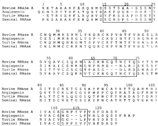
Seminal RNase and RNase A differ greatly in three regions (Figure 1), between residues 16-20, residues 6 2 - 6 6 and residues 111 - 1 1 5 , in addition to a region (residues 28-38) that appears from crystallographic studies to be responsible for the dimeric structure of seminal RNase (Capasso et al., 1983; Mazzarella et al., 1987). The sequences of RNase A and angiogenin differ throughout the alignment, as expected given their greater sequence divergence. However, three regions displaying variability between angiogenin and RNase A, residues 15-24, residues 6 3 - 7 4 and residues 112-115 (where the numbers refer to positions in the sequence for RNase A), overlap the highly variable regions between seminal RNase and RNase A. In all three segments, residues in the digestive RNase are deleted in angiogenin, and the deletion is accompanied by many point mutations in the immediate environment. In the region from residues 63 to 74, a disulfide bond is deleted (Beintema
etal., 1985).
The fact that both angiogenin and seminal RNase differ significantly from RNase A in the same three regions could have many explanations, including trivial ones. For example, the three regions of highest variation, identified by the process mentioned above, are all surface loops in RNase A (Figure 2), and surface loops are known to undergo rapid sequence divergence often regarded as neutral drift (Kimura, 1982). This would normally
Bovine RNase A Anqioqen i n T u r t l e RNase Seminal RNAse 1 5 10 K E T A A A K F E R Q H M D Q D N . 3 R Y T H F L T Q H Y D - E T R Y E K F L R O H V D K E S A A A K F E R Q H M D 20 s A Y S S K P G T P K N S 0 s s A G S P A R A S - S - D P D - S S D S s s R R 5 N -T N 25 Y Y B o v i n e R N a s e A A n g i o g e n i n T u r t l e R N a s e S e m j n a l RNAse 3 0 3 5 4 0 4 5 5 0 C N Q M M K S R N L T K D R C K P V N T F V H E S L A C E S I M R R R G L T S P - C K D I N T F I H G N K R C N Q M M Q R R G M T S P V C K F T N T F V H A S A A C N L M M C C R K M T Q G K C K P V N T F V H E ^ L . A Bovine RNAse A Anqioqen in T u r t l e RNase Seminal RNase 55 6 0 D V Q A V C S Q K N S I K A I C E N K N S I T T V C G S G G D V K A V C S Q K K 65 70 V G T V A N P T C P A C K H S K N R G N G E -G Q -Q T -T N N D N C L L C Y R R Y Q I D Q 7 5 S M S F S Y S K S N S K S T M T S A S F 80 85 90 95 100 105 B o v i n e RNase A S I T D C R E T G S S K Y P N C A Y K T T Q A N K H ] A n g i o g e n l n Q V T T C K L H G G S P W P P C Q Y R A T A G F R N V T u r t l e RNase A L T T C R L Q G G S Q T P N C P Y N A D A S T Q R 1 S e m i n a l RNase R I T D C R E T G S S K Y P N C A Y K T T Q V E K H 1 B o v i n e RNase A A n g i o g e n i n T u r t l e RNase S e m i n a l RNase 110 I V A C E V V A C E R I A C V I V A C G 1 15 G N G G N G G K P -P Y -S V L L V P P P P 1 2 0 V H F D A S V V H L D Q S I F R R P V H Y D K S I V H F D A S V
Fig. 1. Sequence alignment of bovine pancreatic RNase A, human angiogenin, turtle RNase and bovine seminal RNase. Regions of high variability between
angiogenin and RNase A (residues 15-24, 4 9 - 5 3 , 6 3 - 7 4 and 112-116) are boxed.
imply that the structural differences noted in Figure 1 have no significance regarding the biological and catalytic differences observed in the three proteins (Presnell, 1988).
However, the assumption that variation in surface loops is neutral is almost certainly not universally valid (Hill and Hastie, 1987; Laskowski et al., 1987), and it is conceivable that structural variation in these regions may be determined by the different chemical and biological activities of these RNase homologs. Further, the three-way sequence comparison greatly limits the number of experiments that might be attempted based on a comparison of the sequences of angiogenin and RNase alone. Therefore, we have prepared hybrids of RNase A and angiogenin in these three regions.
Materials and methods Materials
Restriction endonucleases, T4 polynucleotide kinase and T4 DNA ligase were from New England Biolabs; other enzymes were from Boehringer-Mannheim. Standard procedures were used to prepare genetic constructions (Seed, 1983), and are described in detail elsewhere (Allemann, 1989). UpA was from Sigma. Oligo-nucleotides were synthesized on an Applied Biosystems model 380A synthesizer using phosphoramidite chemistry and purified either by PAGE on 16% gels or, in the case of short oligo-nucleotides, by TLC (McGeehan and Benner, 1989). DNA sequencing was carried out using a Sequenase DNA sequencing kit. The kit for in vitro translation and brome mosaic virus BMV RNA were from Promega Biotech.
Construction of the genes for the hybrid proteins
Hybrid proteins containing a short segment of angiogenin introduced into RNase A (Figure 1) were designated RA(15 -24), RA(63-74) and RA(112-116). Genes for these mutants were constructed from a synthetic gene for RNase A (Nambiar
et al., 1984, 1987) by modular mutagenesis procedures, taking
R A ( I 12-1 16)
R A ( 15-24)
Fig. 2. Ribbon diagram of the crystal structure of bovine pancreatic RNase A indicating the three regions of high sequence divergence of human angiogenin (the numbers refer to positions in the sequence of RNase A).
advantage of the unique restriction sites placed throughout the gene. Details of the constructions are reported elsewhere (Presnell, 1988; Allemann, 1989). All constructions were verified by total sequence analysis.
Expression of the hybrid proteins
Constructed genes for the mutants were expressed in Escherichia
coli under the control of the lambda PL promoter, using the
expression vector described previously (McGeehan and Benner, 1989). This system yields free RNase bearing an N-terminal methionine residue, as determined by Edman degradation. Control experiments with RNase mutants with and without this methionine (Stackhouse et al., 1990) have failed to detect evidence for an impact of this additional methionine on the behavior of several mutant proteins. Competent E.coli cells (N4830/cI857ts, containing the gene for a temperature-sensitive lambda cl repressor) were transformed with the expression vector
Hybrid of rlbonuclea.se A and angiogenin
by the method of Hanahan (1983); positive transformants were isolated and the genes sequenced. Transcription was induced by heat shock at 42 °C following the method of McGeehan and Benner (1989), and optimal times for harvest (25-30 min) were determined individually for each hybrid by following the course of induction by Western blotting under denaturing conditions using polyclonal antibody raised against denatured RNase A (Burnette, 1981).
Reconstitution and purification of hybrid proteins
The hybrid proteins were reconstituted and purified using a standard protocol (McGeehan and Benner, 1989). Following induction, cells from a 1 1 incubation mixture (OD at 550 nm 1.2) were suspended in 'denaturing buffer' [8 M urea, 40 mM sarcosine, 20 mM Tris-HCl, 20 mM NaCl, 20 mM /3-mercaptoethanol, 1 mM ethylenediaminetetracetic acid (EDTA), pH 7.8] at 4°C and lysed in a French press (14 000 p.s.i.). The lysate was centrifuged, and the supernatant dialyzed (Amicon, cutoff 3500) against 'refolding buffer' [50 mM Tris-HCl, 100 mM NaCl, 1 mM EDTA, 0.2 mM phenyl-methylsulfonyl fluoride (PMSF), 4 mM oxidized glutathione, 4 mM reduced glutathione, 0.02% NaN3, pH 7.6] over a period
of 72 h (two changes, 2 1 each). The dialysate was then loaded onto a sizing column (Sephadex G-509, 120 X 4.5 cm) and the column was eluted (flow rate 5.1 ml/h/cm2) with elution buffer
(50 mM Tris-HCl, 100 mM Nacl, 1 mM EDTA, 0.2 mM PMSF, 0.02% NaN3, pH 7.5). This procedure removed - 8 0 %
of the contaminating protein. The effluent containing the ribonuclease was dialyzed against affinity chromatography buffer (100 mM ammonium acetate, 0.02% NaN3, pH 6.2) and further
purified by affinity chromatography on pUp-agarose (Sigma, 5 ml in a 1 cm diameter column), using 2',3'-CMP in the affinity chromatography buffer as eluant. The proteins thus purified were pure by SDS-PAGE using both silver and Coomassie blue staining, as well as by immunoblotting.
Kinetic analysis of hybrid proteins
Steady-state kinetics were measured using UpA as substrate. The rate of hydrolysis of UpA (100 and 500 /tM) at 25°C was measured in sodium acetate buffer (pH 5.0, 100 mM) by coupling the reaction to the deamination of adenosine catalyzed by adenosine deaminase, which was followed at 265 nm using a molar extinction coefficient of 23 600 M/cm, according to the procedure of Ipata and Felicioli (1968). The concentration of RNase A was determined both by measuring its absorbance at 280 run (absorbance of a 1% solution is 7.3; alternatively, at 278 nm the molar extinction coefficient is 9800) and by the Bio-Rad modification of the Bradford assay (Bradford, 1976). Concentrations of hybrid enzymes were determined by SDS — PAGE using known amounts of RNase A as standards.
Inhibition of the rabbit reticulocyte systems
The ability of various ribonucleases to inhibit the synthesis of proteins in rabbit reticulocyte lysate preparations was determined using a kit from Promega Biotech and BMV mRNA, following the procedure of the manufacturer and St Clair et al. (1987). The lysate was stored at -80°C in aliquots (200 /tl) and used immediately after thawing. Lysate (17.5 /il) in an Eppendorf tube was incubated with a defined amount of RNase A or hybrids in the buffer (ammonium acetate, 100 mM, pH 6.2) in a total volume of 22 /tl at 30°C for 15 min. The mixture was then placed on ice, and human placental ribonuclease inhibitor (PRI; Amer-sham, 25 units in 1 yX water) added. Independent experiments showed that the RNase hybrids were inhibited by PRI. The con-tents were mixed, centrifuged, returned to ice, and then diluted
with a solution (1 y\) containing [35S]methionipe (1440 Ci/mmol,
15 mCi/ml, 0.5 y.\) and 19 unlabeled amin0 acids (Promega, 1 mM each, 0.5 /tl). The mixture was again centrifuged, and translation initiated by adding mRNA (1 /il, 0.5 ng). Translation was allowed to proceed for 60 min at 30°C. Two controls, one lacking message, the other lacking RNase, were run in parallel. Following incubation, an aliquot (1 fA) iof the translation mixture was added to a solution (1 ml) of sodium hydroxide (1 N) and hydrogen peroxide (1.5%), the mixture! incubated (37°C, 10 min), diluted with an ice-cold solution of tjrichloroacetic acid (TCA, 4 ml, 25% w/v), and 2% (w/v) caSamino acids were added. The solution was mixed and placed bn ice for 30 min. The precipitated proteins were collected by filtration (Whatman GF/C filter, 2.4 cm diameter) and the filteri was washed with an ice-cold solution of TCA (10 ml, 8%), and then with acetone (3 ml). The filter was dried by suction,1 transferred to a
scintillation vial, and counted. Alternatively, aliquots of the translation mixture (samples and control in parallel) were run directly on a denaturing polyacrylamide gel, the gels dried, and directly scanned. All results are reported relative to RNase A.
Results and discussion
The preparation and expression of genes coding for the hybrids was uneventful. However, it is worth mentioning that in the refolding buffer used here, RA(63-74) recovers its catalytic activity slowly when compared with the rate with which native RNase A or the other hybrids recovered theirs. This is especially noteworthy as this hybrid lacks one of the four disulfide bonds found in RNase A. As the formation'of disulfide bonds might play an important role in the folding process (Creighton, 1979), the slow folding of RA(63-74) is, worth examining in further detail.
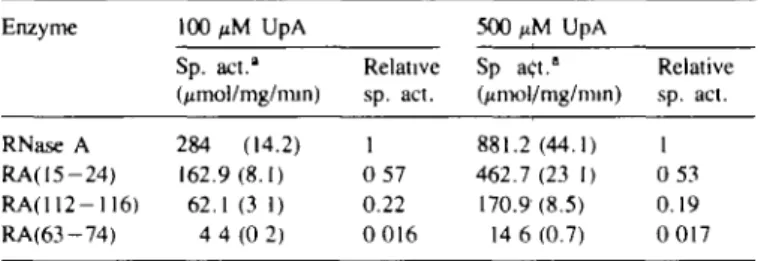
The catalytic activities with UpA as a substrate of the three hybrids, each having a short module of angiogenin introduced into a background of RNase A, show an interesting pattern (Table I). Two of the hybrid proteins, RA(15-24) and RA(112-116) have specific activities 55 and 22 % that of RNase A with UpA as substrate. This diminished activity (by a factor of 2 - 5 ) is within the range normally observed for structurally altered proteins. More notable is the fact that the specific activity of the third hybrid, RA(63-74), is reduced by a factor of 60, < 2% of the specific activity of RNase A.
This result would be uninteresting if the hybrid had generally diminished catalytic activity (due to unfolding, for example).
Table I. Specific activities of RNase A and RNase —angiogenin hybrids with
UpA as substrate
Enzyme 100 UpA 500 UpA
Sp. act." (jimol/mg/min) Relative sp. act. Sp act." (/imol/mg/min) Relative sp. act. RNase A 284 (14.2) 1 881.2(44.1) 1 RA(15-24) 162.9(8.1) 0 57 462.7(23 1) 0 53 RAO 12-116) 62.1(3 1) 0.22 170.9(8.5) 0.19 RA(63-74) 4 4 ( 0 2) 0 016 14 6(0.7) 0 017 "In sodium acetate buffer (100 mM. pH 5.0) at 25°C: estimated standard error is given in parentheses. The reaction was followed
spectrophotometrically at 265 nm in a coupled assay with adenosine deaminase. The concentration of RNase A was determined spectrophotometrically at 280 nm using an extinction coefficient of 9800 M/cm. Concentrations of hybrids were determined by scanning denaturing polyacrylamide gels (15%) using dilutions of RNase A as standards
Therefore, the ability of the hybrids to inhibit in vitro translation (an increase in this activity is a characteristic of angiogenin; St Clair et al., 1987) was measured using the rabbit reticulocyte lysate system. The activities of two of the hybrid proteins, RA(15-24) and RA(112— 116), were comparable or diminished when compared with RNase A (Figure 3 and Table II). However, the third hybrid RA(63 -74), despite its gready reduced catalytic activity against small ribonucleotide substrates, was somewhat better as an inhibitor of protein translation than RNase A (Table II). While the difference in activity between angiogenin and RNase A in this assay is small, the increase in the hybrid's activity as an inhibitor of translation over that of RNase A is about half that of angiogenin itself.
Since these experiments were completed, Harper and Vallee (1989) reported the results of experiments where a segment of angiogenin between positions 58 and 70 is replaced by the homologous segment of bovine pancreatic RNase A (residues 59-73). This experiment is nearly the reciprocal of one of the experiments reported here. Remarkably, the results obtained are also largely reciprocal, in that the catalytic activity against small substrates (e.g. UpA) of the angiogenin containing a short loop of RNase A was - 200-fold greater than that of angiogenin itself. A short segment of RNase A, when inserted into angiogenin, appears to confer RNase-A-l ike catalytic properties on angiogenin, at least with respect to catalytic activity against UpA, and this segment corresponds to the segment of angiogenin reported here that confers angiogenin-like catalytic activity on RNase A when inserted into RNase A.
Thus, the region in RNase A between positions 63 and 74 appears to behave, at least with respect to these details of catalytic activity, as an example of an interchangeable 'module' that influences substrate specificity in an enzyme in a way that is isolated, at least to some degree, from other regions in the protein. Such modules, of great interest to the protein engineer, have not been easy to find, as the analysis of the behavior of most mutants has proved to be complex. Further, this module would not have been easily identified by a simple comparison of the crystal structures of various RNases (Figure 2).
Crystallographic data suggest two sets of interactions between this loop (63 — 74) and the substrate. First, the e-amino group of Lys66 may form a salt bridge with the phosphate group of the substrate on the 5'-hydroxyl group of the ribose bearing the reactive phosphate in oligonucleotide substrates (MacPherson
et al., 1986). This Lys is not present in the hybrid; the only basic
residues present in the new loop near the appropriate region are a His and an Arg.
One might attempt to interpret the kinetic results reported here in terms of a loss of this salt bridge. Naively, however, one might expect this loss to alter most significantly the rate of hydrolysis of substrates that bear a phosphate group at this position. UpA does not have this phosphate; thus, the influence of the structural alterations described here on catalytic activity are the opposite of what one might expect based on this rationale.
Alternatively the structure of the complex of RNase A and a phosphonate analog of UpA shows the e-oxygen group of Gln69 and the 5-oxygen group of Asnl7 forming a binding site for the adenine ring in the UpA substrate through hydrogen bonding with the N(6) nitrogen (Richards and Wyckoff, 1973; Wodak
et al., 1977). This interaction has been proposed to be important
in the ability of RNase A to distinguish between adenine and guanine at this position in the substrate. Gln69 is, of course, missing in RA(63-74), and the Asn corresponding to Asn71 is almost certainly reoriented, given the shortness of the loop.
i
1 2 3 4 5 6 7 8 9 10
Fig. 3. Autoradiography of a 10% polyacrylamide gel of the in \itro
translation products showing the effects of bovine pancreatic RNase A and RA(63 —74) on the translationaJ capacity of the RNA-dependent rabbit reticulocyte lysate system. The rabbit reticulocyte lysate was incubated with the indicated amounts of RNase A and RA(63-74) for 15 mm at 30°C. The ribonucleolytic action of the digestive enzymes was quenched by the addition of PRI, control experiments showed that PRI was a potent inhibitor of RA(63-74). Translation was then initiated by the addition of BMV RNA. Lane 1, no ribonucleolytic enzymes added to preincubation mixture;
lane 2, translation without added MBV RNA, lanes 3 - 6 , preincubation in
the presence of 20, 30, 40 and 50 nM RNase A; lanes 7 - 1 0 , preincubation with 50, 40, 30 and 20 nM RA(63-74).
Table II. Inhibition of translation by RNase A and RNase—angiogenin
hybrids
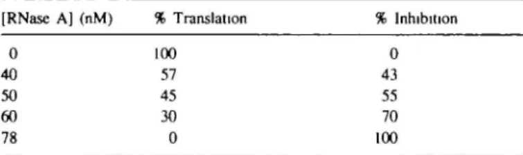
(A) Effect of varying concentrations of RNase A on translational capacity (determined by measuring the TCA-precipitable radioactivity in translation products—see Materials and methods) of rabbit reticulocyte lysate [RNase A] (nM) % Translation % Inhibition
0 40 50 60 78 100 57 45 30 0 0 43 55 70 100
(B) Protein concentrations (nM) of RNase A. RA(15-24), RA{112 — 116). RA(63—74) and angiogenin giving rise to a 50% inhibition of the translational capacity of rabbit reticulocyte lysate
Protein RNase A RA(15-24) RA(1I2-116) RA(63-74) Angiogeninb Protein concentration (nM) giving 50% inhibition" 39 (2.0) 37(1.9) 48 (2.4) 35(1 8) 32 % Inhibition relative to RNase A 100 104 80 111 122
"Value determined by measurement of translation products in a set of experiments at several concentrations of hybrid, as for RNase A in (A). RAO5-24) (15-25 nM). RA(63-74) (20-40 nM) and RA( 112 — 116) (20-40 nM), interpolated [or, in the case of RA(15-23), extrapolated] to the 50% inhibition level Values are the average of three independent measurements; for RA(15—24). of two independent measurements The estimated standard error from independent runs is given in parentheses.
bData from St Clajr el al. (1987)
This might explain the low catalytic activity of this hybrid with UpA as substrate. To explain the fact that activity with larger substrates is apparently retained (and perhaps somewhat
Hybrid of ribonuclea.se A and angiogenin
increased), one must postulate that the lost interaction is compensated by interactions elsewhere between the hybrid and the larger substrate RNA.
What these additional interactions might be remains unclear. Nevertheless, the results reported here show the value of sequence comparisons in planning protein engineering experiments, and that much work remains before the differences in behavior in this class of proteins can be interpreted in terms of specific structural details.
Vescia.S.. TramontaneD., Augusti-Tocco.G. and D'Alessio.G (1980) Cancer
Res., 40, 3740-3744.
WisslerJ.H., Logemann.E., Meyer.H.E., Kruetzfeldt.B . Hoeckel.M . Heilmeyer.L.M.G., Jr (1986) Prondes Biol. Fluids, 34. 525-536. Wodak.S.Y., Liu.M.Y. and WyckofT.H.W. (1977)7. Mol. Biol.. 116. 855-875
Received on April 16, 1991; accepted on July 19, 1991
Acknowledgements
We thank Professor H.Wyckoff for helpful comments concerning interactions in the RNase — phosphorate complex. We are indebted to the Swiss National Science Foundation, the ETH and Sandoz AG for partial support of this work.
References
Allemann.R.K. (1989) ETH Dissertation No. 8804 Barnard E A. (1969) Nature, 221. 340-344.
Blackburn,P and Moore.S. (1982) Enzymes, IS, 317-433.
BeintemaJJ., BroosJ., MeulenbergJ and Schueller.C (1985) Eur. J. Biochem.,
153. 305-312.
Benner.S.A (1987) Chimia, 41. 142-148 Benner.S.A. (1988) FEBS Lett., 233, 225-228.
Benner.S.A. and Allemann.R.K. (1989) Trends Biochem Sci . 14. 396-397. Bradford.M. (1976) Anal. Biochem., 72, 248-254.
Bumette.W.N. (1981) Anal Biochem., 112, 195-203
Capasso.S , Giordano,F., Mattia.C.A.. Mazzarella.L. and Zagari.A. (1983)
Biopolymers, 22, 327-332.
Creighton.T. (1979) J. Mol. Biol , 129, 411-431. D'Alessio.G. and Leone,E (1963) Biochem. J., 89, 7p
FettJ.W., Strydom.D J., Lobb.R.R., Alderman.E.M.. BethuneJ.L.. RiordanJ.F and Vallee.B.L. (1985) Biochemistry. 24, 5480-5486
Folkman.J (1972) Ann Surg., 175.409-416 Hanahan.D. (1983)/ Mol. Biol., 166, 557-580.
Harper.J.W. and Vallee.B.L. (1989) Biochemistry, 28, 1875-1884. Hill.R.E. and Hastie.N.D. (1987) Nature, 326, 9 6 - 9 9 .
Laskowski.M., Jr, Kato.I , Kohr.W.J.. Park.S.J , Tashiro.M and Wahtley.H.E. (1987) Cold Spring Harbor Svmp. Quant. Biol., 52, 545-553
Ipata.P L and Felicioli.R A (1968) FEBS Lett., 1, 2 9 - 3 1 .
Kimura.M (ed ) (1982) Molecular Emlution, Protein Polymorphism, and the
Neutral Theory. Springer-Verlag, Berlin.
Kurachi.K., Davie.E.W., Strydom.D.J . Riordan.J F and Vallee.B L (1985)
Biochemistry. 24, 5494-5499
McGeenan.G M and Benner.S A (1989) FEBS Lett , 247. 5 5 - 5 6 . MacPherson.A . Brayer.G . Cascio.D and Williams,R. (1986) Science, 232.
765-768
Mazzarella.L., Mattia.C.A.. Capasso.S and di Lorenzo,G (1987) Gazz Chim
ltai, 117, 91-97
Matousek.J. (1973) Experientia. 29, 858-859.
Nambiar.K.P., StackhouseJ.. Stauffer.D.M.. Kennedy.W.P , Eldredge.J K and Benner.S.A. (1984) Science. 223, 1299-1301.
Nambiar.K.P.. Stackhouse.J.. Presnell.R.S. and Benner.S.A. (1986) In Schenider.M (ed.), Enzvmes as Catalysts in Organic Synthesis. Riedel. Dor-drecht, pp. 325-329.
Nambiar.K.P.. Stackhouse.J.. Presnell.R S. and Benner.S.A (1987) Eur. J.
Biochem , 163. 6 7 - 7 1
Presnell.S R (1988) Thesis. Harvard University.
Richards.FM and Wyckoff.H W (1973) In Phillips.D C and Richards.F M (eds). Atlas of Molecular Structures in Biology Clarendon Press, Oxford. pp. 000-000
St Clair.D.K., Rybak.S.M.. Riordan.J.F. and Vallee.B.L. (1987) Proc. Nail
Acad.. Sci. USA, 84, 8330-8334
Seed.B. (1983) Nucleic Acids Res.. 11, 2427-2445.
Shapiro.R., Riordan.J F and Vallee.B.L. (1986) Biochemistry, 25, 3527-3532 StackhouseJ., Presnell.S.R., McGeehan.G.M., Nambiar.K.P and Benner.S.A
(1990) FEBS Lett., 262, 104-106.
Strydom.D.J., FettJ.W., Lobb.R R.. Alderman,E M , BethuneJ L., RiordanJ F and Vallee.B.L. (1985) Biochemistry. 24, 5486-5490
Suzuki,S., Parente.A., Fanna.B , Greco.L., La Montagna.R. and Leone.E. (1987)
Biol. Chem. Hoppe-Seyler, 268. 1305-1312.
Taniguchi.T. and Libonati.M (1974) Biochem. Biophvs. Res. Cominun.. 58, 280-286.