Effects of Morphine on the Differentiation and Survival of Developing
Pyramidal Neurons During the Brain Growth Spurt
Horace Massa,* claudia-Marvine Lacoh,†,‡ and Laszlo Vutskits*,†,‡,1
*Department of Anesthesiology, Pharmacology and Intensive Care, University Hospitals of Geneva, 1211 Geneva, Switzerland; †Department of Fundamental Neurosciences, University of Geneva Medical School, 1204 Geneva, Switzerland; and ‡Geneva Neuroscience Center, University of Geneva, Switzerland
1To whom correspondence should be addressed at Department of Anesthesiology, Pharmacology and intensive care, University Hospital of Geneva, 4, rue
Gabrielle-Perret-Gentil, 1211 Geneva 4, switzerland. Fax: +41 22 379 5452. E-mail: [email protected]. Received March 28, 2012; accepted July 21, 2012
Although morphine is frequently administered to treat proce-dural pain in neonates and young children, little is known about the effects of this drug on developing neural circuitry during the brain growth spurt. Here we systematically explored the impact of morphine on neuronal survival and differentiation during the peak synaptogenic period. By focusing on the rat medial prefron-tal cortex, we show that single bolus ip injections of morphine, although it induces deep sedation and analgesia, do not entrain apoptosis in this cortical region either at postnatal day 7 or at post-natal day 15. Iontophoretic single cell injections of Lucifer Yellow followed by semiautomatic neuronal arbor tracing revealed that repeated daily administration of this drug between postnatal days 7 and 15 or 15 and 20 did not interfere with dendritic development of layer 5 pyramidal neurons. Confocal microscopic analysis of dendritic spines at the aforementioned distinct stages of the brain growth spurt demonstrated that neither single bolus nor repeated administration of morphine affected the density of these postsyn-aptic structures. Altogether, these preclinical rodent experimental observations argue against overt neurotoxic effects of morphine exposure during the brain growth spurt.
Key Words: apoptosis; brain; development; dendrite;
mor-phine; synapse.
The opioid analgesic morphine is one of the most commonly prescribed pharmacological agents to treat postoperative pain in children (Duedahl and Hansen, 2007; Tesler et al., 1994). This drug produces analgesia both at the spinal and at the supraspi-nal level through activation of the mu type opioid receptors (Jensen, 1997; Milligan, 2005; Yaksh, 1997). The molecular mechanisms underlying these powerful clinical effects involve the activation of G-protein-activated inwardly rectifying potas-sium channels together with Go protein-coupled inhibition of voltage-gated calcium channels and of the adenylate cyclase signaling cascade (carter and Medzihradsky, 1993; ikeda et al., 2000; Minami and satoh, 1995; Piros et al., 1995). Following morphine-induced activation of the mu receptor, the over-all impact of these signaling pathways on neural circuitry is
decreased neurotransmitter release, and thus, altered excitabil-ity (schoffelmeer et al., 1992).
Neural activity plays a pivotal role in the formation of the central nervous system (chen and Ghosh, 2005; Hensch, 2004). in this context, morphine-induced pharmacological interference with neurotransmitter release could markedly impair physiological activity patterns in the developing brain. This, in turn, may result in altered circuitry assembly, and thereby information processing in the central nervous system. in line with this possibility, experimental evidence indicates that maternal administration of morphine during pregnancy leads to a significant reduction in neuronal packing density and cortical thickness in offspring (sadraie et al., 2008; seatriz and Hammer, 1993), which is correlated with altered sensory-motor processing and learning abilities in these animals (Zagon et al., 1979a,b). Most importantly, human data also suggest a causal link between in utero exposure to opiates and adverse neurodevelopmental outcome (reviewed in Hunt et al., 2008;
Lester and Lagasse, 2010).
Although the impact of chronic maternal opioid exposure on in utero brain development has been widely studied, less is known about the effects of these drugs when administered during the brain growth spurt. This period of intense neur-onal differentiation and synaptic development, during which millions of human infants are exposed to opioids every year worldwide in the context of perioperative or procedural pain management, extends from the last trimester of pregnancy up to the first few years of postnatal life in humans (Huttenlocher and Dabholkar, 1997). in rodents, in contrast, it is limited to a time window between the second and fourth postnatal weeks (De Felipe et al., 1997). Developing neural networks are particu-larly sensitive to external stimuli during the brain growth spurt, and we have recently shown that anesthetics-induced interfer-ence with physiological patterns of neurotransmission during this period can rapidly and lastingly modify neuronal architec-ture (Briner et al., 2010, 2011;De Roo et al., 2009). Given the aforementioned effects of opioids on neurotransmitter systems, Advance Access publication July 27, 2012
© The Author 2012. Published by Oxford University Press on behalf of the Society of Toxicology. All rights reserved. For permissions, please email: [email protected]
the present study aimed to investigate the impact of morphine administration on neuronal survival, differentiation, and synap-togenesis during the brain growth spurt in vivo by focusing on the rat medial prefrontal cortex (mPFc).
MATERIALS AND METHODS
The experimental protocol was conducted according to the guidelines of the swiss Federal Veterinary Office and was approved by the cantonal Animal Welfare committee. Wistar rats (charles River, Arbresle, France) were housed and bred in the animal facilities under a 12-h light/dark cycle and temperature-controlled (22°c ± 2°c) conditions. Food and water were available ad libitum. Every effort was made to minimize the number of animals used and their suf-fering. seven- to twenty-day-old pups were used according to the experimental settings. Each treatment group had a balanced number of pups from each dam, and both newborn male and female pups were used in this study.
Drug administration. Morphine (sintetica, Mendrisio, switzerland) was
ip administered at concentrations of 1 or 10 mg/kg according to the experi-mental protocol. in the control groups, animals received ip injections of physiological saline at equivalent volumes. Propofol was ip administered at a concentration of 50 mg/kg. Because morphine administration rapidly induced important sedation, both control and drug-treated rat pups were separated from their mothers and kept in individual cages on a heating pad to maintain body temperature at 37°c ± 0.5°c for up to 4 h postinjection. in experiments where repeated daily administration of morphine was performed, animals were then replaced with their mothers following this period. in a separate set of experi-ments, we determined the effect of morphine on respiratory frequency and blood gas values. in these animals, morphine-induced analgesia permitted trans-thoracic puncture of the right atrium without any signs of suffering of the pups. Blood gas sample analyses were done with i-sTAT analyzer (Abbott AG, Baar, switzerland) and blood glucose with Bayer Ascensia contour (Bayer Health care, Tarrytown, NY).
Activated caspase-3 immunohistochemistry. Four hours following ip
injections of physiological saline (control), morphine (10 mg/kg) or propofol (50 mg/kg) on PND 7 or PND 15, rat pups received a lethal dose of pentobarb-ital (100 mg/kg ip) and were perfused transcardially with a 4% paraformalde-hyde solution. Brains were then removed, postfixed overnight, and subsequently dehydrated through a serial passage with increasing sucrose concentrations (10, 20, and 30%) at 4°c. Twenty-micrometer-thick coronal sections contain-ing the prefrontal cortical region were then obtained uscontain-ing a cryostate (Leica, Leipzig, Germany). sections were washed in PBs, preincubated 30 min in a 1% Triton, and incubated overnight with an antibody recognizing the activated (cleaved) form of caspase-3 (1:500; cell signaling, Beverly, MA). sections were then rinsed in PBs, incubated overnight with an Alexa-conjugated sec-ondary antibody (1:1000; Molecular Probes, carlsbad, cA), and mounted and coverslipped using immunomount (Thermo scientific, Pittsburgh, PA).
Iontophoretic post hoc single cell injections. Animals were sacrificed at
defined time points by an ip overdose of pentobarbital (100 mg/kg) and per-fused transcardially with a 4% paraformaldehyde and 0.125% glutaraldehyde solution (pH 7.4). Brains were then removed and postfixed for 2 h in 4% para-formaldehyde. Three-hundred-micrometer–thick coronal sections were then cut on a vibratome in ice-cold PBs (pH 7.4). coronal sections were prestained for 30 min with methylene blue, which enables the visualization of neuronal somata, mounted into an injection chamber, and placed on the fixed stage of a Zeiss microscope equipped with a micromanipulator. Layer 5 pyramidal neu-rons were loaded iontophoretically with a 5% Lucifer Yellow solution (sigma Aldrich, st Louis, MO), using sharp micropipettes with a negative current of 70 nA. Loading time per cell was 4 min, and 6–9 cells were injected per slice; we used two slices per animal and three animals per experimental group. To reveal Lucifer Yellow injected neurons, brain sections were incubated 48 h with an anti-Lucifer-Yellow antibody (1:5000; sigma Aldrich, schnelldorf, Germany)
at room temperature in a PBs solution containing sucrose (5%), bovine serum albumin (2%), Triton (1%), and azide (0.1%). slices were washed thrice for 20 min in PBs and incubated with an Alexa-conjugated secondary antibody (1:1000; Molecular Probes) for 48 h. slices were then mounted and cover-slipped using immunomount (Thermo scientific).
Analysis of neuronal cytoarchitecture. Only pyramidal neurons lying
within layer 5 of the mPFc with proper filling of distal dendritic tips were included into the analysis. Reconstruction of the three-dimensional basal dendritic arbor structure was performed using a computer-based Neurolucida system (Microbrightfield, Williston, VT) with a 40× objective on a Nikon microscope (Nikon corporation, Tokyo, Japan). in each experimental group, total dendritic length, number of branching points, and sholl’s distribution for both basal dendritic arbors were quantified by an observer blind to the experimental conditions. An LsM 510 meta confocal microscope (carl Zeiss, Göttingen, Germany) equipped with a 63× oil-immersion objective was used for dendritic spine analysis. spine analysis was performed on acquired stacks of images using a homemade plug-in written for Osirix software (Pixmeo, Geneva, switzerland). This plug-in allows precise spine quantification, indi-vidual tagging, and measurement in three dimensions by scrolling through the z-axis. We defined spines as structures emerging from the dendrites that were longer than 0.4 µm and for which we could distinguish an enlargement at the tip (spine head). spine head diameters were measured at their largest width in xy-axis on the z-image corresponding to the central axis of the spine head.
Note that for illustration purposes, images presented in figures are maxi-mum intensity projections of z stacks with volume rendering, further treated with a Gaussian blur filter.
Statistics. All statistics are presented as the mean ± sD. Normality was
tested for each distribution (D’Agostino and Pearson test), and α was set to 5% for all tests. For multiple comparisons, statistical significance was deter-mined using one-way ANOVA followed by Bonferroni’s post hoc tests (Prism software version 5.0a, GraphPad inc., La Jolla, cA). Where appropriate, two-tailed student’s t-test was performed. p < 0.05 was considered as statistically significant.
RESULTS
Effect of Morphine Administration on Physiological Parameters
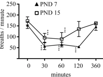
in previous experimental studies, evaluating the effects of morphine on brain development, the maximum ip-administered dose of this drug was 10 mg/kg. Because one important side effect of morphine administration is mu receptor–mediated respiratory depression, in a first set of experiments we evaluated how this dosing regimen affects physiological parameters. To this aim, we focused on two distinct developmental time points of the brain growth spurt: postnatal days 7 and 15. single-dose morphine (10 mg/kg) administration at these stages induced the loss of the righting reflex within 30 min, an effect that persisted up to 90 min postinjection. Also, this dosing regimen induced profound analgesia for at least 6 h when evaluated with the tail clamp test. Respiratory monitoring following single-dose ip administration of 10 mg/kg morphine revealed a highly significant depression of respiratory frequency in the two age groups when evaluated 30 min following injection (PND 7: −63 ± 10%, from 153 ± 15 to 57 ± 16 breaths/min, p < 0.0001; PND 15: −45 ± 12%, from 173 ± 19 to 95 ± 20 breaths/min, p < 0.0001) (Fig. 1). in 7-day-old animals, this respiratory depression persisted up to 120 min following morphine injection
and normalized when assessed at 6 h postadministration. in contrast, respiratory frequency was comparable with control values 120 min following morphine exposure in 15-day-old pups (Fig. 1). Blood gas sampling at defined time intervals following morphine administration revealed that the observed respiratory depression was accompanied with mild respiratory acidosis in both age groups (Table 1).
single-dose morphine (10 mg/kg) administration does not induce apoptosis in the prefrontal and somatosensory cortices during the brain growth spurt.
A large number of drugs that interfere with physiological pat-terns of neurotransmission, including general anesthetics, have been shown to induce developmental stage-dependent apop-tosis when administered during the brain growth spurt (Loepke and soriano, 2008). To our knowledge, no such data in the lit-erature is available regarding morphine exposure. Thus, in a next series of experiments, a single dose of 10 mg/kg morphine was ip administered to rat pups at PND 7 or at PND 15, and the effect of this treatment paradigm on apoptosis in the mPFc was evaluated 4 h postinjection using an antibody against the cleaved (i.e., activated) form of caspase-3. As seen in Figure 2, the number of cleaved caspase-3 positive cells was comparable between control and morphine-exposed pups either at PND 7 or at PND 15. As a positive control to define specificity of these findings, we exposed pups from the same litters to propofol (50 mg/kg ip) because this drug has been previously demon-strated to rapidly induce neuroapoptosis in the developing brain at PND 7 (cattano et al., 2008). in line with these observations, we also found that propofol rapidly induced highly significant (p = 0.0002) increase in the number of cleaved caspase-3 posi-tive cells in the mPFc at PND 7 but not at PND 15 (Fig. 2). To
assess the effect of morphine exposure on neuronal cell death in other cortical regions, we also evaluated neuroapoptosis in the somatosensory cortex of PND 7 and PND 15 animals. As seen in Figure 2, although the extent of naturally occurring cell death at PND 7 in this cortical region is higher than that in the mPFc, propofol but not morphine still induced significant cell death. Altogether, these data strongly suggest that single-dose morphine exposure, despite the accompanying respiratory depression and mild respiratory acidosis, does not trigger apop-tosis during the brain growth spurt.
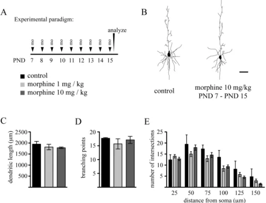
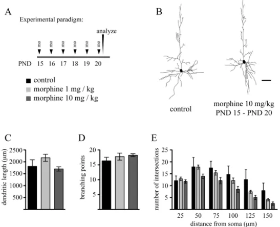
Repeated Exposure to Morphine During the Brain Growth Spurt Does Not Impair Dendritic Arbor Differentiation We have previously shown that dendritic arbor of layer 5 pyramidal neurons in the mPFc undergoes marked differentiation during the early postnatal period (Briner et al., 2011). Given that the most intense phase of arbor development during the brain growth spurt occurs around the second postnatal week (Juraska, 1982; Petit et al., 1988; Uylings et al., 1994), we thus investigated how exposure of rat pups to morphine during this period affects dendritic growths. To this end, rat pups received daily ip injections of morphine (either 1 or 10 mg/kg) from PND 7 to PND 15, and the effects of this treatment on basal dendritic arbor architecture of layer 5 pyramidal neurons in the mPFc was evaluated at PND 15. As seen in Figure 3, this experimental paradigm did not induce any significant changes in morphological parameters describing dendritic arborization pattern. Because ontogeny of mu receptor expression has been reported to be different between early and later stages of the brain growth spurt (Kent et al., 1981; Kornblum et al., 1987), we next explored the impact of morphine exposure on neuronal differentiation at later developmental stages by daily exposing young pups to this drug between PND 15 and PND 20. similar to exposure at earlier developmental stages, we found no effect of morphine on dendritic arbor development during this time period (Fig. 4). Altogether, these results strongly suggest that exposure to morphine during the brain growth spurt does not impair dendritic arbor development of these layer 5 pyramidal cells.
FIG. 1. Effect of morphine administration on respiratory frequency.
Morphine (10 mg/kg) was ip injected either on PND or on PND 15, and the effect of this treatment paradigm on respiratory frequency was monitored up to 360 min postinjection. Data are expressed as mean ± sD, n = 4–8 animals at each time point in each age group. One-way ANOVA with Bonferroni’s post hoc tests were used for searching statistical differences between time points in each age group. ***p < 0.001 compared with the initial respiratory frequency (i.e., at 0 min) in each age group.
TABLE 1
Physiological Data in 7- and 15-Day-Old Animals Following ip Single Bolus Administration of Morphine
pH PcO2 (kPA) Glucose (mg/dl)
PND 7 0 min 7.39 ± 0.04 6.63 ± 0.94 107 ± 13 30 min 7.22 ± 0.02 10.31 ± 0.45 180 ± 20 60 min 7.27 ± 0.1 10.02 ± 2.17 182 ± 32 120 min 7.22 ± 0.01 7.56 ± 1.60 66 ± 25 PND 15 0 min 7.38 ± 0.05 5.63 ± 1.22 76 ± 7 30 min 7.22 ± 0.02 9.61 ± 0.30 147 ± 6 60 min 7.23 ± 0.02 10.04 ± 0.44 136 ± 14 120 min 7.36 ± 0.02 7.89 ± 0.44 142 ± 8 Note. Data depict mean ± sD values of venous blood samples at distinct time points following morphine (10 mg/kg) administration. n = 3 animals per time points in each age groups.
Effects of Morphine on Dendritic Spinogenesis During Development
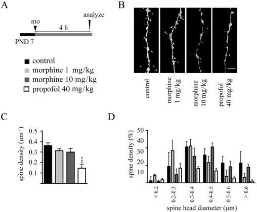
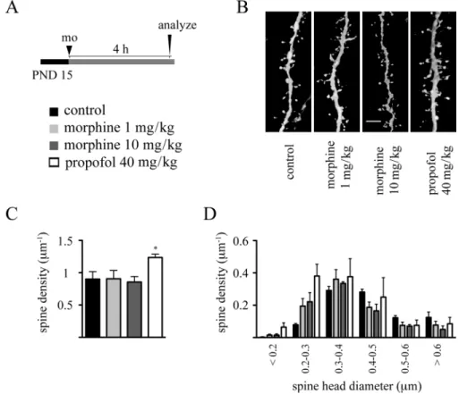
Dendritic spines represent primary postsynaptic sites of excitatory synaptic inputs to pyramidal neurons, and it is now well established that studying densities and morphology of these structures provide us valuable information on morphofunctional characteristics of neural networks (Bhatt et al., 2009; Holtmaat and svoboda, 2009). in order to gain insights into the impact of morphine on dendritic spine development, we evaluated the effects of both acute and repeated exposures to this drug on dendritic spine development. Because we have previously shown that dendritic spines can undergo rapid, within hours, remodeling following drug exposure (Briner et al., 2010, 2011; De Roo et al., 2009), PND 7 rat pups were ip injected with either 1 or 10 mg/kg morphine, and the effect of this treatment on dendritic spines of layer 5 pyramidal neurons in the mPFc was evaluated 4 h later. As seen in Figure 5, although single bolus propofol (40 mg/kg ip) rapidly decreased dendritic spine densities, single-dose morphine exposure affected neither dendritic spine densities nor their morphology, as evaluated by the analysis of spine head diameter, at this developmental stage. similar results were obtained following single-dose morphine exposure (1 and 10 mg/kg) at PND 15, where propofol increased
spine densities (Fig. 6). Altogether, these data strongly suggest that single-dose morphine exposure does not impair, at least acutely, dendritic spines during the brain growth spurt.
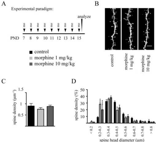
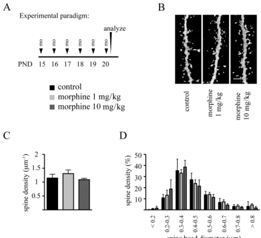
We next analyzed the impact of chronic morphine exposure on dendritic spines using the aforementioned repeated drug exposure protocols between PND 7 and PND 15 and between PND 15 and PND 20. As seen in Figures 7 and 8, despite the par-ticularly intense phase of spine development during these peri-ods (Briner et al., 2011), neither of these paradigms impaired dendritic spine densities. Analysis of spine head diameters following the PND 7 to PND 15 exposure revealed a statisti-cally significant increase in the percentage of spines present-ing with smaller head diameters either with 1 or with 10 mg/kg morphine exposure, and this was paralleled with a significant reduction in the number of middle-size spines at either doses of this drug (Fig. 7D). in contrast, no such changes were observed in the PND 15 to PND 20 exposed animals (Fig. 8D).
DISCUSSION
The present study demonstrates that morphine exposure does not alter neuronal arbor differentiation and excitatory synaptogenesis of pyramidal neurons during the brain growth
FIG. 2. Morphine does not induce caspase-3 activation in the cerebral cortex during the brain growth spurt. Rat pups received single ip injection of morphine
(10 mg/kg) or propofol (50 mg/kg) either on PND 7 or on PND 15, and the number of cleaved caspase-3 positive cells in the mPFc (A, c) and in the somatosensory cortex (B, D) was evaluated 4 h post injection. Data are expressed as mean ± sD, n = 4 animals at each drug regimen in each age group. One-way ANOVA with Bonferroni’s post hoc tests was used for searching statistical differences between experimental conditions in each age group. ***p < 0.001 compared with the control, non-drug-exposed animals in each age group. ssc, somatosensory cortex.
spurt. To our knowledge, these investigations provide the first in vivo data regarding the impact of this drug on morphofunctional parameters describing neuronal networks in the cerebral cortex during this intense phase of early postnatal development. We first show that single-dose morphine administration at two distinct stages of the brain growth spurt, although results in sedation, analgesia, and respiratory depression accompanied by mild respiratory acidosis, does not induce apoptosis in the cerebral cortex. By focusing on dendritic arbor development of layer 5 pyramidal neurons, we then demonstrate that repeated daily exposure to morphine during either the early or the later stages of the early postnatal period does not impair gross dendritic arbor development of these cells. Analysis of dendritic spines following single-dose morphine exposure using confocal microscopy reveals no effect of this drug on the structures representing postsynaptic sites of excitatory synaptic contacts on neurons. chronic administration of morphine during the early phase of the brain growth spurt results in an increase in the proportion of spines with smaller head diameters, whereas spine density remains unaltered. Finally, no effect of chronic morphine administration on dendritic spines was found when applied during later stages of the brain growth spurt. Altogether, these results are in agreement with and further extend recent preclinical observations arguing against
potential neurotoxic effects of opioid exposure during the early postnatal period.
We investigated the effects of morphine exposure during the brain growth spurt. The rationale behind this choice was that although morphine is frequently administered during this period in human clinical practice (Duedahl and Hansen, 2007;
Tesler et al., 1994), available knowledge on whether and how this drug may influence brain maturation at these developmen-tal stages remains scarce. Opioid receptors, along with their endogenous peptide ligands, are present from early stages of development in peripheral organs and in the developing brain. in mice, transcripts of opioid receptors are detected in the gut epithelium as early as embryonic day 9.5 (E 9.5), while μ, κ, and δ receptor mRNAs are present in multiple brain regions from E 12 (Zhu et al., 1998). Regarding more specifically the development of the cerebral cortex, earlier ligand binding stud-ies along with more recent works using in situ hybridization indicate important spatiotemporal differences in opioid recep-tor ontogeny.
in rodents, μ opioid receptor mRNA can be detected as early as E 12 in the deep neuroepithelium of the cortical plate, and expression is also present later on in the developing cortical subplate (Tong et al., 2000; Zhu et al., 1998). in the rat neocortex, μ receptor transcripts are first detected at E16
FIG. 3. Repeated daily administration of morphine does not impair dendritic growth during the early phase of the brain growth spurt. (A) Experimental
protocol. (B) Representative Neurolucida reconstructions of Lucifer Yellow–injected pyramidal neurons in layer 5 of the mPFc on PND 15 from control and morphine-exposed animals. Quantitative analysis of (c) basal dendritic lengths, (D) branching points, and (E) sholl distribution in layer 5 pyramidal neurons from control and morphine-exposed animals. Data are expressed as mean ± sD, n = 4 animals at each drug regimen. One-way ANOVA with Bonferroni’s post hoc tests was used for searching statistical differences between experimental conditions. scale bar: 50 μm.
with a predominance of expression in deep cortical layers (Tong et al., 2000). μ receptor densities have been reported to decrease during the first few days of early postnatal life in rodents, and this period is then followed by a progressive several fold increase that reaches steady state around the end of the first postnatal month (Kent et al., 1981; Kornblum et al., 1987; spain et al., 1985; Tong et al., 2000). Although further in vivo studies are needed to determine precise subcellular localization of μ receptors, in vitro observations using dissociated cortical cultures from newborn rodents indicate that these receptors are predominantly expressed on dendritic spines of pyramidal neurons (Liao et al., 2005). δ opioid receptors are first detected in the rat telencephalon around E 21; they are predominantly localized to cortical layer 2/3 and 5, and their distribution remains similar up to adult ages (Georges et al., 1998). κ receptor expression in the developing mouse cerebral cortex becomes detectable at E 17.5 (Zhu et al., 1998), but expression of this opioid receptor subtype in the postnatal cerebral cortex remains lower compared with that of μ and δ receptors (Georges et al., 1998).
Given the important socioeconomic impact of maternal drug abuse (Hunt et al., 2008; Lester and Lagasse, 2010), a large
number of experimental research studies targeted the impact of in utero opioid exposure on brain development. in this regard, early observations following prenatal exposure to methadone or morphine revealed decreased brain size and associated behavioral, cognitive, and motor deficits in offspring (seatriz and Hammer, 1993; Zagon and McLaughlin, 1977; Zagon et al., 1979a,b). More recent data further extend these obser-vations by indicating impaired dendritic growth and synaptic plasticity in juvenile offspring of morphine-exposed pregnant rats (Mei et al., 2009; Niu et al., 2009). in contrast to stimu-lation of opiod signaling pathways, naltrexone-induced block-ade of opiate receptors in utero results in increased brain size, neurogenesis, and more elaborated neuronal arbor in newborn pups (Hauser et al., 1987; Reznikov et al., 1999; Zagon and McLaughlin, 1983).
To gain insights into how administration of morphine impacts on the developing cerebral cortex during the brain growth spurt, we primarily focused on layer 5 pyramidal neurons of the developing rat mPFc. in rodents, this brain region is considered as the functional equivalent of the human frontal cortex (Uylings et al., 2003), a cortical structure that is implicated in higher order cognitive and emotional functions
FIG. 4. Repeated daily administration of morphine does not impair dendritic growth during the later phase of the brain growth spurt. (A) Experimental
proto-col. (B) Representative Neurolucida reconstructions of Lucifer Yellow–injected pyramidal neurons in layer 5 of the mPFc on PND 20 from control and morphine-exposed animals. Quantitative analysis of (c) basal dendritic lengths, (D) branching points, and (E) sholl distribution in layer 5 pyramidal neurons from control and morphine-exposed animals. Data are expressed as mean ± sD, n = 4 animals at each drug regimen. One-way ANOVA with Bonferroni’s post hoc tests was used for searching statistical differences between experimental conditions. scale bar: 50 μm.
(Tsujimoto, 2008). importantly, considerable densities of opioid receptors have been reported in this cortical region of which layer 5 pyramidal neurons form the major output source (Adams, 2009; Kornblum et al., 1987; Leriche et al., 2007;
Mansour et al., 1987; Martin-schild et al., 1999). Because the intense phase of neuronal differentiation and circuitry development spans over a time scale of weeks in rodents, we focused on two developmental time points, representing two functionally distinct period of the brain growth spurt, and explored the impact of morphine on neuronal survival, differentiation, and synaptogenesis. As an early stage, we chose the time point of PND 7 because this developmental period corresponds to the beginning of the brain growth spurt, when dendritic spine and synapse densities in the cerebral cortex are low (Briner et al., 2011; Juraska, 1982) and GABAA receptor– mediated neurotransmission is primarily excitatory due to high intracellular chloride concentrations (Blaesse et al., 2009). in contrast, synaptic density is several fold higher and actively ongoing on PND 15 (Briner et al., 2011; Juraska, 1982), and GABAergic signaling is already of inhibitory nature (Blaesse et al., 2009). Also, as discussed above, receptor binding and in situ hybridization studies indicate important differences in the temporal expression pattern of opioid receptor subtypes during
the brain growth spurt (Kent et al., 1981; Kornblum et al., 1987; spain et al., 1985; Tong et al., 2000). The ensemble of these observations thus strongly suggests that the sensitivity of developing neural systems to morphine exposure could be different at different stages of early postnatal life, raising thereby the need to target multiple time points in experimental studies.
We found that single bolus administration of morphine does not induce cell death in the prefrontal and somatosensory cortices at PND 7 and at PND 15. These results are in line with previous observations demonstrating that subcutaneous administration of a unique dose of morphine (10 mg/kg) on PND 3 did not induce apoptosis in rat pups (Black et al., 2008). in line, no apoptosis was detected in the spinal cord of rat pups following lumbar intrathecal injections of this drug in the early postnatal period (Westin et al., 2010). in fetal guinea pigs, at a developmental stage comparable with our and these aforementioned rodent studies, a 4-h-long iv infusion of the opioid fentanyl at high concentrations, aimed to maintain anesthesia, did not induce apoptosis (Rizzi et al., 2008). Data from adult mice also suggest the lack of proapoptotic effect of morphine (Emeterio et al., 2006). Although these observations argue against short-term morphine exposure–induced neurotoxicity, future work
FIG. 5. single-dose morphine administration on PND 7 does not affect dendritic spine density and morphology. (A) Experimental protocol. (B) Representative
volume rendering confocal reconstructions of dendritic shafts from control and morphine-exposed animals. (c) Quantitative analysis of dendritic spine densities on second-order basal dendritic shafts. (D) Frequency distribution histogram of spine head diameters in control and morphine-treated groups. Data are expressed as mean ± sD, n = 3 animals at each drug regimen. One-way ANOVA with Bonferroni’s post hoc tests was used for searching statistical differences between experimental conditions. A total of 1467 spines for controls, 1383 spines for the 1 mg/kg morphine group, 1448 spines for the 10 mg/kg morphine group, and 1349 spines for the propofol (40 mg/kg) group were counted to determine spine density. scale bar: 4 μm.
is needed to determine whether other cortical and subcortical regions, where opioid systems are also expressed, can be affected by morphine exposure during development. in this context, it is important to note that some subcortical regions, such as the mesostriatal system, express significantly higher amounts of opioid receptors than the cerebral cortex (Georges et al., 1998), and evaluating the impact of neonatal morphine exposure on the development of these brain regions should be thus of future interest. it is also important to underline that we found temporary respiratory depression and mild acidosis in animals following drug exposure, a condition that was often questioned to be a cause of increased neuroapoptosis following exposure to general anesthetics during the brain growth spurt (soriano et al., 2005). Although in this study we cannot formally exclude that morphine protects against the potentially deleterious effects of transient respiratory depression in the developing brain, we believe that our data also suggest that transient mild respiratory acidosis per se does not induce apoptosis in the cerebral cortex in the early postnatal period.
One important focus of the present study was to investi-gate how morphine impacts on the intense dendritic develop-ment during the brain growth spurt. indeed, because the causal
relationship between dendritic geometry and neuronal firing properties is well established (Hines and carnevale, 2001; van Ooyen et al., 2002), neuronal apoptosis is not the sole histologi-cal parameter to be considered in evaluating potentially adverse drug effects on neuronal development. Earlier works reported morphine-induced impairment of dendritic arbor development when this drug was repeatedly administered in the pre- or peri-natal period (Mei et al., 2009; Ricalde and Hammer, 1990). chronic morphine administration was also found to decrease dendritic arbor complexity in the adult brain (Hu et al., 2008; Li et al., 2007). Results of our study, however, reveal that chronic morphine administration affects dendritic growth neither at early nor at later stages of the brain growth spurt, thereby posing additional arguments against potential neurotoxic effects of the drug during this period. The most plausible explanation of dis-crepancies between our and the aforementioned results concern the differences in the developmental stage at which morphine was administered. Alternatively, this drug might have cortical layer–specific effects on pyramidal neurons. indeed, although we focused on layer 5 pyramidal neurons, emphasis has been placed on cortical layer 2/3 principal cells in those other studies. We cannot completely exclude potential technical confounders.
FIG. 6. single-dose morphine administration on PND 15 does not affect dendritic spine density and morphology. (A) Experimental protocol. (B) Representative
volume rendering confocal reconstructions of dendritic shafts from control and morphine-exposed animals. (c) Quantitative analysis of dendritic spine densities on second-order basal dendritic shafts. (D) Frequency distribution histogram of spine head diameters in control and morphine-treated groups. Data are expressed as mean ± sD, n = 3 animals at each drug regimen. One-way ANOVA with Bonferroni’s post hoc tests was used for searching statistical differences between experimental conditions. A total of 4123 spines for controls, 4889 spines for the 1 mg/kg morphine group, 5269 spines for the 10 mg/kg morphine group, and 6152 spines for the propofol (40 mg/kg) group were counted to determine spine density. scale bar: 4 μm.
Here, we used iontophoretic Lucifer Yellow single cell injec-tions, a method that is generally considered as a gold standard to fully label neuronal arbor architecture. in contrast, Golgi-stained randomly labeled neurons were analyzed by the other investigators mentioned herein. Finally, it is important to state that the fact that our results do not reveal a significant impact of morphine exposure on neuronal arbor does not obligatorily mean the absence of such an effect. indeed, our data represent results obtained from four animals (5 to 8 neurons analyzed per animal) in each experimental group. Power analysis reveals that the statistical power of these experiments remains under 50% and at least 26 animals per experimental group would be needed to increase this power up to 80%. clearly, additional studies are needed to further investigate the effects of morphine on neuronal architecture.
in addition to spatial branching topology of dendritic arbor, microscopic surface irregularities, including dendritic spine density, shape, and distribution are also major determinants of neural responses (Arellano et al., 2007; schikorski and stevens, 2001). Mu opioid receptors are expressed on dendritic spines (Liao et al., 2005), and chronic administration of morphine
in the perinatal period has been reported to decrease dendritic spine density (Ricalde and Hammer, 1990). More recent in vitro data demonstrate that this is mainly due to the decreased stabilization of newly formed spines, an effect mediated via the modulation of NeuroD activity (Liao et al., 2005; Zheng et al., 2010). Our data demonstrate that single-dose morphine administration does not impair dendritic spine density and morphology. Moreover, we found no effect on spine density when rat pups were repetitively exposed to this drug during either the early or the later stages of the peak synaptogenic period. These observations thus suggest a lack of effect of morphine on synaptogenesis during the brain growth spurt. Nevertheless, an important caveat to these results is that they represent post hoc observations. Because dendritic spines undergo intense turnover during the most intense phase of synaptogenesis (Holtmaat and svoboda, 2009; Lendvai et al., 2000), we cannot this exclude that morphine affects the rate of this turnover, the formation, stabilization, or the elimination of spines. in fact, although we observed no change in spine densities following chronic exposure to morphine during the early phases of the brain growth spurt, analysis of dendritic
FIG. 7. Effects of repeated daily administration of morphine during the early phase of the brain growth spurt on dendritic spine density and morphology.
(A) Experimental protocol. (B) Representative volume rendering confocal reconstructions of dendritic shafts from control and morphine-exposed animals. (c) Quantitative analysis of dendritic spine densities on second-order basal dendritic shafts. (D) Frequency distribution histogram of spine head diameters in control and morphine-treated groups. Data are expressed as mean ± sD, n = 3 animals at each drug regimen. One-way ANOVA with Bonferroni’s post hoc tests was used for searching statistical differences between experimental conditions. A total of 4123 spines for controls, 4060 spines for the 1 mg/kg morphine group, and 3601 spines for the 10 mg/kg morphine group were counted to determine spine density. scale bar: 4 μm; ***p < 0.001 compared with control group.
spine head diameters revealed a significantly higher number of small spines in the morphine group compared with control littermates. in light of available in vitro data (Liao et al., 2005), one plausible explanation would be that chronic morphine exposure during this early phase of synaptogenesis would impede the stabilization of newly formed spines and that this would lead to an increased turnover and thus a higher percentage of immature spine in this experimental group. Further studies will be required to shed light on this issue.
The clinical relevance of our observations remains to be established. To our belief, we provide here the first in vivo study regarding the effects of morphine exposure during the brain growth spurt. According to works on extrapolating brain development from experimental species to humans, the developmental period situated between the first and third weeks of postnatal life in rat pups corresponds to a period extending from the third trimester of pregnancy up to the first few years of life in humans (clancy et al., 2001, 2007). We thus consider that experiments conducted around PND 7 would have preclinical relevance regarding morphine exposure to premature children,
whereas drug exposure in PND 15 animals might reflect a developmental stage somewhere during the first few years of life in humans. Extrapolation of experimental results is further complicated by the fact that even a single dose–induced few-hour-long effect might reflect exposure to weeks or months in the human time scale (Anand and soriano, 2004). Nevertheless, given the lack of any morphological effects that might suggest neurotoxicity of morphine in our experimental paradigms, we believe that our results bring preclinical arguments in favor of the relative safety of this drug during the brain growth spurt. These results are of particular interest in view of the large amount of data suggesting that exposure to the majority of general anesthetics rapidly induces experimental neurotoxicity during this developmental period (Loepke and soriano, 2008). To our knowledge, and despite the fact that opioids constitute one of the main pharmacological approaches to alleviate pain in neonates (Anand, 2001) and in children (Duedahl and Hansen, 2007; Tesler et al., 1994), there are sparse data on long-term neurocognitive and behavioral effects of neonatal morphine exposure. Morphine administration in very premature neonates
FIG. 8. Repeated daily administration of morphine during the later phase of the brain growth spurt does not affect dendritic spine density and morphology.
(A) Experimental protocol. (B) Representative volume rendering confocal reconstructions of dendritic shafts from control and morphine-exposed animals. (c) Quantitative analysis of dendritic spine densities on second-order basal dendritic shafts. (D) Frequency distribution histogram of spine head diameters in control and morphine-treated groups. Data are expressed as mean ± sD, n = 3 animals at each drug regimen. One-way ANOVA with Bonferroni’s post hoc tests was used for searching statistical differences between experimental conditions. A total of 4802 spines for controls, 7147 spines for the 1 mg/kg morphine group, and 5442 spines for the 10 mg/kg morphine group were counted to determine spine density. scale bar: 4 μm.
did not seem to have any adverse effects on intelligence, motor function, or behavior when these children are assessed at 5–6 years of age (MacGregor et al., 1998). A recent follow-up study among 5-year-old children who have previously been involved in a placebo-controlled trial on the effects of morphine on pain and neurologic outcome demonstrated significantly lower overall intelligence quotient score compared with the placebo group; these differences, however, disappeared following correction for potential confounding factors (de Graaf et al., 2011). Nevertheless, scores on one iQ subtest, “visual analysis,” were significantly negatively related to having received morphine, warranting longer term follow-up in these children by focusing on higher order neurocognitive function (de Graaf et al., 2011).
in conclusion, this study demonstrates that morphine expos-ure during the most intense period of neuronal differentiation and synaptogenesis does not induce apoptosis in the mPFc, a cortical region of utmost importance in mediating higher order cognitive functions. importantly, this drug did not affect dendritic differentiation of layer 5 pyramidal neurons in this cortical region and had no impact on dendritic spine dens-ities of these cells. These experimental observations, in line and together with recent preclinical data (Black et al., 2008;
Emeterio et al., 2006; Rizzi et al., 2008; Westin et al., 2010), suggest the lack of overt neurotoxic effects of this drug during the brain growth spurt.
FUNDING
swiss National science Foundation (31003A-130625 to L.V.); De Reuter Foundation (subside 501 to L.V.).
REFERENCES
Adams, J. D., Jr. (2009). chemical interactions with pyramidal neurons in layer 5 of the cerebral cortex: control of pain and anxiety. Curr. Med. Chem. 16, 3476–3479.
Anand, K. J., and soriano, s. G. (2004). Anesthetic agents and the immature brain: Are these toxic or therapeutic? Anesthesiology 101, 527–530. Anand, K. J., and international Evidence-Based Group for Neonatal Pain.
(2001). consensus statement for the prevention and management of pain in the newborn. Arch. Pediatr. Adolesc. Med. 155, 173–180.
Arellano, J. i., Benavides-Piccione, R., Defelipe, J., and Yuste, R. (2007). Ultrastructure of dendritic spines: correlation between synaptic and spine morphologies. Front. Neurosci. 1, 131–143.
Bhatt, D. H., Zhang, s., and Gan, W. B. (2009). Dendritic spine dynamics. Annu. Rev. Physiol. 71, 261–282.
Black, A. M., Pandya, s., clark, D., Armstrong, E. A., and Yager, J. Y. (2008). Effect of caffeine and morphine on the developing pre-mature brain. Brain Res. 1219, 136–142.
Blaesse, P., Airaksinen, M. s., Rivera, c., and Kaila, K. (2009). cation-chloride cotransporters and neuronal function. Neuron 61, 820–838.
Briner, A., De Roo, M., Dayer, A., Muller, D., Habre, W., and Vutskits, L. (2010). Volatile anesthetics rapidly increase dendritic spine density in the rat medial prefrontal cortex during synaptogenesis. Anesthesiology 112, 546–556.
Briner, A., Nikonenko, i., De Roo, M., Dayer, A., Muller, D., and Vutskits, L. (2011). Developmental stage-dependent persistent impact of propofol anes-thesia on dendritic spines in the rat medial prefrontal cortex. Anesthesiology
115, 282–293.
carter, B. D., and Medzihradsky, F. (1993). Go mediates the coupling of the mu opioid receptor to adenylyl cyclase in cloned neural cells and brain. Proc. Natl. Acad. Sci. U.S.A. 90, 4062–4066.
cattano, D., Young, c., straiko, M. M., and Olney, J. W. (2008). subanesthetic doses of propofol induce neuroapoptosis in the infant mouse brain. Anesth. Analg. 106, 1712–1714.
chen, Y., and Ghosh, A. (2005). Regulation of dendritic development by neu-ronal activity. J. Neurobiol. 64, 4–10.
clancy, B., Darlington, R. B., and Finlay, B. L. (2001). Translating develop-mental time across mammalian species. Neuroscience 105, 7–17.
clancy, B., Finlay, B. L., Darlington, R. B., and Anand, K. J. (2007). Extrapolating brain development from experimental species to humans. Neurotoxicology 28, 931–937.
De Felipe, J., Marco, P., Fairén, A., and Jones, E. G. (1997). inhibitory synap-togenesis in mouse somatosensory cortex. Cereb. Cortex 7, 619–634. de Graaf, J., van Lingen, R. A., simons, s. H., Anand, K. J., Duivenvoorden, H.
J., Weisglas-Kuperus, N., Roofthooft, D. W., Groot Jebbink, L. J., Veenstra, R. R., Tibboel, D., et al. (2011). Long-term effects of routine morphine infu-sion in mechanically ventilated neonates on children’s functioning: Five-year follow-up of a randomized controlled trial. Pain 152, 1391–1397. De Roo, M., Klauser, P., Briner, A., Nikonenko, i., Mendez, P., Dayer, A., Kiss,
J. Z., Muller, D., and Vutskits, L. (2009). Anesthetics rapidly promote synap-togenesis during a critical period of brain development. PLoS ONE 4, e7043. Duedahl, T. H., and Hansen, E. H. (2007). A qualitative systematic review of
morphine treatment in children with postoperative pain. Paediatr. Anaesth.
17, 756–774.
Emeterio, E. P., Tramullas, M., and Hurlé, M. A. (2006). Modulation of apop-tosis in the mouse brain after morphine treatments and morphine withdrawal. J. Neurosci. Res. 83, 1352–1361.
Georges, F., Normand, E., Bloch, B., and Le Moine, c. (1998). Opioid receptor gene expression in the rat brain during ontogeny, with special reference to the mesostriatal system: An in situ hybridization study. Brain Res. Dev. Brain Res. 109, 187–199.
Hauser, K. F., McLaughlin, P. J., and Zagon, i. s. (1987). Endogenous opioids regulate dendritic growth and spine formation in developing rat brain. Brain Res. 416, 157–161.
Hensch, T. K. (2004). critical period regulation. Annu. Rev. Neurosci. 27, 549–579. Hines, M. L., and carnevale, N. T. (2001). NEURON: A tool for
neuroscien-tists. Neuroscientist 7, 123–135.
Holtmaat, A., and svoboda, K. (2009). Experience-dependent structural syn-aptic plasticity in the mammalian brain. Nat. Rev. Neurosci. 10, 647–658. Hu, F., Li, G., Liang, Z., Yang, Y., and Zhou, Y. (2008). The morphological
changes of pyramidal and spiny stellate cells in the primary visual cortex of chronic morphine treated cats. Brain Res. Bull. 77, 77–83.
Hunt, R. W., Tzioumi, D., collins, E., and Jeffery, H. E. (2008). Adverse neu-rodevelopmental outcome of infants exposed to opiate in-utero. Early Hum. Dev. 84, 29–35.
Huttenlocher, P. R., and Dabholkar, A. s. (1997). Regional differences in syn-aptogenesis in human cerebral cortex. J. Comp. Neurol. 387, 167–178. ikeda, K., Kobayashi, T., Kumanishi, T., Niki, H., and Yano, R. (2000).
involvement of G-protein-activated inwardly rectifying K (GiRK) channels in opioid-induced analgesia. Neurosci. Res. 38, 113–116.
Jensen, T. s. (1997). Opioids in the brain: supraspinal mechanisms in pain control. Acta Anaesthesiol. Scand. 41(1 Pt 2), 123–132.
Juraska, J. M. (1982). The development of pyramidal neurons after eye opening in the visual cortex of hooded rats: A quantitative study. J. Comp. Neurol.
Kent, J. L., Pert, c. B., and Herkenham, M. (1981). Ontogeny of opiate recep-tors in rat forebrain: Visualization by in vitro autoradiography. Brain Res.
254, 487–504.
Kornblum, H. i., Hurlbut, D. E., and Leslie, F. M. (1987). Postnatal develop-ment of multiple opioid receptors in rat brain. Brain Res. 465, 21–41. Lendvai, B., stern, E. A., chen, B., and svoboda, K. (2000).
Experience-dependent plasticity of dendritic spines in the developing rat barrel cortex in vivo. Nature 404, 876–881.
Leriche, M., cote-Vélez, A., and Méndez, M. (2007). Presence of pro-opi-omelanocortin mRNA in the rat medial prefrontal cortex, nucleus accum-bens and ventral tegmental area: studies by RT-PcR and in situ hybridization techniques. Neuropeptides 41, 421–431.
Lester, B. M., and Lagasse, L. L. (2010). children of addicted women. J. Addict. Dis. 29, 259–276.
Li, Y., Wang, H., Niu, L., and Zhou, Y. (2007). chronic morphine exposure alters the dendritic morphology of pyramidal neurons in visual cortex of rats. Neurosci. Lett. 418, 227–231.
Liao, D., Lin, H., Law, P. Y., and Loh, H. H. (2005). Mu-opioid receptors modulate the stability of dendritic spines. Proc. Natl. Acad. Sci. U.S.A. 102, 1725–1730.
Loepke, A. W., and soriano, s. G. (2008). An assessment of the effects of gen-eral anesthetics on developing brain structure and neurocognitive function. Anesth. Analg. 106, 1681–1707.
MacGregor, R., Evans, D., sugden, D., Gaussen, T., and Levene, M. (1998). Outcome at 5-6 years of prematurely born children who received morphine as neonates. Arch. Dis. Child. Fetal Neonatal Ed. 79, F40–F43.
Mansour, A., Khachaturian, H., Lewis, M. E., Akil, H., and Watson, s. J. (1987). Autoradiographic differentiation of mu, delta, and kappa opioid receptors in the rat forebrain and midbrain. J. Neurosci. 7, 2445–2464.
Martin-schild, s., Gerall, A. A., Kastin, A. J., and Zadina, J. E. (1999). Differential distribution of endomorphin 1- and endomorphin 2-like immu-noreactivities in the cNs of the rodent. J. Comp. Neurol. 405, 450–471. Mei, B., Niu, L., cao, B., Huang, D., and Zhou, Y. (2009). Prenatal morphine
exposure alters the layer ii/iii pyramidal neurons morphology in lateral sec-ondary visual cortex of juvenile rats. Synapse 63, 1154–1161.
Milligan, G. (2005). Opioid receptors and their interacting proteins. Neuromolecular Med. 7, 51–59.
Minami, M., and satoh, M. (1995). Molecular biology of the opioid receptors: structures, functions and distributions. Neurosci. Res. 23, 121–145. Niu, L., cao, B., Zhu, H., Mei, B., Wang, M., Yang, Y., and Zhou, Y. (2009).
impaired in vivo synaptic plasticity in dentate gyrus and spatial memory in juve-nile rats induced by prenatal morphine exposure. Hippocampus 19, 649–657. Petit, T. L., LeBoutillier, J. c., Gregorio, A., and Libstug, H. (1988). The
pat-tern of dendritic development in the cerebral cortex of the rat. Brain Res.
469, 209–219.
Piros, E. T., Prather, P. L., Loh, H. H., Law, P. Y., Evans, c. J., and Hales, T. G. (1995). ca2+ channel and adenylyl cyclase modulation by cloned mu-opioid receptors in GH3 cells. Mol. Pharmacol. 47, 1041–1049.
Reznikov, K., Hauser, K. F., Nazarevskaja, G., Trunova, Y., Derjabin, V., and Bakalkin, G. (1999). Opioids modulate cell division in the germinal zone of the late embryonic neocortex. Eur. J. Neurosci. 11, 2711–2719.
Ricalde, A. A., and Hammer, R. P., Jr. (1990). Perinatal opiate treatment delays growth of cortical dendrites. Neurosci. Lett. 115, 137–143.
Rizzi, s., carter, L. B., Ori, c., and Jevtovic-Todorovic, V. (2008). clinical anesthesia causes permanent damage to the fetal guinea pig brain. Brain Pathol. 18, 198–210.
sadraie, s. H., Kaka, G. R., sahraei, H., Dashtnavard, H., Bahadoran, H., Mofid, M., Nasab, H. M., and Jafari, F. (2008). Effects of maternal oral administration of morphine sulfate on developing rat fetal cerebrum: A mor-phometrical evaluation. Brain Res. 1245, 36–40.
schikorski, T., and stevens, c. F. (2001). Morphological correlates of function-ally defined synaptic vesicle populations. Nat. Neurosci. 4, 391–395. schoffelmeer, A. N., Van Vliet, B. J., De Vries, T. J., Heijna, M. H., and Mulder,
A. H. (1992). Regulation of brain neurotransmitter release and of adenylate cyclase activity by opioid receptors. Biochem. Soc. Trans. 20, 449–453. seatriz, J. V., and Hammer, R. P., Jr. (1993). Effects of opiates on neuronal
development in the rat cerebral cortex. Brain Res. Bull. 30, 523–527. soriano, s. G., Anand, K. J., Rovnaghi, c. R., and Hickey, P. R. (2005). Of
mice and men: should we extrapolate rodent experimental data to the care of human neonates? Anesthesiology 102, 866–868.
spain, J. W., Roth, B. L., and coscia, c. J. (1985). Differential ontogeny of multiple opioid receptors (mu, delta, and kappa). J. Neurosci. 5, 584–588. Tesler, M. D., Wilkie, D. J., Holzemer, W. L., and savedra, M. c. (1994).
Postoperative analgesics for children and adolescents: Prescription and administration. J. Pain Symptom Manage. 9, 85–95.
Tong, Y., chabot, J. G., shen, s. H., O’Dowd, B. F., George, s. R., and Quirion, R. (2000). Ontogenic profile of the expression of the mu opioid receptor gene in the rat telencephalon and diencephalon: An in situ hybridization study. J. Chem. Neuroanat. 18, 209–222.
Tsujimoto, s. (2008). The prefrontal cortex: Functional neural development during early childhood. Neuroscientist 14, 345–358.
Uylings, H. B., Groenewegen, H. J., and Kolb, B. (2003). Do rats have a pre-frontal cortex? Behav. Brain Res. 146, 3–17.
Uylings, H. B., van Pelt, J., Parnavelas, J. G., and Ruiz-Marcos, A. (1994). Geometrical and topological characteristics in the dendritic development of cortical pyramidal and non-pyramidal neurons. Prog. Brain Res. 102, 109–123.
van Ooyen, A., Duijnhouwer, J., Remme, M. W., and van Pelt, J. (2002). The effect of dendritic topology on firing patterns in model neurons. Network
13, 311–325.
Westin, B. D., Walker, s. M., Deumens, R., Grafe, M., and Yaksh, T. L. (2010). Validation of a preclinical spinal safety model: Effects of intrathecal mor-phine in the neonatal rat. Anesthesiology 113, 183–199.
Yaksh, T. L. (1997). Pharmacology and mechanisms of opioid analgesic activ-ity. Acta Anaesthesiol. Scand. 41(1 Pt 2), 94–111.
Zagon, i. s., and McLaughlin, P. J. (1977). Morphine and brain growth retarda-tion in the rat. Pharmacology 15, 276–282.
Zagon, i. s., and McLaughlin, P. J. (1983). increased brain size and cellular con-tent in infant rats treated with an opiate antagonist. Science 221, 1179–1180. Zagon, i. s., McLaughlin, P. J., and Thompson, c. i. (1979a). Learning ability
in adult female rats perinatally exposed to methadone. Pharmacol. Biochem. Behav. 10, 889–894.
Zagon, i. s., McLaughlin, P. J., and Thompson, c. i. (1979b). Development of motor activity in young rats following perinatal methadone exposure. Pharmacol. Biochem. Behav. 10, 743–749.
Zheng, H., Zeng, Y., chu, J., Kam, A. Y., Loh, H. H., and Law, P. Y. (2010). Modulations of NeuroD activity contribute to the differential effects of morphine and fentanyl on dendritic spine stability. J. Neurosci. 30, 8102–8110.
Zhu, Y., Hsu, M. s., and Pintar, J. E. (1998). Developmental expression of the mu, kappa, and delta opioid receptor mRNAs in mouse. J. Neurosci. 18, 2538–2549.