Review
Role of gap junctions in the propagation of the cardiac action potential
Stephan Rohr*
Department of Physiology, University of Bern, Bu¨hlplatz 5, CH-3012 Bern, Switzerland Received 1 October 2003; received in revised form 26 November 2003; accepted 28 November 2003
Time for primary review 26 days
Abstract
Gap junctions play a pivotal role for the velocity and the safety of impulse propagation in cardiac tissue. Under physiologic conditions, the specific subcellular distribution of gap junctions together with the tight packaging of the rod-shaped cardiomyocytes underlies anisotropic conduction, which is continuous at the macroscopic scale. However, when breaking down the three-dimensional network of cells into linear single cell chains, gap junctions can be shown to limit axial current flow and to induce ‘saltatory’ conduction at unchanged overall conduction velocities. In two- and three-dimensional tissue, these discontinuities disappear due to lateral averaging of depolarizing current flow at the activation wavefront. During gap junctional uncoupling, discontinuities reappear and are accompanied by slowed and meandering conduction. Critical gap junctional uncoupling reduces conduction velocities to a much larger extent than does a reduction of excitability, which suggests that the safety for conduction is higher at any given conduction velocity for gap junctional uncoupling. In uniformly structured tissue, gap junctional uncoupling is accompanied by a parallel decrease in conduction velocity. However, this is not necessarily the case for non-uniform structures like tissue expansion where partial uncoupling paradoxically increases conduction velocity and has the capacity to remove unidirectional conduction blocks. Whereas the impact of gap junctions on impulse conduction is generally assessed from the point of view of cell coupling among cardiomyocytes, it is possible that other cell types within the myocardium might be coupled to cardiomyocytes as well. In this context, it has been shown that fibroblasts establish successful conduction between sheets of cardiomyocytes over distances as long as 300 Am. This might not only explain electrical synchronization of heart transplants but might be of importance for cardiac diseases involving fibrosis. Finally, the intriguing fact that sodium channels are clustered at the intercalated disc recently rekindled the provocative question of whether gap junctions alone are responsible for impulse propagation or whether electric field mechanisms might account for conduction as well. Whereas computer simulations show the feasibility of conduction in the absence of gap junctional coupling, a definite answer to this question must await further investigations into the biophysical properties of the intercalated disc.
D 2004 European Society of Cardiology. Published by Elsevier B.V. All rights reserved.
Keywords: Heart; Impulse propagation; Gap junction; Tissue structure; Sodium channels
1. Introduction
Half a century ago, Weidmann[1]published his pioneer-ing electrophysiological study which showed that cardiac Purkinje fibers behaved electrically similarly to axons. Two years after this work, however, electron microscopic obser-vations of cardiac tissue led Sjo¨strand and Andersson[2]to conclude that myocardial tissue is not, as was previously thought, an anatomical syncytium but that each cardiomyo-cyte is surrounded by a contiguous cell membrane. Based on both electrophysiological studies of the cable properties of Purkinje fibers and the observation that radioactive potassium
readily spread along the tissue, this apparent contradiction between electrophysiological findings and cellular ultrastruc-ture was resolved by postulating the presence of low-resis-tance pathways interconnecting the cardiomyocytes [3,4]. These findings and observations in other tissues led to the establishment of the concept of ‘gap junctions’ as the funda-mental principle underlying impulse transmission and sig-naling molecule exchange among adjacent cells in a variety of tissues. It is the goal of this short review to summarize recent findings related to the role of gap junctions in the process of the spread of cardiac excitation on the cellular level based on both experimental studies and computer simulations and to briefly touch the controversial issue of whether gap junctions are the ‘truth and nothing but the truth’ for electrical impulse transmission between cardiomyocytes. For a review of the important effects of gap junctional remodeling and
0008-6363/$ - see front matterD 2004 European Society of Cardiology. Published by Elsevier B.V. All rights reserved. doi:10.1016/j.cardiores.2003.11.035
* Tel.: +41-31-631-87-46; fax: +41-31-631-46-11. E-mail address: [email protected] (S. Rohr).
genetic manipulations of connexin expression on impulse propagation in the heart, the reader is referred to the respec-tive chapters of this spotlight issue.
2. General aspects of the role of gap junctions in propagation
The working myocardium can be regarded as a three-dimensional network of coupled excitable elements. In this network, the velocity and safety of the spread of excitation is dependent on both active and passive properties of the individual elements and on the connectivity of the network. Among passive properties, gap junctions play a pivotal role because they ultimately determine how much depolarizing current passes from excited to non-excited regions of the network. Thereby, they act as important determinants of the speed and safety of this process. A second important factor defining the passive properties of the network consists of the specific cellular architecture of cardiac tissue: whereas pro-gressive uncoupling in a uniform network will result in a slowing of conduction velocity following the square root relationship, this does not necessarily have to be the case for non-uniform networks, i.e., for tissue structures where the size of a given excited region supplying depolarizing current (‘current source’) is ill matched to the amount of depolarizing current necessary to excite the regions ahead (‘current sink’). A long known example for such a source-to-sink mismatch is represented by the Purkinje – fiber – ventricular junction where a small source (Purkinje fiber) is coupled to a large sink (mass of ventricular tissue)[5 – 7]. Depending on the size of the mismatch, this results in either local conduction delays or unidirectional conduction blocks at the junction during anterograde conduction (Purkinje – fiber!ventricle). In con-trast to the effects of gap junctional coupling on propagation in uniform tissue, partial uncoupling in such non-uniform tissue structures can actually be accompanied by a paradox-ical increase in safety and velocity of conduction. Because of these marked differences of the effect of gap junctional coupling on conduction in uniform vs. non-uniform tissue, the two issues are discussed separately.
3. Gap junctions and impulse propagation in uniform tissue during normal coupling
Under physiological conditions, a given cardiomyocyte in the adult working myocardium is electrically coupled to an average of f11 adjacent cells with gap junctions being predominantly localized at the intercalated discs at the ends of the rod shaped cells [8]. This particular subcellular distribution of gap junctions is a main determinant of anisotropic conduction in the heart even though, as shown recently, cell size on its own also significantly modifies the characteristics of transverse conduction [9](for review, cf.
Ref. [10]). It is generally accepted that macroscopic impulse
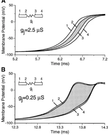
conduction along the main fiber axis of cardiac tissue is continuous because of the low resistance coupling by gap junctions. However, when abstracting from the three-dimen-sional network to a chain of single cardiomyocytes, the results of the computer simulations shown inFig. 1Asuggest that propagation is actually saltatory and is composed of rapid excitation of individual cells followed by a transjunc-tional conduction delay[11,12]. When both gap junctional resistance and the resistivity of the myoplasm are set to levels resulting in macroscopic conduction velocities of f50 cm/s, both conduction times along the entire cell soma and trans-junctional propagation delays are f100 As. Whereas, at first glance, this difference seems to be moderate, one has to keep in mind that the gap junctional cleft is roughly 4 orders of magnitude shorter than the cell itself resulting in apparent local ‘conduction velocities’ of f1 m/s along the cell and 0.1 mm/s across the junction. As shown in Fig. 1B, this saltatory type of conduction becomes highly accentuated during a tenfold reduction of gap junctional conductance (macroscopic conduction velocities of f20 cm/s). In this case, cell somata are excited in a virtually simultaneous manner due to the cellular confinement of depolarizing
Fig. 1. Computer simulation of microscopic impulse propagation. (A) Action potential upstrokes recorded during impulse propagation between two adjacent cells at the sites indicated in the insert. Under conditions of normal gap junctional coupling, the transjunctional conduction delay is roughly equal to the myoplasmic conduction times of the cells. (B) During a tenfold reduction of gap junctional coupling, myoplasmic conduction times are abbreviated whereas transjunctional conduction times are substantially increased. Redrawn with modifications fromRef. [12].
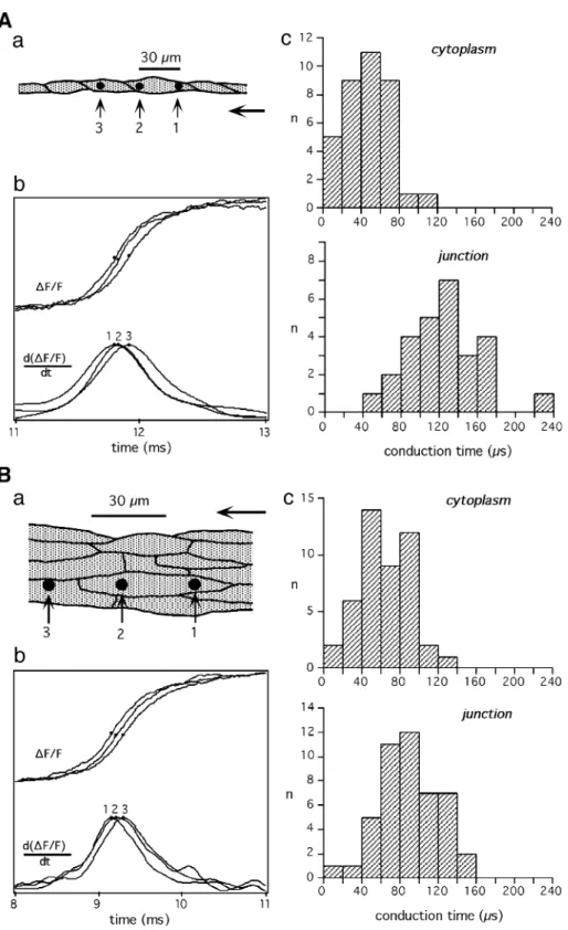
current, whereas transjunctional conduction delays substan-tially increase to 500 As. Experimentally, the question of microscopically discontinuous conduction in cardiac tissue has been addressed by combining patterned growth of cultured cardiomyocytes [13,14] and optical recording of transmembrane voltage with subcellular resolution[15]. As shown inFig. 2, simultaneous optical recordings of action potential upstrokes in a single cell chain of cultured neonatal rat ventricular cardiomyocytes with subcellular resolution (15 Am) indicated the presence of an activation delay at the contact site between two end-to-end abutted cardiomyocytes, whereas conduction within individual cells was rapid and continuous [16]. Quantitative aspects of this phenomenon are shown in Fig. 3. In the case of single cell chains of cardiomyocytes (panel A), the difference between mean cytoplasmic (38 As) and transjunctional (118 As) conduction time as measured with detectors spaced 30 Am apart indi-cated the presence of a transjunctional activation delay of 80 As[17]. Assuming an average cell length of 60 Am, trans-junctional delays and cytoplasmic conduction times were of roughly equal magnitude, thus being in close agreement with the aforementioned computer simulation study. In contrast to the findings in single cell chains, the characteristic of conduction at the microscopic scale change drastically in strands several cells wide(Fig. 3B). There, mean cytoplas-mic conduction times increased to 57 As, whereas transjunc-tional conduction times simultaneously decreased to 89 As, resulting in significantly reduced cell-to-cell propagation delays. This suggests that, in uniform multicellular tissue, conduction delays across longitudinally abutted cells largely disappear at the expense of increased cytoplasmic
conduc-tion times. This can be explained by the presence of lateral gap junctional coupling, which serves to average slight local advances/delays of the activation wavefront among laterally apposed cells. This lateral averaging, which can be expected to be even more pronounced in three-dimensional tissue, ultimately results in largely continuous conduction along the main fiber axis in intact uniform tissue.
4. Gap junctions and impulse propagation in uniform tissue during progressive uncoupling
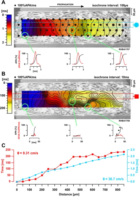
Whereas, as noted above, propagation in uniform multi-cellular tissue under normal conditions is continuous, this changes during progressive uncoupling. As shown inFig. 4, severe gap junctional uncoupling in a 250 Am wide cell strand not only drastically reduced conduction velocity from 36.7 to 0.3 cm/s, but further resulted in a meandering activation wavefront [18]. Meandering is induced by the presence of islands of completely uncoupled cells which causes the activation wavefront to follow a ‘zig-zag’ path of activation similar to that occurring in infarcted[19]and non-uniform anisotropic tissue [20]. Also at the cellular level, impulse propagation under these conditions is highly dis-continuous as illustrated in Fig. 5. Whereas conduction is fast (43 cm/s) and continuous under control conditions(Fig. 5A), severe uncoupling (Fig. 5B) causes action potential upstrokes to rise in clusters with large inter-cluster activa-tion delays which result in very slow overall conducactiva-tion velocities of 1.1 cm/s(Fig. 5Bb). Moreover, when mapping the clustered action potential upstrokes to the cellular structure of the preparation(Fig. 5Bc), it becomes obvious that activation during severe uncoupling jumps from one region consisting of one or a few cardiomyocytes to the next and that completely uncoupled cells force the activation wavefront to meander. Thus, irrespective of scaling, mean-dering activation seems to be a basic principle governing impulse propagation during reduced gap junctional coupling in cardiac tissue.
When comparing minimal conduction velocities achieved by either a reduction of active membrane properties (f20 cm/s)[21 – 23]or by a reduction of gap junctional coupling (f1 cm/s; cf. data above), the fact that conduction velocities can be an order of magnitude slower during uncoupling suggests that this is a ‘safer way’ to reduce velocity than a decrease in excitability. The reason for this behavior has been analyzed in detail in computer simulation studies by Shaw and Rudy [12,24], which compared the characteristics of conduction slowing during a reduction of either excitability or gap junctional coupling. In order to analyze the robustness of conduction under these conditions, they defined the ‘safety factor’ for conduction as the ratio of the amount of charge produced by a given membrane patch during activa-tion to the charge consumed during the activaactiva-tion process. By this definition, conduction fails when the safety factor drops below 1 and becomes increasingly stable as it rises
Fig. 2. Characteristics of impulse propagation on the subcellular scale. (A) Cellular architecture of the optically mapped region of a chain of single cardiomyocytes during impulse propagation from left to right (end-to-end abutted cardiomyocytes shown in two shades of gray). Squares indicate the positions of individual photodetectors. (B) Simultaneously recorded action potential upstrokes indicate a local delay between detectors 5 and 7, i.e., in the region of overlap of the light and dark gray shaded cells. Redrawn with permission fromRef. [16].
Fig. 3. Differences in intra- and intercellular activation delays between single cell chains and strands several cells wide. (A) Single cell chains: (Aa) Schematic drawing of the cellular architecture of a chain of single cardiomyocytes. Three equally spaced photodetectors (black discs) were placed such that they recorded activation (corresponding to a fractional change in fluorescence of the voltage sensitive dye, (DF/F)) either within cells (1!2; ‘cytoplasm’) or between cells (2!3; ‘junction’). (Ab) Optically recorded action potential upstrokes (above) and their derivatives (below) of a measurement during propagation from right to left. (Ac) Histograms of interdetector conduction times indicate coexistence of fast cytoplasmic and delayed transjunctional activation. (B) Multiple cell wide strands: (panels as in (A)). In the case of strands several cells wide, differences between cytoplasmic and transjunctional conduction times are substantially reduced indicating nearly continuous conduction along the preparation. Redrawn with modifications fromRef. [17].
above 1. As shown inFig. 6A, a reduction of excitability, which was modeled by reducing sodium channel conduc-tance, was accompanied by a decrease of conduction veloc-ity from 54 to 17 cm/s before conduction failed. At the same time, the safety factor for conduction fell from f1.6 to 1. As expected for a decrease in sodium channel conductance, the reduction of conduction velocity was paralleled by a ften-fold decrease of maximal upstroke velocities(Fig. 6B). In contrast to these effects, a reduction of intercellular coupling showed marked differences(Fig. 7). First, whereas progres-sive uncoupling also induced a monotonic decrease of
conduction velocity, slowest conduction velocities reached before block (0.26 cm/s) were over an order of magnitude below those obtained during a reduction of excitability. Moreover, both maximal upstroke velocities and the safety factor for conduction showed a biphasic behavior with a substantial initial increase during progressive uncoupling. For both parameters, maxima close to twice control values were reached at conduction velocities in the few centimeters per second range corresponding to a f15-fold reduced level of gap junctional coupling. This transient increase of max-imal upstroke velocities and of the safety factor of
conduc-Fig. 4. Characteristics of conduction during severe gap junctional uncoupling in multiple cell wide strands. (A) Under control conditions, activation along a 250-Am wide cell strand is continuous as indicated by the regular spacing of isochrones and the monophasic action potential upstrokes shown below for three selected regions. (B) During severe gap junctional uncoupling, activation invades the preparation in a meandering fashion, which is due to the presence of electrically insulated, i.e., completely uncoupled regions (cross-hatched). Typically, optically recorded action potential upstrokes at this spatial resolution (50 Am) are multiphasic, suggesting the presence of discontinuities of conduction at the single cell level. (C) Whereas activation is continuous and fast during normal gap junctional coupling (36.7 cm/s, blue), the velocity is reduced by approximately 2 orders of magnitude during severe uncoupling (0.31 cm/s, red). Redrawn with modifications fromRef. [18].
tion above control values is due to the fact that, with decreasing gap junctional conductance, the sodium inward current is increasingly confined to individual cells because less current is lost downstream. On the other hand, the
decrease in the safety factor for conduction and maximal upstroke velocity at very low levels of gap junctional coupling is caused by the highly reduced axial current flow downstream, which causes long subthreshold charging of the
cells ahead and, concomitantly, a progressive inactivation of sodium channels.
In conclusion, whereas impulse propagation under phys-iologic conditions along single cell chains of cardiomyocytes is saltatory due to the recurrent increases in axial resistance at the sites of gap junctional coupling, this feature is lost in intact multicellular tissue due to lateral gap junctional coupling which serves to average local small differences in activation times of individual cardiomyocytes at the excita-tion wavefront. In multicellular tissue, saltatory conducexcita-tion only reappears under conditions of critical gap junctional uncoupling. There it leads to a functional unmasking of the cellular structure and induces ultra-slow and meandering
conduction, which is well known to be a key ingredient in arrhythmogenesis. In both experiments and computer simu-lations, partial gap junctional uncoupling was shown to result in conduction velocities, which are over an order of magnitude slower than those obtained during a maximal reduction of excitability. The only feature of the character-istics of impulse propagation during severe uncoupling in computer simulation studies not reproduced routinely by experiments so far (for exception, cf.Ref. [25]) concerns the transient increase in maximal upstroke velocity. This is most probably due to the lack of specific uncoupling agents available, because increases in maximal upstroke velocities accompanying a reduction in conduction velocity have been found in mice with connexin43 null mutations[26].
Fig. 6. Computer simulation of the effects of a gradual reduction of membrane excitability on the characteristics of impulse propagation. (A) Dependence of conduction velocity and of the safety factor for conduction on membrane excitability. (B) Dependence of maximal upstroke velocity and maximal sodium inward current on membrane excitability. Redrawn with modifications fromRef. [12].
Fig. 7. Computer simulation of the effects of a gradual reduction of gap junctional coupling on the characteristics of impulse propagation. (A) Dependence of conduction velocity and of the safety factor for conduction on the degree of intercellular coupling. (B) Dependence of maximal upstroke velocity and maximal sodium inward current on the degree of intercellular coupling. Redrawn with modifications fromRef. [12].
Fig. 5. Effects of critical gap junctional uncoupling on impulse propagation characteristics at the cellular level. (A) Normal coupling: (Aa) Phase contrast picture of a 55-Am wide cell strand with white circles indicating the positions of individual photodetectors. (Ab) Evenly spaced isochrones of activation indicate fast and continuous activation at an overall velocity of 43 cm/s. (B) Critical gap junctional uncoupling: (Ba) Phase contrast picture of a 55-Am wide preparation with rings indicating the positions of photodetectors. (Bb) During propagation from left to right under conditions of severe gap junctional uncoupling, action potential upstrokes rise in clusters with large inter-cluster activation delays indicating a highly discontinuous type of conduction (average conduction velocity=1.1 cm/s). (Bc) Schematic representation of the path of activation and its dependence on the cellular tissue architecture. Regions showing quasi-simultaneous activation are numbered according to the cluster numbers indicated in (Bb). The hatched region indicates a completely uncoupled cell showing no electrical activation. Redrawn with modifications fromRef. [18].
5. Gap junctions and impulse propagation in non-uniform tissue
As pointed out above, the general principle of ‘less gap junctional coupling cimpaired conduction’ as found in uniform tissue architectures does not necessarily apply to
impulse propagation along non-uniform structures as repre-sented, e.g., by an abrupt tissue expansion. Such tissue structures induce propagation delays in anterograde direc-tion due to the presence of a source-to-sink mismatch. If the mismatch is large enough, unidirectional conduction blocks ensue. Computer simulations of the characteristics of
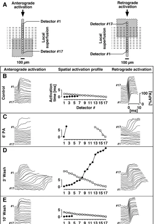
prop-Fig. 8. Induction of successful bi-directional conduction across a tissue expansion by partial gap junctional uncoupling. (A) Schematic drawing of the experimental layout. A tissue expansion was locally superfused with the gap junctional uncoupler palmitoleic acid and impulse propagation characteristics across the expansion were assessed optically by detectors (black rings) along the central axis during anterograde (left) and retrograde (right) conduction. (B) Action potential upstrokes recorded under control conditions indicate decremental conduction during anterograde and normal conduction during retrograde stimulation, i.e., unidirectional conduction block. (C) Complete gap junctional uncoupling results in bi-directional conduction blocks at the boundaries of the superfusion. (D) During gradual re-coupling, successful bi-directional conduction is established. (E) At the end of the washout period, normal gap junctional conductance is reestablished as indicated by fast retrograde conduction and anterograde conduction block. Redrawn with modifications fromRef. [29].
agation across such expansions suggested that, contrary to intuition, partial gap junctional uncoupling might improve conduction and remove unidirectional conduction blocks
[27,28]. This prediction has been investigated with patterned growth cell cultures as illustrated in Fig. 8 [29]. The preparations consisted of narrow strands of cardiomyocytes, which merged with a large rectangular cell sheet(Fig. 8A). The widths of the strands were chosen such that unidirec-tional conduction block occurred under control conditions
(Fig. 8B). Initially, complete gap junctional uncoupling of the strand and the expansion was induced by local super-fusion with palmitoleic acid(Fig. 8C). Thereafter, washout was started which resulted in a gradual increase of gap junctional coupling during which successful bi-directional conduction could be observed within a limited time window
(Fig. 8D). After reaching normal levels of gap junctional coupling, unidirectional conduction blocks were reestab-lished (Fig. 8E). This illustrates that partial gap junctional uncoupling has the potential to remove unidirectional con-duction blocks. This counter-intuitive behavior can be explained by differences in the dimensionality of the effect of gap junctional uncoupling on the source (strand) and the sink (expansion). Partial uncoupling of the essentially linear source will reduce its size in a linear proportional manner whereas equal uncoupling of the two-dimensional sink will reduce the size thereof following a square function. This overproportional reduction of the sink improves the source-to-sink mismatch up to the point of successful anterograde conduction. As can be expected for a gradual change in the balance between the source and the sink, overall conduction velocity in anterograde direction across a tissue expansion changes in a biphasic manner during progressive re-cou-pling. This is shown inFig. 9where the gradual increase in gap junctional conductance is initially accompanied by an increase and then by a paradoxical decrease in overall anterograde conduction velocity. How can this be explai-ned? At the beginning of the establishment of successful anterograde conduction, conduction velocities are initially in the range of those observed in linear cell strands (cf. above). This suggests that conduction at these very low levels of gap junctional conductance is primarily determined by the de-gree of cell-to-cell coupling and not by the source-to-sink mismatch represented by the expansion. Accordingly, increases of gap junctional conductance led initially to an increase in conduction velocity. At the same time, however, the parallel increase in the size of the sink became more important and, ultimately, resulted in the paradoxical situa-tion that overall conducsitua-tion velocity decreased even though gap junctional conductance increased. Therefore, in contrast to the situation in uniform cell structures where a decrease in gap junctional coupling is invariably accompanied by a decrease of conduction velocity, overall conduction velocity across sites where planar wavefronts change to curved wavefronts (expansion, isthmus), initially show an increase which is followed by a decrease only during severe uncoupling.
Thus, whereas gap junctional uncoupling in general leads to an impairment of conduction and thereby contributes significantly to the generation of arrhythmias, there is another side to this coin for the case of discontinuous tissue architectures. In such structures, which are likely to be present in the border zone of healed infarcts [30] or in fibrotic tissue of the aged and/or hypertrophied myocardium
[31], partial gap junctional uncoupling might actually re-move unidirectional conduction blocks and therefore elimi-nate one of the key ingredients of reentrant arrhythmias. Accordingly, while improvement of gap junctional coupling in structurally diseased heart might be expected to act in an anti-arrhythmogenic manner by reducing the incidence of slowly conducting pathways, it might at the same time provoke arrhythmias by unmasking potential regions of unidirectional conduction blocks.
6. Gap junctional gating: is there an effect on impulse conduction?
One of the prominent biophysical features of gap junctions is their time- and voltage-dependent inactivation. Even though the time constants for inactivation (f150 ms at a transjunctional voltage gradient of 100 mV) [32]
are extremely slow compared to transjunctional activation delays encountered during normal action potential propa-gation (f30 As for multicellular strands, cf. above), recent computer simulations suggested that, under
con-Fig. 9. Development of macroscopic conduction velocity across a tissue expansion during a gradual increase of gap junctional conductance (same experimental protocol as inFig. 8). ‘xb’ indicates presence of bi-directional
conduction block, ‘xu’ presence of unidirectional conduction block in
anterograde direction. Note decrease of conduction velocity after 6V of washout despite further increase of gap junctional conductance.
ditions of severe uncoupling, junctional conduction times slightly increase when using a dynamic model of gap junctions and that conduction blocks occur at lower levels of resting transjunctional conductance than those found using a static model [33]. In another recent study using transfected neuroblastoma cells, inactivation kinetics of connexin43 were studied by imposing an action potential clamp instead of a rectangular voltage pulses on one of the cells [34]. These experiments showed that, following the peak of the action potential, junctional conductance decreased within 25 ms to 58% of control. Although these kinetics are faster than those reported earlier, the compar-ison of these inactivation times to transjunctional conduc-tion times observed during steady state propagaconduc-tion under conditions of severe uncoupling (<5 ms)[18]suggests that gap junctional gating has only a small effect on overall conduction velocities. In addition to the effects of gap junctional gating on depolarizing current flow in the ortho-dromic direction, it would be interesting to know whether gating possibly affects the trailing part of activation, i.e., the repolarization. While it is tempting to speculate that partial inactivation might last into the repolarization phase, thereby channeling depolarizing current from the activation wave-front in the orthodromic direction, this mechanism is unlike-ly to exist because gap junctional conductance will rapidunlike-ly recover during repolarization due to the reversal of polarity
[35]and the decrease in size of the transjunctional voltage
[34].
Thus, whereas gating possibly influences the degree of ultra-slow conduction to some extent during severe uncou-pling, the role thereof during normal propagation is likely to be insignificant. However, because gating is dependent on a variety of factors such as the expression system used, trans-junctional voltages present, and the type of gap junctions present (isoforms, connexon composition (homomeric, het-eromeric) and connexon coupling (homotypic, heterotypic)) definitive answers to this question will require further studies, which take into account transjunctional voltage differences as they occur across a given junction during propagated activity.
7. Gap junctional coupling between cardiomyocytes and non-excitable cells
In the heart, approximately half of the cells consist of non-myocardial cells, among which fibroblasts constitute the largest fraction [36]. This number can be expected to increase as a result of cardiac diseases leading to fibrosis. Whereas the formation of excessive collagen sheets, which act as electrical insulators, has been recognized for a long time as being a cause for discontinuous conduction and the occurrence of arrhythmias [37], the question of whether the cellular constituents of fibrotic tissue, i.e., the fibro-blasts, might affect impulse propagation directly by form-ing gap junctions with cardiomyocytes has been addressed
only recently. It has been known for several decades that individual fibroblasts of cardiac origin can establish gap junctional communication with adjacent cardiomyocytes in culture [38,39]. In this context, it was shown that impulse propagation in monolayer cardiomyocyte cultures can be modified by grafting a layer of fibroblasts transfected with the voltage gated potassium channel Kv1.3 over them[40]. This co-culture resulted in conduction blocks in the car-diomyocyte monolayer, which were reversed upon appli-cation of specific blockers of the potassium channel. Moreover, it was shown in cell culture that fibroblasts adjacent to cardiomyocytes induce a decrease in maximal upstroke velocity [41] or a local slowing of conduction
[42]. Whereas these findings illustrate that fibroblasts in intimate contact with cardiomyocytes can influence the electrophysiological behavior of the latter via gap junc-tions, the question arises whether such interactions might also occur over longer distances, i.e., whether fibroblasts are capable of relaying electrical activation between dis-parate sheets of cardiomyocytes. This issue was recently investigated with a cell culture model where patterned growth strands of cardiomyocytes were interrupted over defined distances by fibroblasts of cardiac origin [43]. The results of one of these experiments are shown in Fig. 10. In this particular preparation, the fibroblast insert had a length of 134 Am and propagating action potentials were elicited at 2 Hz on the left. As indicated by the optically recorded transmembrane voltage signals during propagated activity in Fig. 10C, action potential upstrokes were monotonically rising in the region of the cardiomyocytes, whereas they showed ‘double-humps’ in the region of the fibroblasts due to bidirectional electrotonic interaction with the proximal and the delayed activated distal cardiomyo-cyte strand. As shown by immunocytochemistry, this electrotonic interaction was based on the presence of both connexin43 and connexin45. As indicated by the plot of activation times along the preparation (Fig. 10D), the fibroblast insert induced a local propagation delay of 30 ms. This delay became as long as 68 ms for the longest inserts supporting impulse transmission (f300 Am) result-ing in apparent local ‘conduction velocities’ as low as 2.2 mm/s. Whereas it is tempting to speculate that such extremely slow conduction might be instrumental in the generation of arrhythmias in fibrotic hearts, studies with cell cultures have to be interpreted cautiously in regard to extrapolation of the results to intact tissue because there is as yet no firm proof of gap junctional coupling between cardiomyocytes and fibroblasts in-vivo. In contrast, a thorough investigation of this question found no evidence for robust gap junctional coupling in healthy intact tissue
[44]. This raises the question whether fibroblasts in culture might undergo a phenotype switch to so-called myofibro-blasts, which enables them to form gap junctions with cardiomyocytes. The conversion from fibroblasts into myofibroblasts, which are characterized by the expression of a-smooth muscle actin [45], has been described for
different types of fibroblasts in culture. In intact hearts, it was shown that fibroblasts convert into myofibroblast after a local loss of cardiomyocytes[46]. Most interestingly, this conversion has been described to be accompanied by the expression of connexin43 in the case of breast cancer stroma cells[47] and myofibroblasts derived from corneal fibroblasts [48]. If such a conversion of fibroblasts into connexin expressing myofibroblasts should also occur in the heart under pathophysiological conditions such as myocardial infarction[36], this would raise the interesting hypothesis that the ensuing coupling of non-excitable cells to cardiomyocytes might lead to very slow conduction and, thus, might constitute a possibly important new arrhyth-mogenic mechanism.
In conclusion, the observation that cardiomyocytes read-ily form functional gap junctions with heterogeneous cells and that this coupling supports the spread of excitation over extended distances may have implications beyond electrical interactions with fibroblasts as presented above. In partic-ular, given the recent interest in using stem cells as a therapeutic approach for the diseased heart, the promiscu-ous gap junctional coupling is a prerequisite both for permitting orderly excitation sequences in the regions of grafted cells and for the intercellular exchange of signaling molecules.
8. Gap junctions, the ‘‘truth and nothing but the truth’’ for impulse propagation?
Whereas all of the above evidence stresses the importance of gap junctional coupling for impulse propagation under both physiologic and pathophysiologic conditions, the ob-servation that the main ion channels underlying fast conduc-tion, i.e., the sodium channels, are clustered at intercalated discs[49 – 53]is intriguing. If one were to design a cardio-myocyte, one would probably not plan to insert sodium channels at the intercalated disc because, among other reasons, they would face a highly restricted extracellular space which could be expected to be subject to large fluctuations in ion concentration and, consequently, in ad-verse changes in electrochemical driving forces. On the other hand and as formulated many years ago by Sperelakis and Mann[54], the fact that space is restricted at the intercalated disc could also act in favor of impulse propagation. These authors postulated an electric field mechanism of impulse transfer, which is based on the idea that activation of sodium channels located at the intercalated disc results in a negative shift of the cleft potential between a given activated cardi-omyocyte and a neighboring quiescent cardicardi-omyocyte. This negative shift of the cleft potential reduces the transmem-brane potential ‘seen’ by the sodium channels of the
post-Fig. 10. Impulse transmission along cardiac fibroblasts. (A) Phase contrast picture of the preparation consisting of an 80-Am wide strand of cardiomyocytes interrupted over a distance of 134 Am by cardiac fibroblasts. (B) Staining with anti-myomesin antibodies shows the boundaries of the cardiomyocyte strands. (C) Optically recorded action potential upstrokes along the preparation show monotonically rising signals in the region of cardiomyocytes (black) and biphasic upstrokes in the region of fibroblasts (gray). Signals from the distal cardiomyocyte strand show pronounced subthreshold depolarization. (D) Activation times along the preparation indicate a fibroblast-induced conduction delay of f30 ms. Redrawn with modifications fromRef. [43].
junctional membrane and, hence, brings this region to threshold with subsequent rapid activation of the entire cell
[55]. Sperelakis and McConnell[51]further suggested that this mechanism might be supported by rapid potassium accumulation in the cleft during activation of the pre-junc-tional cell which would induce a depolarization of the post-junctional membrane cell to threshold. Obviously, this elec-tric field mechanism is critically dependent on the radial shunt resistance of the intercellular cleft which has (i) to assume a value high enough to permit the build-up of a local extracellular negativity in the cleft region and (ii) which, at same time, must permit establishment of a local circuit current large enough as to depolarize the quiescent cell. Under the assumptions of appropriate parameters, Sperelakis and McConnell showed in computer simulations that their model consisting of linear chains of single cardiomyocytes permits successful impulse propagation with conduction velocities up to ~ 40 cm/s (for review, cf.Ref. [51]).
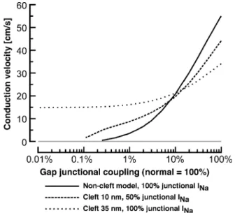
Recently, the interplay between the effects of sodium channel clustering, cleft potentials and gap junctional cou-pling on impulse propagation have been further investigated by using a linear strand of cardiomyocytes represented by the Luo – Rudy model[52]. The effects of different combinations of cleft widths and degrees of sodium channel partitioning on conduction velocities achieved during progressive uncou-pling are shown inFig. 11. Under the assumption of a cleft width of 35 nm and sodium channels being present exclu-sively at the intercalated disc, these simulations showed that conduction velocities were substantially slower than those obtained with a non-cleft model during normal gap
junc-tional coupling. This was explained by the occurrence of large negative cleft-potentials during activation, which in-duced a rapid and largely overshooting response of pre- and post-junctional membranes and, therefore, resulted in an attenuation of the sodium inward current and a concomitant slowing of conduction. With decreasing gap junctional coupling, the difference in conduction velocities between standard model and cleft-model became smaller until, at coupling levels <20%, the cleft-model performed increas-ingly better and supported conduction velocities of 15 cm/s even during a 10,000-fold reduction of gap junctional coupling. Whereas this finding of conduction in the virtual absence of gap junctional coupling points in the same direction as earlier findings by Sperelakis and colleagues,
Fig. 11 also illustrates that only moderate changes in the parameters (cleft width 35!10 nm, sodium channel parti-tioning 100%!50%) resulted in a behavior which was only slightly different from the standard model. Whereas these studies show the possibility of conduction in the absence of gap junctional coupling, it is not clear whether these findings are relevant for intact tissue. Whereas clustering of sodium channels at the intercalated discs is undisputed for both intact cardiac tissue and cell cultures, there are no experimental data available regarding actual radial cleft resistances in cardiac tissue. Because a direct determination of the radial cleft resistance is out of reach of present experimental methods, an exact morphometric assessment of the three-dimensional architecture of the cleft between two adjoining cardiomyocytes might indirectly permit one to obtain an approximate estimate thereof. However, even if such studies should reinforce the computer simulation studies, there remains a number of open questions which are difficult to reconcile with the concept that electric field mechanisms alone are significantly involved in cardiac impulse conduc-tion: (i) in the absence of substantial gap junctional coupling, space constants should be much shorter than what was generally reported in the past (for a critical discussion of this issue, cf. Ref. [51]); (ii) the electric field model cannot readily explain calcium inward current based conduction which occurs even though these ion channels are not clustered at the intercalated disc; (iii) it is difficult to understand how the experimentally firmly established effects of source-to-sink mismatches on impulse propagation could be explained by an electric field model in the absence of gap junctional coupling.
In conclusion, at least for now, it seems safe to conclude that gap junctions are ‘‘the truth and nothing but the truth’’ for impulse propagation in cardiac tissue. Nevertheless, the clustering of sodium channels at the intercalated disc together with the presence of Na – K-ATPases at the same location [56] remains a highly intriguing and interesting fact. It indicates that this part of the sarcolemma, even though facing a highly restricted extracellular space, might exhibit a specific function. However, answers as to the exact nature and relevance of this function will have to await further studies.
Fig. 11. Dependence of conduction velocity on gap junctional coupling for 3 different models of cleft configuration and partitioning of sodium channels. Solid line: model with no cleft-effects but clustering of all sodium channels at intercalated disc. Dashed line: model with 10-nm wide cleft and even partitioning of the sodium channels between the intercalated disc and the surface sarcolemma. Punctate line: model with a 35-nm wide cleft and clustering of all sodium channels at intercalated disc. Redrawn with modifications fromRef. [52].
9. Conclusions
Gap junctional coupling plays a crucial role for impulse propagation in cardiac tissue. It is well established that a reduction of gap junctional conductance as occurring, e.g., during ischemia is importantly contributing to arrhythmo-genesis because of the resulting reduction of conduction velocities, which is more than an order of magnitude larger than that observed during a reduction of excitability. At the same time, however, partial gap junctional uncoupling has the capacity to remove unidirectional conduction blocks and therefore acts, at least over a certain range of partial uncou-pling, in an anti-arrhythmogenic fashion. This suggests that electrical uncoupling is not inevitably followed by arrhyth-mias. Rather, it ultimately depends on the balance between pro- and antiarrhythmic effects of uncoupling whether a certain reduction of axial current flow in the setting of non-uniform tissue structures can give rise to reentrant excitation. A second possibly pro-arrhythmic mechanism caused by gap junctions in the diseased heart might be related to heteroge-neous cell coupling between cardiomyocytes and fibroblasts. Whereas it has been shown in cell culture that fibroblasts are able to relay electrical activation with substantial delays over appreciable distances, it remains to be shown whether gap junctional coupling between heterogeneous cell populations is indeed present in the diseased heart. Finally, since the observation of a clustering of sodium channels at the inter-calated disc, the hypothesis that there might exist gap-junction independent mechanisms of impulse propagation in cardiac tissue has received renewed attention. Whereas computer simulations show the feasibility of such concepts, their validation in intact cardiac tissue will depend on the development of adequate experimental approaches.
Acknowledgements
I would like to thank Giedrius Gaudesius and Michele Miragoli for helpful discussions regarding the manuscript. This work was supported by the Swiss National Science Foundation (grant # 3100-64914) and by the Swiss University Conference.
Note added in proof
With the reference to in vitro findings of electrotonic coupling between heterogeneous cells in the heart (Section 7), it has been shown very recently that gap junctional coupling exists between fibroblasts and cardiomyocytes both in the sinoatrial node[57]and in the myocardium[58].
References
[1] Weidmann S. The electrical constants of Purkinje fibres. J Physiol (Lond) 1952;118:348 – 60.
[2] Sjo¨strand FS, Andersson E. Electron microscopy of the intercalated disc of cardiac muscle tissue. Experientia 1954;10.
[3] Weidmann S. The diffusion of radiopotassium across intercalated disks of mammalian cardiac muscle. J Physiol (Lond) 1966;187:323 – 42. [4] Weidmann S. Electrical constants of trabecular muscle from
mamma-lian heart. J Physiol (Lond) 1970;210:1041 – 54.
[5] Mendez C, Mueller WJ, Urguiaga X. Propagation of impulses across the Purkinje fiber-muscle junctions in the dog heart. Circ Res 1970; 36:135 – 50.
[6] Overholt ED, Joyner RW, Veenstra RD, Rawling D, Wiedmann R. Unidirectional block between Purkinje and ventricular layers of pap-illary muscle. Am J Physiol 1984;247:H584 – 95.
[7] Joyner RW. Effects of the discrete pattern of electrical coupling on propagation through an electrical syncytium. Circ Res 1982;50: 192 – 200.
[8] Saffitz JE, Kanter HL, Green KG, Tolley TK, Beyer EC. Tissue-specific determinants of anisotropic conduction velocity in canine atrial and ventricular myocardium. Circ Res 1994;74:1065 – 70. [9] Spach MS, Heidlage JF, Dolber PC, Barr RC. Electrophysiological
effects of remodeling cardiac gap junctions and cell size: experimen-tal and model studies of normal cardiac growth. Circ Res 2000;86: 302 – 11.
[10] Kle´ber AG, Janse MJ, Fast VG. Normal and abnormal conduction in the heart. Handbook of physiology. The Heart, vol. 1. New York (NY): Oxford Univ. Press; 2001. p. 455 – 530.
[11] Rudy Y, Quan W. Propagation delays across cardiac gap junctions and their reflection in extracellular potentials: a simulation study. J Car-diovasc Electrophysiol 1991;2:299 – 315.
[12] Shaw RM, Rudy Y. Ionic mechanisms of propagation in cardiac tissue: roles of the sodium and L-type calcium currents during re-duced excitability and decreased gap-junction coupling. Circ Res 1997;81:727 – 741.
[13] Rohr S, Scho¨lly DM, Kleber AG. Patterned growth of neonatal rat heart cells in culture. Morphological and electrophysiological charac-terization. Circ Res 1991;68:114 – 30.
[14] Rohr S, Flu¨ckiger-Labrada R, Kucera JP. Photolithographically de-fined deposition of attachment factors as a versatile method for pat-terning the growth of different cell types in culture. Pflugers Arch-Eur J Physiol 2003;446:125 – 32.
[15] Rohr S, Kucera JP. Optical recording system based on a fiber optic image conduit: assessment of microscopic activation patterns in car-diac tissue. J Biophys 1998;75:1062 – 75.
[16] Rohr S, Salzberg BM. Discontinuities in action potential propagation along chains of single ventricular myocytes in culture: multiple site optical recording of transmembrane voltage (MSORTV) suggests propagation delays at the junctional sites between cells. Biol Bull Mar Biol Lab 1992;183:342 – 3.
[17] Fast VG, Kle´ber AG. Microscopic conduction in cultured strands of neonatal rat heart cells measured with voltage-sensitive dyes. Circ Res 1993;73:914 – 25.
[18] Rohr S, Kucera JP, Kle´ber AG. Slow conduction in cardiac tissue: I. Effects of a reduction of excitability versus a reduction of electrical coupling on microconduction. Circ Res 1998;83:781 – 94.
[19] De Bakker JMT, Van Capelle FJL, Janse MJ, et al. Slow conduction in the infarcted human heart—zigzag course of activation. Circle 1993; 88:915 – 26.
[20] Spach MS, Dolber PC, Heidlage JF. Influence of the passive aniso-tropic properties on directional differences in propagation following modification of the sodium conductance in human atrial muscle. A model of reentry based on anisotropic discontinuous propagation. Circ Res 1988;62:811 – 32.
[21] Kle´ber AG, Janse MJ, Wilms-Schopmann FJG, Wilde AAM, Coronel R. Changes in conduction velocity during acute ischemia in ventricular myocardium of the isolated porcine heart. Circle 1986;73:189 – 98. [22] Hisatome I, Arita M. Effects of catecholamines on the residual sodium
channel dependent slow conduction in guinea pig ventricular muscles under normoxia and hypoxia. Cardiovasc Res 1995;29:65 – 73.
[23] Kagiyama Y, Hill JL, Gettes LS. Interaction of acidosis and increased extracellular potassium on action potential characteristics and conduc-tion in guinea pig ventricular muscle. Circ Res 1982;51:614 – 23. [24] Shaw RM, Rudy Y. Electrophysiologic effects of acute myocardial
ischemia. A mechanistic investigation of action potential conduction and conduction failure. Circ Res 1997;80:124 – 38.
[25] Cole WC, Piccone JB, Sperelakis N. Gap junction uncoupling and discontinuous propagation in the heart. Biophys J 1988;53:809 – 18. [26] Vaidya D, Tamaddon HS, Lo CW, et al. Null mutation of connexin 43
causes slow propagation of ventricular activation in the late stages of mouse embryonic development. Circ Res 2001;88:1196 – 202. [27] Fast VG, Kle´ber AG. Block of impulse propagation at an abrupt tissue
expansion: evaluation of the critical strand diameter in 2- and 3-di-mensional computer models. Cardiovasc Res 1995;30:449 – 59. [28] Leon LJ, Roberge FA. Directional characteristics of action potential
propagation in cardiac muscle. A model study. Circ Res 1991;69: 378 – 95.
[29] Rohr S, Kucera JP, Fast VG, Kle´ber AG. Paradoxical improvement of impulse conduction in cardiac tissue by partial cellular uncoupling. Science 1997;275:841 – 4.
[30] Ursell PC, Gardner PI, Albala A, Fenoglio Jr JJ, Wit AL. Structural and electrophysiological changes in the epicardial border zone of canine myocardial infarcts during infarct healing. Circ Res 1985; 56:436 – 51.
[31] Spach MS, Dolber PC. Relating extracellular potentials and their derivatives to anisotropic propagation at a microscopic level in human cardiac muscle. Evidence for electrical uncoupling of side-to-side fiber connections with increasing age. Circ Res 1986;58: 356 – 71.
[32] Yao J, Hussain W, Patel P, Peters NS, Boyden PA, Wit AL. Remod-eling of gap junctional channel function in epicardial border zone of healing canine infarcts. Circ Res 2003;92:437 – 43.
[33] Henriquez AP, Vogel R, Muller-Borer BJ, Henriquez CS, Weingart R, Cascio WE. Influence of dynamic gap junction resistance on impulse propagation in ventricular myocardium: a computer simulation study. Biophys J 2001;81:2112 – 21.
[34] Xianming L, Crye M, Veenstra RD. Regulation of connexin43 gap junctional conductance by ventricular action potentials. Circ Res 2003;93:e63 – 73.
[35] Bukauskas FF, Elfgang C, Willecke K, Weingart R. Biophysical prop-erties of gap junction channels formed by mouse connexin40 in in-duced pairs of transfected human HeLa cells. Biophys J 1995;68: 2289 – 98.
[36] Manabe I, Takayuki S, Nagai R. Gene expression in fibroblasts and fibrosis. Circ Res 2002;91:1103 – 13.
[37] Spach MS, Boineau JP. Microfibrosis produces electrical load varia-tions due to loss of side-to-side cell connecvaria-tions: a major mechanism of structural heart disease arrhythmias. Pacing Clin Electrophysiol 1997; 20:397 – 413.
[38] Hyde A, Blondel B, Matter A, Cheneval JP, Filloux B, Girardier L. Homo- and heterocellular junctions in cell cultures: an electrophysio-logical and morphoelectrophysio-logical study. Prog Brain Res 1969;31:283 – 311. [39] Goshima K. Synchronized beating of and electrotonic transmission between myocardial cells mediated by heterotypic strain cells in monolayer culture. Exp Cell Res 1969;58:420 – 6.
[40] Feld Y, Melamed-Frank M, Kehat I, Tal D, Marom S, Gepstein L. Electrophysiological modulation of cardiomyocytic tissue by trans-fected fibroblasts expressing potassium channels: a novel strategy to manipulate excitability. Circle 2002;105:522 – 9.
[41] Rohr S, Salzberg BM. Multiple site optical recording of transmem-brane voltage in patterned growth heart cell cultures: assessing elec-trical behavior, with microsecond resolution, on a cellular and subcellular scale. Biophys J 1994;67:1301 – 15.
[42] Fast VG, Darrow BJ, Saffitz JE, Kle´ber AG. Anisotropic activa-tion spread in heart cell monolayers assessed by high-resoluactiva-tion optical mapping: role of tissue discontinuities. Circ Res 1996;79: 115 – 27.
[43] Gaudesius G, Miragoli M, Thomas SP, Rohr S. Coupling of cardiac electrical activity over extended distances by fibroblasts of cardiac origin. Circ Res 2003;93:421 – 8.
[44] De Maziere AMGL, Van Ginneken ACG, Wilders R, Jongsma HJ, Bouman LN. Spatial and functional relationship between myocytes and fibroblasts in the rabbit sinoatrial node. J Mol Cell Cardiol 1992; 24:567 – 78.
[45] Swynghedauw B. Molecular mechanisms of myocardial remodeling. Physiol Rev 1999;79:215 – 62.
[46] Weber K. Cardiac interstitium. In: Poole-Wilson P, Colucci W, Massie B, Chatterjee K, Coats A, editors. Heart failure. New York (NY): Churchill Livingstone; 1997. p. 13 – 31.
[47] Jamieson S, Going JJ, D’Arcy R, George WD. Expression of gap junction proteins connexin 26 and connexin 43 in normal human breast and in breast tumours. J Pathol 1998;184:37 – 43.
[48] Spanakis SG, Petridou S, Masur SK. Functional gap junctions in cor-neal fibroblasts and myofibroblasts. Invest Ophthalmol Vis Sci 1998; 39:1320 – 8.
[49] Cohen SA. Immunocytochemical localization of rH1 sodium channels in adult rat heart atria and ventricle. Presence in terminal intercalated discs. Circle 1994;94:3083 – 6.
[50] Rohr S, Flu¨ckiger R, Cohen SA. Immunocytochemical localization of sodium and calcium channels in cultured neonatal rat ventricular cardiomyocytes. Biophys J 1999;76:A366 [Tu-Pos515, abstract]. [51] Sperelakis N, McConnell K. An electric field mechanism for
trans-mission of excitation from cell to cell in cardiac muscles and smooth muscles. Res Adv Biomed Eng 2001;2:39 – 66.
[52] Kucera J, Rohr S, Rudy Y. Localization of sodium channels in intercalated disks modulates cardiac conduction. Circ Res 2002;91: 1176 – 82.
[53] Maier SKG, Westenbroek RE, Schenkmann KA, Feigl EO, Scheuer T, Catterall WA. An unexpected role for brain-type sodium channels in coupling of cell surface depolarization to contraction in the heart. Proc Natl Acad Sci U S A 2002;99:4073 – 8.
[54] Sperelakis N, Mann JE. Evaluation of electric field changes in the cleft between excitable cells. J Theor Biol 1977;64:71 – 96. [55] Sperelakis N. An electric field mechanism for transmission of
excita-tion between myocardial cells. Circ Res 2002;91:985 – 7 [Editorial]. [56] Petrecca K, Atanasiu R, Grinstein S, Orlowski J, Shrier A. Subcellular
localization of the Na+/H+ exchanger NHE1 in rat myocardium. Am J Physiol (Heart Circ Physiol) 1999;276:H709 – 17.
[57] Camelliti P, Green CR, LeGrice I, Kohl P. Fibroblast network in rabbit sinoatrial node. Structural and functional identification of ho-mogeneous and heterogeneous cell coupling. Circ Resdoi:10.1161/ 01.RES.0000122382.19400.14
[58] Goldsmith EC, Hoffman A, Morales MO, Potts JD, Price RL, McFad-den A, Borg TK. Organization of fibroblasts in the heart. Dev Dyn 2004 (in press).