Nephrol Dial Transplant (2014) 29: iv45–iv54 doi: 10.1093/ndt/gfu217
Full Review
Genetic diseases of renal phosphate handling
Carsten A. Wagner, Isabel Rubio-Aliaga, Jürg Biber and Nati Hernando
Institute of Physiology and Center for Integrative Human Physiology, University of Zurich, Zurich, Switzerland Correspondence and offprint requests to: Carsten A. Wagner; E-mail: Wagnerca@access.uzh.ch
A B S T R AC T
Renal control of systemic phosphate homeostasis is critical as evident from inborn and acquired diseases causing renal phos-phate wasting. At least three transport proteins are responsible for renal phosphate reabsorption: IIa (SLC34A1), NAPI-IIc (SLC34A3) and PIT-2 (SLC20A2). These transporters are highly regulated by various cellular mechanisms and factors in-cluding acid–base status, electrolyte balance and hormones such as dopamine, glucocorticoids, growth factors, vitamin D3, para-thyroid hormone and fibroblast growth factor 23 (FGF23). Whether renal phosphate wasting is caused by inactivating mu-tations in the NAPI-IIa transporter is controversial. Mumu-tations in the NAPI-IIc transporter cause hereditary hypophosphatemic rickets with hypercalciuria. Besides the primary inherited defects, there are also inherited defects in major regulators of phosphate homeostasis that lead to alterations in phosphate handling. Autosomal dominant hypophosphatemic rickets is due to FGF23 mutations leading to resistance against its own degradation. Similarly, inactivating mutations in the PHEX gene, which causes FGF23 inactivation, cause X-linked hypophospha-temia due to renal phosphate losses. In contrast, mutations in galactosamine:polypeptide N-acetyl-galactosaminyltransferase, responsible for O-glycosylation of FGF23, or in klotho, a cofac-tor for FGF23 signalling result in hyperphosphatemia. Acquired syndromes of renal phosphate wasting, hypophosphatemia and osteomalacia (tumour-associated osteomalacia) can be due to the excessive synthesis or release of phosphaturic factors (FGF23, FGF-7, MEPE and sFRP4) from mesenchymal tumours.
Keywords: bone, FGF23, kidney, phosphate, PTH
IN TRO DU CT IO N
About 85% of total phosphate is deposited in bone and∼10% in soft tissues, the remaining 2–3% is found in serum,
constituting a freely exchangeable and tightly regulated phos-phate pool. Serum phosphos-phate levels in adults are maintained in a narrow range of ∼0.8–1.45 mmol/L (2.4–4.5 mg/dL). Deviations as found in patients with hypophosphatemia or hyperphosphatemia can cause severe disturbances of cellular and organ function such as ATP depletion, rickets, osteomal-acia, anaemia or excessive tissue calcifications, nephrolithiasis, arteriosclerosis and increased risk of cardiovascular morbidity and mortality [1–3].
Phosphate homeostasis is the product of intestinal uptake of phosphate (in the range of 0.9–1 g/day) and reabsorption of phosphate from urine (in the range of 60–80%). Both intes-tinal uptake and renal reabsorption are mediated by sodium-dependent phosphate cotransporters (Na+/Pi-cotransporters) belonging to the SLC20 and SLC34 gene families of solute car-riers [4,5]. Their activity and expression is tightly regulated by a large variety of hormones and metabolic factors adjusting uptake and excretion to short and long-term body require-ments. Inborn errors are caused by mutations in phosphate transporters and in several genes that encode factors directly and indirectly involved in their regulation. The identification of these genes and elucidation of their functions have greatly contributed to our present understanding of how phosphate homeostasis is achieved. This review will give a brief overview over physiological function and regulation of renal phosphate transporters. For more extensive reviews, the reader is referred to a number of recent reviews and book chapters [5–9]. The second part of this review will discuss mutations in several genes that directly or indirectly participate in (renal) control of phosphate balance.
General aspects of renal phosphate handling
About 80% offiltered phosphate is reabsorbed from urine under normal dietary phosphate intake by the earlier convoluted parts of the proximal tubule of mostly juxtamedullary nephrons [4]. Phosphate absorption is mediated by sodium-dependent
cotransporters for inorganic phosphate (Pi) located in the brush border membrane.
Three members of the Type II Na+-dependent phosphate cotransporter family have been identified and classified in the SLC34 solute carrier gene family [4]. SLC34A1 (NAPI-IIa) is predominantly expressed in kidney mediating ∼70–80% of overall phosphate reabsorption as suggested by a Slc34a1 de fi-cient mouse model [10]. Another member of this family, SLC34A3 (NAPI-IIc), is also almost exclusively found in kidney [11]. SLC34A2 (NAPI-IIb) is mostly expressed in small intestine and in a number of other organs including testis, lung, liver and lactating mammary glands [12].
Also, sodium-dependent phosphate transporters from the SLC20 family are expressed in the kidney. PIT-2 (SLC20A2) is localized in the brush border membrane of the proximal tubule whereas the exact site of expression of PIT-1 in kidney is unclear [13]. PIT-2 is widely expressed in most tissues. Its exact role in renal phosphate reabsorption has not been clarified but simultaneous genetic deletion of NaPi-IIa and NaPi-IIc in mice reduced renal phosphate reabsorption by >90%, suggesting that its contribution to overall phosphate reabsorption may be minor [14]. PIT-2 is regulated by dietary phosphate intake, acid–base status and parathyroid hormone (PTH) [15,16]. Mutations in PIT-2 have been described in pa-tients with idiopathic basal ganglia calcification [17].
SLC34A1 (NaPi-IIa) and SLC34A3 (NaPi-IIc)
The human SLC34A1 gene lies on chromosome 5q35 [18], is ∼13 kb in length and consists of 13 exons and 12 introns [19]. Recently, a model for the protein structure of NAPI-IIa was suggested based on homology modelling and detailed structure–function studies [20].
The human SLC34A3 gene spans ∼5 kb containing 13 exons and lies on chromosome 9q34 [21,22]. Human NAPI-IIc contains 599 amino acids [21, 22]. NaPi-IIc expression in mouse is stimulated in response to low-dietary phosphate intake [23]. Also, dietary Pi-responsive elements ( potential TFE-3-binding sites) are located at−1846 and −1822 [23].
The transport functions of SLC34A transporters and their structure–function relationship have been extensively studied elucidating important features such as cotransport mode, elec-trogenicity or voltage and pH-dependency [4]. Collectively, these data demonstrate that NAPI-IIa transports three sodium ions together with one divalent phosphate ion (HPO42 ) per
transport cycle. This transport mode is electrogenic [24, 25]. The transporter homodimerizes but the functional unit is a monomer [26,27]. The Type IIc cotransporter transports only two sodium ions per phosphate and is electroneutral, indicat-ing that divalent phosphate is its preferred substrate [28].
Regulation of renal phosphate reabsorption
A variety of factors affects renal phosphate handling as listed in Table 1[4,29]. Alterations in the number of phos-phate transporter molecules in the brush border membrane re-present the main mechanism leading to changes in urinary phosphate excretion [29]. The cellular and molecular mechan-isms, by which NaPi-IIa and NaPi-IIc are regulated have been
extensively studied for PTH and changes in dietary phosphate intake (for review, [4,5]).
PTH rapidly induces phosphaturia within minutes by de-creasing apical phosphate transport activity. Activation of PLC by apical PTH receptors leads to protein kinase C dependent stimulation of ERK1/2 kinases and internalization of NaPi-IIa [29–31]. NaPi-IIa, apical PTH receptors and PLCβ1 are orga-nized in a macromolecular complex via the PDZ-scaffold protein NHE regulating factor 1 (NHERF1) [32–34]. In con-trast, basolateral PTH receptors are linked to adenylate cyclase, protein kinase A (PKA) and ERK1/2 [31]. NaPi-IIa is not phosphorylated but dissociates from NHERF1, which is phosphorylated in response to PTH [35,36]. A similar mech-anism has been proposed for fibroblast growth factor 23 (FGF23)- and dopamine-induced endocytosis of NaPi-IIa [4].
The regulation of renal phosphate reabsorption by FGF23 is mediated by several mechanisms: FGF23 reduces circulating levels of active 1,25(OH)2 vitamin D3by reducing expression of the activating enzyme CYP27B1 and by increasing expres-sion of the inactivating enzyme CYP24A1 [37]. Lower active vitamin D3 reduces the stimulation of renal and intestinal phosphate (re)absorption. Moreover, renal phosphate re-absorption is directly reduced by FGF23 through downregula-tion of NaPiIIa and NaPiIIc. Two alternative but not exclusive pathways have been proposed. FGF23 binds to FGF1c recep-tors in the distal convoluted tubule, the main site of klotho ex-pression in kidney leads to ERK1/2 phosphorylation and the distal convoluted tubule then signals to the proximal tubule to internalize NaPi-IIa and NaPi-IIc [38]. Alternatively, FGF23 may bind to FGF23 receptors in the proximal tubule and lead to phosphorylation of NHERF1 via Sgk1 [39]. Also, a direct regulation of NaPiIIa activity by klotho without FGF23 has been demonstrated where klotho causes cleavage and inactiva-tion of NaPi-IIa in the brush border membrane and subse-quent internalization [40].
An acute reduction in NaPi-IIa transporters is mediated by the internalization from the apical brush border membrane via the same route as receptor-mediated endocytosis, requir-ing megalin and involvrequir-ing clathrin-coated vesicles and early endosomes [41,42]. Consequently, NaPi-IIa is routed to lyso-somes for degradation [43, 44]. The regulation of NaPi-IIc has been studied in less detail: in response or high phosphate IIc downregulation occurs but is slower than for NaPi-IIa [16,45].
Table 1. Factors affecting phosphate reabsorption in the proximal tubule
Factors that decrease Pi reabsorption Factors that increase Pi
reabsorption Acidosis
High-dietary Pi intake Parathyroid hormone/cAMP Atrial natriuretic peptide/cGMP Glucocorticoids
Dopamine
Fibroblast growth factor 23 MEPE Frizzled-related protein 4 Alkalosis Low-dietary Pi intake Parathyroidectomy Thyroid hormone 1α,25(OH)2D3 Growth hormone(s) Insulin-like growth factor
For explanation and references see text.
FU
LL
RE
VI
Renal phosphate reabsorption closely matches dietary phosphate intake being high during dietary phosphate restric-tion and decreasing with high phosphate intake [11, 46–48]. In animals lacking PTH and/or 1,25(OH)2-vitamin D3, the adaptive response of NaPi-IIa and NaPi-IIc is partially pre-served, indicating that other factors may be responsible for sensing and mediating changes in dietary phosphate intake [49–51].
P R I M A RY I N H E RI T E D D E F E C T S I N R E N A L P H O S P H ATE H A N D L I N G
SLC34A1 (NAPI-IIa): hypophosphatemic
nephrolithiasis/osteoporosis-1 and Fanconi renotubular syndrome-2
Two reports addressed patients with mutations in the SLC34A1/NAPI-IIa gene. In thefirst report, two patients with urolithiasis or bone demineralization and persistent idiopathic hypophosphatemia with lower maximal renal phosphate re-absorption (TmP/GFR) were presented [52]. Single nucleotide changes resulting in missense mutations (A48F and V147M) were detected. These mutations were found only on one allele per patient suggesting a dominant effect [52]. Expression of the mutant NAPI-IIa transporters in oocytes has demonstrated reduced phosphate transport activity, a dominant negative effect on wild-type NAPI-IIa cotransporters, and reduced af-finity for phosphate [52]. However, these data were controver-sially discussed. SLC34A1 mutations were not confirmed in other kindreds with similar symptoms, relatives of patients had not been genetically investigated and the expression assay showed only low expression even for the wild-type transporter rendering a kinetic analysis very difficult. Moreover, expres-sion of the reported mutant NAPI-IIa cotransporters in Xenopus oocytes and the renal OK cell line did not show any evidence for altered subcellular localization, levels of expres-sion or reduced substrate affinity [53], but showed a lower transport activity. Yet, this decrease cannot fully explain the massive phosphaturia observed. Therefore, it was suggested that the nucleotide changes may represent only polymorph-isms. A similar conclusion was derived from screening a large number of pedigrees of calcium stone formers with low TmP/ GFR [54]. Even though a number of mutations including an N-terminal deletion of seven amino acids (91del7) in the NAPI-IIa gene were found, no correlation with the clinical phenotype could be established. Two of the mutations had lower phosphate transport activity after expression in Xenopus oocytes, most likely due to decreased membrane abundance. However, the reduction in transport rates was mild and im-portantly, only one allele was found to be mutated in stone forming patients. The interpretation that these mutations may represent rather relatively common polymorphisms was further supported by the fact that these gene alterations were also found in several subjects with normal renal phosphate ex-cretion. Thus, polymorphisms in the NAPI-IIa gene may be relatively common and their relevance for renal phosphate handling may require further clarification (Table2).
A second report described siblings from a consanguineous Arab Israeli family with hypophosphatemic rickets, multiple fractures and stunted growth [55]. The presence of a Fanconi-like syndrome was suggested, but hallmarks of a generalized proximal tubule dysfunction are not evident from data re-ported. The patients suffered from hypercalciuria due to ele-vated serum 1,25(OH)2D3 levels. Hypophosphatemia was refractory to vitamin D therapy. Genetic analysis detected a homozygous in-frame duplication of 21 bp leading to inser-tion of seven addiinser-tional amino acids at posiinser-tion G154-V160. Heterozygous carriers of the mutation were clinically normal suggesting that the mutation did not exert a dominant negative effect. Expression of the mutant protein in Xenopus oocytes abolished all phosphate-induced currents whereas expression in renal OK cells indicated failure of the mutant protein to reach the plasma membrane.
Thus, homozygous mutations in SLC34A1 may be respon-sible for renal phosphate wasting but whether this is only the case for this specific mutation and may be due to toxic effects of the mutant protein for proximal tubule functions or whether mutations in this phosphate transporter can provide evidence for the biological and clinical importance in human kidney function remains to be established (Table3).
SLC34A3 (NAPI-IIc): hereditary hypophosphatemic rickets with hypercalciuria
Hereditary hypophosphatemic rickets with hypercalciuria (HHRH) (OMIM #241530) is caused by mutations in the SLC34A3 gene encoding for NAPI-IIc [21, 22]. The disease wasfirst described in a Bedouin family [56] and is character-ized by renal phosphate wasting causing hypophosphatemia and secondary rickets, with elevated serum alkaline phosphat-ase. Serum levels of 1,25(OH2)-vitamin D3are high, PTH and FGF23 are low, resulting in enhanced intestinal calcium absorp-tion and subsequent renal excreabsorp-tion (hypercalciuria) promoting nephrolithiasis or nephrocalcinosis in some patients [21,22,56, 57]. Long-term phosphate supplementation appears to reverse the clinical and biochemical abnormalities in HHRH.
The SLC34A3 gene is localized on chromosome 9q34. Ob-served mutations in HHRH include frame shifts in the open reading frame with subsequent translation of sequences unre-lated to NaPiIIc and premature termination, missense muta-tions, intronic deletions and nucleotide changes in a putative splice site [21,22]. Inheritance follows an autosomal recessive pattern [21,22]. Some compound heterozygous patients carry-ing two different missense mutations were also identified [21]. However, carriers with only one allele mutated show normal phosphate balance and excretion but borderline or elevated renal calcium excretion [21,22].
Since NAPI-IIc mutations are associated with a severe renal phosphate leak, NAPI-IIc may have a more important role in human kidney function than in rodents. NAPI-IIc function in man remains important during adulthood in contrast to rodents where NaPi-IIc may play an important role only during growth [11]. Accordingly, two mouse models of Slc34a3 deficiency show only mild hypercalcuria without hy-perphosphaturia or even no symptoms in the case of a kidney-specific gene deletion [58,59].
FU
LL
RE
VI
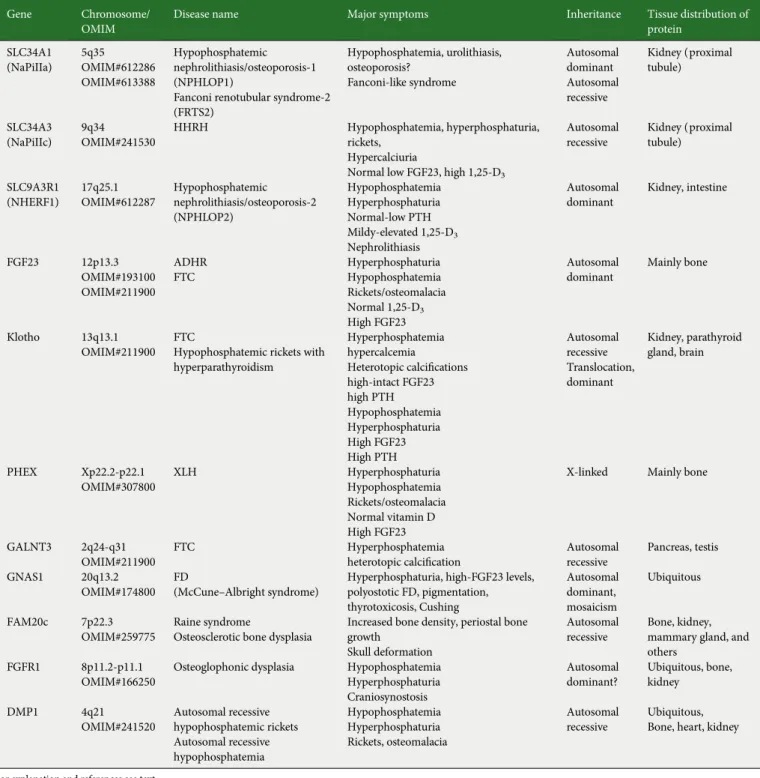
Table 2. Genes/diseases causing altered renal phosphate handling
Gene Chromosome/
OMIM
Disease name Major symptoms Inheritance Tissue distribution of
protein SLC34A1 (NaPiIIa) 5q35 OMIM#612286 OMIM#613388 Hypophosphatemic nephrolithiasis/osteoporosis-1 (NPHLOP1)
Fanconi renotubular syndrome-2 (FRTS2) Hypophosphatemia, urolithiasis, osteoporosis? Fanconi-like syndrome Autosomal dominant Autosomal recessive Kidney (proximal tubule) SLC34A3 (NaPiIIc) 9q34 OMIM#241530 HHRH Hypophosphatemia, hyperphosphaturia, rickets, Hypercalciuria
Normal low FGF23, high 1,25-D3
Autosomal recessive Kidney (proximal tubule) SLC9A3R1 (NHERF1) 17q25.1 OMIM#612287 Hypophosphatemic nephrolithiasis/osteoporosis-2 (NPHLOP2) Hypophosphatemia Hyperphosphaturia Normal-low PTH Mildy-elevated 1,25-D3 Nephrolithiasis Autosomal dominant Kidney, intestine FGF23 12p13.3 OMIM#193100 OMIM#211900 ADHR FTC Hyperphosphaturia Hypophosphatemia Rickets/osteomalacia Normal 1,25-D3 High FGF23 Autosomal dominant Mainly bone Klotho 13q13.1 OMIM#211900 FTC
Hypophosphatemic rickets with hyperparathyroidism Hyperphosphatemia hypercalcemia Heterotopic calcifications high-intact FGF23 high PTH Hypophosphatemia Hyperphosphaturia High FGF23 High PTH Autosomal recessive Translocation, dominant Kidney, parathyroid gland, brain PHEX Xp22.2-p22.1 OMIM#307800 XLH Hyperphosphaturia Hypophosphatemia Rickets/osteomalacia Normal vitamin D High FGF23
X-linked Mainly bone
GALNT3 2q24-q31 OMIM#211900
FTC Hyperphosphatemia
heterotopic calcification Autosomalrecessive
Pancreas, testis
GNAS1 20q13.2 OMIM#174800
FD
(McCune–Albright syndrome) Hyperphosphaturia, high-FGF23 levels,polyostotic FD, pigmentation, thyrotoxicosis, Cushing Autosomal dominant, mosaicism Ubiquitous FAM20c 7p22.3 OMIM#259775 Raine syndrome
Osteosclerotic bone dysplasia
Increased bone density, periostal bone growth
Skull deformation
Autosomal recessive
Bone, kidney, mammary gland, and others
FGFR1 8p11.2-p11.1 OMIM#166250
Osteoglophonic dysplasia Hypophosphatemia Hyperphosphaturia Craniosynostosis Autosomal dominant? Ubiquitous, bone, kidney DMP1 4q21 OMIM#241520 Autosomal recessive hypophosphatemic rickets Autosomal recessive hypophosphatemia Hypophosphatemia Hyperphosphaturia Rickets, osteomalacia Autosomal recessive Ubiquitous, Bone, heart, kidney
For explanation and references see text.
Table 3. Acquired forms of renal phosphate wasting
Gene/factor Chromosome/OMIM Disease Symptoms Occurrence Tissue distribution
sFRP4 7p14-p13
OMIM606570
TIO Hypophosphatemia Hyperphosphaturia Osteomalacia
Spontaneous? Tumour bone
FGF7 15q15-q21.1 OMIM148180 TIO Hypophosphatemia Hyperphosphaturia Osteomalacia Spontaneous? Tumour MEPE 4q21.1 OMIM605912 TIO Hypophosphatemia Hyperphosphaturia Osteomalacia
Spontaneous? Tumour, bone
FU
LL
RE
VI
SLC9A3R1 (NHERF1): Na+/H+exchanger regulating factor 1: hypophosphatemic nephrolithiasis/
osteoporosis-2
NHERF1 mutations were detected in eight patients with mildly reduced TmP/GFR, slightly lower serum phosphate levels, nephrolithiasis in four of eight patients, lower bone mineral den-sity in one patient, normal to low PTH and calcium values and 1,25(OH)2D3 just above the normal range in all patients. Four NHERF1 mutations (E68A, L110V, R153Q and E225K) were found. All patients were heterozygous [60,61]. The mechanisms by which mutant NHERF1 may cause phosphaturia may be site specific. In the case of the L110V, R153Q and E225K mutations, PTH- and cAMP-dependent inhibition of phosphate transport may be increased, whereas the E68A mutation may reduce the stability of the NaPiIIa transporter in the plasma membrane leading, in all cases, to lower transport activities [60,61].
D E F E C T S IN R E N A L P H O S P H ATE H A N D LI N G S E CON D A RY TO E X T R A R E N A L IN H E R I T E D DE FECTS
Fibroblast growth factor 23
FGF23 is primarily expressed in osteocytes [62–64]. Syn-thesis and release of FGF23 from bone are increased during hyperphosphatemia or high intake of phosphate [9, 65]. The major functions of FGF23 are the inhibition of renal phos-phate reabsorption by downregulation of IIa and NAPI-IIc expression and activity and by reducing the synthesis of active vitamin D3 through lowering expression of the renal CYP27B1 (1,25-alpha hydroxylase) and increasing CYP24A1 (1,24,25 hydroxylase). Lower concentrations of vitamin D3 de-crease intestinal phosphate absorption. In addition, FGF23 suppresses PTH secretion. The effects of FGF23 on phosphate homeostasis are mediated byfibroblast growth factor receptor 1c (FGFR1c) and FGFR4 receptors and require klotho as an obligatory co-ligand converting the low-affinity receptors into high-affinity FGF23 receptors [9,66].
Synthesis of FGF23 in osteocytes is regulated by a number of factors including PTH, 1,25 vitamin D3 and a cascade of pro-teins consisting of PHEX, 7B2/PC2, BMP1 and Dentin matrix protein 1 (DMP1). PTH and 1,25 vitamin D3transcriptionally stimulate FGF23 production by mechanisms involving PKA/ Wnt and the VDR receptor, respectively [67,68]. Activation of this cascade increases proteolytic processing of DMP1 into N-and C-terminal fragments, from which the latter is proposed to blunt the transcription of FGF23 (for review, see [69]).
FGF23 is degraded by C-terminal cleavage most likely by subtilisin-like proprotein convertases [70]. FGF23 possesses a recognition site for cleavage by subtilisin-like proprotein con-vertases consisting of an RXXR motif at position 176. A recent report suggested that the PC2 proprotein convertase, com-plexed with its chaperon 7B2, may mediate this process [71].
FGF23-activating mutations: autosomal dominant hypophosphatemic rickets
Autosomal dominant hypophosphatemic rickets (ADHR) (OMIM # 193100) is characterized by rickets, hypophosphatemia,
hyperphosphaturia, fatigue, bone pain and lower bone de-formities in face of inappropriately low or normal vitamin D3 levels. ADHR is caused by mutations destroying the RXXR cleavage motif mentioned above, causing excessive levels of active FGF23 [72]. Similarly, injection of mice with wild-type Fgf23 or Fgf23R179Q caused ADHR-like symptoms in mice. Mutant Fgf23 caused hypophosphatemia, hyperphosphaturia, reduced intestinal phosphate absorption and suppressed serum vitamin D3levels. In serum, high levels of mutant Fgf23 were detected, whereas wild-type Fgf23 was degraded [73]. Transgenic mice for Fgf23R176Q display typical ADHR symp-toms and show signs of secondary hyperparathyroidism [74]. Thus, ADHR is caused by mutations in a motif important for cleavage and degradation of FGF23 resulting in excessive FGF23 signalling.
Loss-of-function FGF23 mutations: familial tumoral calcinosis
Familial tumoral calcinosis (FTC) (OMIM # 211900) is the mirror image of phosphate wasting diseases such as ADHR, tumour-induced osteomalacia (TIO) and X-linked hypopho-sphatemia (XLH). FTC is characterized by hyperphosphate-mia, reduced renal phosphate excretion, ectopic calcifications and inappropriately normal or elevated 1,25(OH2) vitamin D3 levels. In a subset of FTC patients, a homozygous 211A-G transition in the FGF23 gene was identified, resulting in the substitution at an evolutionarily conserved serine to glycine (S71G) [75]. This mutation may cause loss of function [75]. Patients have abnormally low-FGF23 plasma concentrations. Mutant FGF23 protein expressed in vitro was not secreted and retained intracellularly [75].
Fibrous dysplasia (McCune–Albright syndrome)
Fibrous dysplasia (FD) (OMIM #174800) is caused by non-inherited genetic somatic activating missense mutations in the α-subunit of the stimulatory G protein, Gs[76,77]. FD is char-acterized by abnormalities in bone (monostotic or polyostotic FD), in skin ( pigmentation) and in the endocrine system (thyrotoxicosis, pituitary gigantism and Cushing syndrome). The severity of disease and particularly the association with skin and endocrine symptoms shows a wide variability. Renal phosphate wasting is detected in∼50% of patients [78]. FGF23 levels are elevated and caused by a large mass of FGF23-producing cells infibrous bone lesions. Abnormal high-FGF23 levels are caused by an increase in FGF23-producing cells but not by abnormal production of FGF23 per se [62,79].
Klotho: FTC and hypophosphatemic rickets with hyperparathyrodism
Mutations in the alpha-klotho (KL) gene can be another reason for FTC besides mutations in galactosamine:polypep-tide N-acetyl-galactosaminyltransferase (GALNT3) or loss-of-function mutations in FGF23. A single patient with a missense mutation was identified [80]. The mutation is predicted to affect the glucosidase domain of klotho, which is involved in the regulation of TRPV5 calcium channels [81]. Moreover, reduced expression and glycosylation of klotho was demon-strated, impairing the ability to interact with FGF23 and the
FU
LL
RE
VI
FGF1R receptor. Accordingly, the patient had hyperphospha-temia, increased vitamin D3and PTH levels and ectopic calci-fications of vessels.
The mirror image was described in another single case of a balanced translocation between chromosomes 13 and 9 at a position on chromosome 13 adjacent to the alpha-klotho gene. The translocation massively increased circulating klotho levels leading to hypophosphatemia, hyperphosphaturia, hy-percalcaemia, elevated PTH and FGF23 and low vitamin D3 [82]. The elevated FGF23 levels may be explained by a feed-back loop, by which circulating alpha klotho stimulates bone FGF23 expression [83].
DMP1: autosomal recessive hypophosphatemic rickets
Patients with mutations in DMP1 suffer from hypopho-sphatemia due to hyperphosphaturia with inappropriately normal levels of 1,25(OH)2 vitamin D3 [84, 85]. DMP1 belongs to the large SIBLING family (small integrin-binding ligand, N-linked glycoproteins) of extracellular matrix proteins and is mainly co-expressed with FGF23 in bone. The similar phenotype of patients with XLH and autosomal recessive hy-pophosphatemia with rickets suggested a link to FGF23. Indeed, in patients with loss of intact DMP1, FGF23 levels in serum were normal or elevated [84,85]. DMP1 (in particular the C-terminal fragment) blocks the transcription of FGF23; therefore, its absence may result in the release of this negative control leading to higher FGF23 production. The hypopho-sphatemia in DMP1 deficient patients causes severe rickets in children or osteomalacia in adults [84,85], which is character-ized by abnormal amounts of osteoid indicating defective min-eralization and maturation of bone [85]. Similarly, a mouse model deficient for DMP1 shows hypophosphatemia, hyper-phosphaturia, rickets, retarded skeletal growth with abnormal mineralization, disturbed lacunocanicular organization and defective osteoblast to osteocyte differentiation [85].
Tumour-induced osteomalacia
TIO is a rare paraneoplastic syndrome mostly associated with mesenchymal tumours releasing (a) phosphaturic factor (s). Symptoms include renal phosphate wasting causing hy-pophosphatemia, osteomalacia and abnormal vitamin D metabolism [86]. Surgical removal of the tumour reverses symptoms. In contrast to syndromes of hyperparathyroidism or humoral hypercalcaemia of malignancy, the plasma con-centrations of calcium, PTH and PTH-related protein are in the normal range. Several proteins, such as FGF23, sFRP4, FGF-7 and matrix extracellular phospho-glycoprotein (MEPE), have been identified that are produced and secreted from tumours from patients with TIO. Some of these proteins have been shown to regulate phosphate transport in vitro and/ or in vivo.
Secreted frizzled-related protein-4 (sFRP-4) is highly upre-gulated in tumour tissue from patients with renal phosphate wasting [87] and inhibits phosphate transport in the renal OK cell line as well as in vivo [87,88]. However, mice lacking sfrp4 do not show any abnormalities of systemic phosphate balance [89]. Thus, the relevance of sFRP4 for phosphate homeostasis remains to be further clarified.
MEPE: oncogenic hypophosphatemia
MEPE, a glycosylated protein of about 60 kDa, was initially cloned from tumour tissue obtained from a patient with onco-genic hypophosphatemia [90]. Bone cells (osteoblasts, osteo-cytes and odontoblasts) are the major source of MEPE. MEPE-like DMP1 is another member of the SIBLING family of extracellular matrix proteins involved in bone regulation. Injection of MEPE into intact mice results in hypophosphate-mia, hyperphosphaturia and mild increases in circulating 1,25 (OH)2 vitamin D3 levels [91]. Moreover, implantation of MEPE-producing CHO cells into nude mice caused renal phosphate wasting, whereas MEPE deficient mice have higher bone density [92]. The interactions between MEPE and other hormones regulating phosphate homeostasis and handling require more investigation.
PHEX: XLH rickets
XLH rickets (OMIM #307800) is the most common herit-able form of rickets with a prevalence of∼1 in 20 000. The disease is characterized by hypophosphatemia due to excessive renal phosphate wasting leading to rickets, lower extremities deformity, short stature, bone and joint pain, enthesopathy and dental abscesses. Vitamin D3 levels are inappropriately normal or even low [93]. The disorder is inherited in a domin-ant manner. Positional cloning identified the PHEX gene (PHosphate regulating gene with homology to Endopeptidases on the X chromosome) in XLH patients [93]. Several mouse models of XLH are available: Hyp, Gy and Ska1 mice resem-bling XLH and which were later shown to have mutations in the mouse Phex homologue [94–96]. The X-linked Hyp mouse model demonstrates a defect in proximal tubular phosphate absorption, decreased expression of NaPi-IIa and NaPi-IIc [97–99]. Serum FGF23 levels are highly elevated in XLH pa-tients [100]. Crossing of hypophosphatemic Hyp mice with hyperphosphatemic Fgf23 deficient mice produced hyperpho-sphatemic mice that showed exactly the same phenotype as Fgf23 null mice, indicating that both mutations affect the same system and that FGF23 may act downstream of PHEX [101]. Observations in Hyp and FGF23 null mice indicate that PHEX and FGF23 may regulate each other’s expression levels and that loss of PHEX may lead to higher expression levels of FGF23 [63, 65]. Although originally proposed, PHEX seems not to mediate direct cleavage of FGF23 [63, 70]. Instead, PHEX may activate the PC2 proprotein convertase by promot-ing the transcription of its 7B2 chaperon [71]. Transfection of osteoblast with 7B2.PC2 promoted cleavage of FGF23, whereas depletion of 7B2 mRNA reduced FGF23 cleavage and increased its transcription. The activated FGF23 transcription seems to result from an impaired proteolytic processing of DMP1 (see above). In agreement with this model, the mRNA levels of 7B2 are reduced in bones from Hyp mice [71].
UDP-N-acetyl-α-DGALNT3: FTC
Homozygous loss-of-function mutations of the UDP-N-acetyl-α-DGALNT3, a glycosyltransferase involved in
mucin-type O-glycosylation, underlie also FTC (OMIM # 211900) [102,103]. However, patients carrying only one mutated allele
FU
LL
RE
VI
appear to also have mild symptoms, leading to the initial de-scription of FTC as an autosomal dominant disease [104]. Because inactivating mutations in FGF23 also cause FTC and FGF23 has some O-glycosylation sites within the subtilisin-recognition sites, it had been speculated that GALNT3 is crit-ical for FGF23 gylcosylation and full function. Accordingly, in vitro secretion of FGF23 from CHO cells deficient in O-glycosylation is impaired, and cotransfection of GALNT3 markedly increases O-glycosylation and secretion of FGF23. Thus, GALNT3 may play an important role in FGF23 secre-tion by mediating its O-glycosylasecre-tion [105]. This function may also explain how loss of function mutations in either FGF23 or GALNT3 can produce the same disease, FTC.
Fibroblast growth factor receptor 1: osteoglophonic dysplasia
Osteoglophonic dysplasia (OMIM #166250) is caused by mutations in the FGFR1 that result in a gain-of-function and activation of the receptor [106]. Patients suffer from craniosy-nostosis, prominent supraorbital ridge and depressed nasal bridge, as well as from rhizomelic dwarfism and nonossifying bone lesions. Several missense mutations in the FGFR1 gene were identified in four patients. Three patients were hypopho-sphatemic due to massive renal phosphate wasting [106]. One patient had inappropriately high-FGF23 levels, two patients had high 1,25(OH)2vitamin D3levels. As two related patients carried the Y372C FGFR1 mutation. The activity of the mutant receptor was highly increased suggesting that overactivity of the FGF1 receptor is responsible for this disease [106]. The FGFR1 receptor mediates the downregulation of NAPI-IIa and NAPI-IIc by FGF23, whereas the effects of FGF23 on vitamin D3metabolism may involve the FGFR4 receptor [66].
FAM20C: Raine syndrome (osteosclerotic bone dysplasia)
Raine syndrome is caused by mutations in the protein kinase FAM20C, which resides in the Golgi apparatus and is secreted [107, 108]. Patients develop generalized higher bone density with characteristic changes and enhancement of the ossification of the skull, cerebral calcification and in some cases hypoplastic lungs. The disease is often lethal in thefirst weeks after birth but patients with longer survival reaching into puberty have been described. The renal phenotype of pa-tients with Raine syndrome has not been reported in detail. Targets phosphorylated by FAM20C are casein, osteoproteger-in, DMP1 and MEPE, members of the SIBLING family. In Fam20c deficient mice, hypophosphatemic rickets was ob-served possibly due to dysregulation of FGF23 (which is very high) due to lack of DMP1 phosphorylation [109].
CON CL U SIO N A N D O UT LOO K
Renal and extrarenal control of systemic phosphate homeo-stasis requires a complicated network of regulatory factors, phosphate transporters in kidney, intestine and bone and intracellular protein–protein interactions. Rare human genetic disorders and mouse genetics have greatly contributed to our
current understanding of (renal) phosphate homeostasis. Im-portantly, population-based genome-wide association studies identified SLC34A1 and FGF23 as important determinants of plasma phosphate levels [110]. Moreover, the highly elevated FGF23 levels in patients with various types of chronic kidney disease and the potential link between FGF23 and cardiovas-cular disease in these patients underline even further the bio-logical importance of this homeostatic system for health and disease. Nevertheless, our understanding of key elements of this system is incomplete and we require deeper insights on how organs and cells sense phosphate and how changes in sys-temic phosphate levels can trigger disease. The identification of key players through genetics may also pave the way for ther-apies targeting disturbed phosphate balance in both rare in-herited disorders as well as in more common diseases such as chronic kidney disease.
AC K N OW L E D G E M E N T S
Work in the laboratories of the authors has been supported by the Swiss National Science Foundation (SNSF) as part of the National Center for Competence in Research NCCR Kidney. CH, the European Union (FP7 frame work project EUReNomics) and the Zurich Center for Integrative Human Physiology (ZIHP).
CON FL I C T O F I N T E R E S T S TATE M E N T
The results presented in this paper have not been published previously in whole or part.
R E F E R E N C E S
1. Knochel JP. Clinical and physiologic phosphate disturbances. In: Seldin DW, Giebisch GH (eds). The Kidney. 3rd edn. Philadelphia: Lippincott Williams & Wilkins, 2000, pp. 1905–1934
2. Tonelli M. Serum phosphorus in people with chronic kidney disease: you are what you eat. Kidney Int 2013; 84: 871–873
3. Razzaque MS. Phosphate toxicity and vascular mineralization. Contrib Nephrol 2013; 180: 74–85
4. Wagner CA, Hernando N, Forster IC et al. The SLC34 family of sodium-dependent phosphate transporters. Pflugers Arch 2014; 466: 139–153 5. Biber J, Hernando N, Forster I. Phosphate transporters and their
func-tion. Annu Rev Physiol 2013; 75: 535–550
6. Forster IC, Hernando N, Biber J et al. Phosphate transporters of the SLC20 and SLC34 families. Mol Aspects Med 2013; 34: 386–395 7. Miyamoto K, Haito-Sugino S, Kuwahara S et al. Sodium-dependent
phosphate cotransporters: lessons from gene knockout and mutation studies. J Pharm Sci 2011; 100: 3719–3730
8. Marks J, Debnam ES, Unwin RJ. Phosphate homeostasis and the renal-gastrointestinal axis. Am J Physiol Renal Physiol 2010; 299: F285–F296 9. Bergwitz C, Juppner H. Regulation of phosphate homeostasis by PTH,
vitamin D, and FGF23. Annu Rev Med 2010; 61: 91–104
10. Beck L, Karaplis AC, Amizuka N et al. Targeted inactivation of Npt2 in mice leads to severe renal phosphate wasting, hypercalciuria, and skeletal abnormalities. Proc Natl Acad Sci USA 1998; 95: 5372–5377
11. Segawa H, Kaneko I, Takahashi A et al. Growth-related renal type II Na/Pi cotransporter. J Biol Chem 2002; 277: 19665–19672
FU
LL
RE
VI
12. Hilfiker H, Hattenhauer O, Traebert M et al. Characterization of a murine type II sodium-phosphate cotransporter expressed in mamma-lian small intestine. Proc Natl Acad Sci USA 1998; 95: 14564–14569 13. Villa-Bellosta R, Ravera S, Sorribas V et al. The Na+-Pi cotransporter
PiT-2 (SLC20A2) is expressed in the apical membrane of rat renal prox-imal tubules and regulated by dietary Pi. Am J Physiol Renal Physiol 2009; 296: F691–F699
14. Segawa H, Onitsuka A, Furutani J et al. Npt2a and Npt2c in mice play distinct and synergistic roles in inorganic phosphate metabolism and skeletal development. Am J Physiol Renal Physiol 2009; 297: F671–F678
15. Nowik M, Picard N, Stange G et al. Renal phosphaturia during metabolic acidosis revisited: molecular mechanisms for decreased renal phosphate reabsorption. Pflugers Arch 2008; 457: 539–549
16. Picard N, Capuano P, Stange G et al. Acute parathyroid hormone differ-entially regulates renal brush border membrane phosphate cotranspor-ters. Pflugers Arch 2010; 460: 677–687
17. Wang C, Li Y, Shi L et al. Mutations in SLC20A2 link familial idiopathic basal ganglia calcification with phosphate homeostasis. Nat Genet 2012; 44: 254–256
18. Kos CH, Tihy F, Econs MJ et al. Localization of a renal sodium-phosphate cotransporter gene to human chromosome 5q35. Genomics 1994; 19: 176–177
19. Hartmann CM, Hewson AS, Kos CH et al. Structure of murine and human renal type II Na+-phosphate cotransporter genes (Npt2 and NPT2). Proc Natl Acad Sci USA 1996; 93: 7409–7414
20. Fenollar-Ferrer C, Patti M, Knopfel T et al. Structural fold and binding sites of the human Na(+)-phosphate cotransporter NaPi-II. Biophys J 2014; 106: 1268–1279
21. Bergwitz C, Roslin NM, Tieder M et al. SLC34A3 mutations in patients with hereditary hypophosphatemic rickets with hypercalciuria predict a key role for the sodium-phosphate cotransporter NaP(i)-IIc in maintain-ing phosphate homeostasis. Am J Hum Genet 2006; 78: 179–192 22. Lorenz-Depiereux B, Benet-Pages A, Eckstein G et al. Hereditary
hypo-phosphatemic rickets with hypercalciuria is caused by mutations in the sodium-phosphate cotransporter gene SLC34A3. Am J Hum Genet 2006; 78: 193–201
23. Ohkido I, Segawa H, Yanagida R et al. Cloning, gene structure and dietary regulation of the type-IIc Na/Pi cotransporter in the mouse kidney. Pflugers Arch 2003; 446: 106–115
24. Busch AE, Wagner CA, Schuster A et al. Properties of electrogenic Pi
transport by a human renal brush border Na+/P
itransporter. J Am Soc
Nephrol 1995; 6: 1547–1551
25. Virkki LV, Forster IC, Biber J et al. Substrate interactions in the human type IIa sodium-phosphate cotransporter (NaPi-IIa). Am J Physiol Renal Physiol 2005; 288: F969–F981
26. Köhler K, Forster IC, Lambert G et al. The functional unit of the renal type IIa Na+/P
i cotransporter is a monomer. J Biol Chem 2000; 275:
26113–26120
27. Gisler SM, Kittanakom S, Fuster D et al. Monitoring protein-protein interactions between the mammalian integral membrane transport-ers and PDZ-interacting partntransport-ers using a modified split-ubiquitin membrane yeast two-hybrid system. Mol Cell Proteomics 2008; 7: 1362–1377
28. Bacconi A, Virkki LV, Biber J et al. Renouncing electroneutrality is not free of charge: switching on electrogenicity in a Na+-coupled phosphate cotransporter. Proc Natl Acad Sci USA 2005; 102: 12606–12611 29. Murer H, Hernando N, Forster I et al. Proximal tubular phosphate
re-absorption: molecular mechanisms. Physiol Rev 2000; 80: 1373–1409 30. Traebert M, Völkl H, Biber J et al. Luminal and contraluminal action of
1–34 and 3–34 PTH peptides on renal type IIa Na-P(i) cotransporter. Am J Physiol Renal Physiol 2000; 278: F792–F798
31. Bacic D, Schulz N, Biber J et al. Involvement of the MAPK-kinase pathway in the PTH mediated regulation of the proximal tubule type IIa Na+/P
icotransporter in mouse kidney. Pflügers Arch 2003; 446: 52–60
32. Capuano P, Bacic D, Roos M et al. Defective coupling of apical PTH receptors to phospholipase C prevents internalization of the Na+-phosphate cotransporter NaPi-IIa in Nherf1-deficient mice. Am J Physiol Cell Physiol 2007; 292: C927–C934
33. Mahon MJ, Donowitz M, Yun CC et al. Na+/H+exchanger regulatory
factor 2 directs parathyroid hormone 1 receptor signalling. Nature 2002; 417: 858–861
34. Weinman EJ, Cunningham R, Wade JB et al. The role of NHERF-1 in the regulation of renal proximal tubule sodium-hydrogen exchanger 3 and sodium-dependent phosphate cotransporter 2a. J Physiol 2005; 567: 27–32
35. Deliot N, Hernando N, Horst-Liu Z et al. PTH treatment induces dis-sociation of NaPi-IIa/NHERF1 complexes. Am J Physiol Cell Physiol 2005; 289: C159–C167
36. Weinman EJ, Biswas RS, Peng G et al. Parathyroid hormone inhibits renal phosphate transport by phosphorylation of serine 77 of sodium-hydrogen exchanger regulatory factor-1. J Clin Invest 2007; 117: 3412–3420 37. Martin A, David V, Quarles LD. Regulation and function of the FGF23/
klotho endocrine pathways. Physiol Rev 2012; 92: 131–155
38. Farrow EG, Davis SI, Summers LJ et al. Initial FGF23-mediated signaling occurs in the distal convoluted tubule. J Am Soc Nephrol 2009; 20: 955–960
39. Andrukhova O, Zeitz U, Goetz R et al. FGF23 acts directly on renal prox-imal tubules to induce phosphaturia through activation of the ERK1/2-SGK1 signaling pathway. Bone 2012; 51: 621–628
40. Hu MC, Shi M, Zhang J et al. Klotho: a novel phosphaturic substance acting as an autocrine enzyme in the renal proximal tubule. FASEB J 2010; 24: 3438–3450
41. Bacic D, Lehir M, Biber J et al. The renal Na+/phosphate cotransporter NaPi-IIa is internalized via the receptor-mediated endocytic route in re-sponse to parathyroid hormone. Kidney Int 2006; 69: 495–503
42. Bachmann S, Schlichting U, Geist B et al. Kidney-specific inactivation of the megalin gene impairs trafficking of renal inorganic sodium phos-phate cotransporter (NaPi-IIa). J Am Soc Nephrol 2004; 15: 892–900 43. Keusch I, Traebert M, Lötscher M et al. Parathyroid hormone and
dietary phosphate provoke a lysosomal routing of the proximal tubular Na/Pi-cotransporter type II. Kidney Int 1998; 54: 1224–1232
44. Pfister MF, Ruf I, Stange G et al. Parathyroid hormone leads to the lyso-somal degradation of the renal type II Na/Pi cotransporter. Proc Natl Acad Sci USA 1998; 95: 1909–1914
45. Miyamoto KI, Segawa H, Ito M et al. Physiological regulation of renal sodium-dependent phosphate cotransporters. Japan J Physiol 2004; 54: 93–102
46. Trohler U, Bonjour JP, Fleisch H. Renal tubular adaptation to dietary phosphorus. Nature 1976; 261: 145–146
47. Levi M, Lötscher M, Sorribas V et al. Cellular mechanisms of acute and chronic adaptation of rat renal Pitransporter to alterations in dietary Pi.
Am J Physiol 1994; 267: F900–F908
48. Bourgeois S, Capuano P, Stange G et al. The phosphate transporter NaPi-IIa determines the rapid renal adaptation to dietary phosphate intake in mouse irrespective of persistently high FGF23 levels. Pflugers Arch 2013; 465: 1557–1572
49. Brautbar N, Walling MW, Coburn JW. Interactions between vitamin D deficiency and phosphorus depletion in the rat. J Clin Invest 1979; 63: 335–341
50. Capuano P, Radanovic T, Wagner CA et al. Intestinal and renal adapta-tion to a low-Pi diet of type II NaPi cotransporters in vitamin D recep-tor- and 1alphaOHase-deficient mice. Am J Physiol Cell Physiol 2005; 288: C429–C434
51. Trohler U, Bonjour JP, Fleisch H. Inorganic phosphate homeostasis. Renal adaptation to the dietary intake in intact and thyroparathyroidec-tomized rats. J Clin Invest 1976; 57: 264–273
52. Prie D, Huart V, Bakouh N et al. Nephrolithiasis and osteoporosis asso-ciated with hypophosphatemia caused by mutations in the type 2a sodium-phosphate cotransporter. N Engl J Med 2002; 347: 983–991 53. Virkki LV, Forster IC, Hernando N et al. Functional characterization of
two naturally occurring mutations in the human sodium-phosphate co-transporter type IIa. J Bone Miner Res 2003; 18: 2135–2141
54. Lapointe J-Y, Tessier J, Paquette Y et al. NPT2a gene variation in calcium nephrolithiasis with renal phosphate leak. Kidney Int 2006; 69: 2261–2267
55. Magen D, Berger L, Coady MJ et al. A loss-of-function mutation in NaPi-IIa and renal Fanconi’s syndrome. N Engl J Med 2010; 362: 1102–1109
FU
LL
RE
VI
56. Tieder M, Modai D, Samuel R et al. Hereditary hypophosphatemic rickets with hypercalciuria. New Eng J Med 1985; 312: 611–617 57. Tieder M, Modai D, Shaked U et al.‘Idiopathic’ hypercalciuria and
her-editary hypophosphatemic rickets: two phenotypical expressions of a common genetic defect. New Eng J Med 1987; 316: 125–129
58. Segawa H, Onitsuka A, Kuwahata M et al. Type IIc sodium-dependent phosphate transporter regulates calcium metabolism. J Am Soc Nephrol 2009; 20: 104–113
59. Myakala K, Motta S, Murer H et al. Renal-specific and inducible deple-tion of NaPi-IIc/Slc34A3, the cotransporter mutated in HHRH, does not affect phosphate or calcium homeostasis in mice. Am J Physiol Renal Physiol 2014; 306: F833–F843
60. Courbebaisse M, Leroy C, Bakouh N et al. A new human NHERF1 muta-tion decreases renal phosphate transporter NPT2a expression by a PTH-independent mechanism. PLoS ONE 2012; 7: e34764
61. Karim Z, Gerard B, Bakouh N et al. NHERF1 mutations and responsive-ness of renal parathyroid hormone. N Engl J Med 2008; 359: 1128–1135 62. Riminucci M, Collins MT, Fedarko NS et al. FGF-23 infibrous dysplasia
of bone and its relationship to renal phosphate wasting. J Clin Invest 2003; 112: 683–692
63. Liu S, Guo R, Simpson LG et al. Regulation offibroblastic growth factor 23 expression but not degradation by PHEX. J Biol Chem 2003; 278: 37419–37426
64. Mirams M, Robinson BG, Mason RS et al. Bone as a source of FGF23: regulation by phosphate? Bone 2004; 35: 1192–1199
65. Perwad F, Azam N, Zhang MY et al. Dietary and serum phosphorus regulatefibroblast growth factor 23 expression and 1,25-dihydroxyvita-min D metabolism in mice. Endocrinology 2005; 146: 5358–5364 66. Hu MC, Shiizaki K, Kuro-o M et al. Fibroblast growth factor 23 and
klotho: physiology and pathophysiology of an endocrine network of mineral metabolism. Annu Rev Physiol 2013; 75: 503–533
67. Lavi-Moshayoff V, Wasserman G, Meir T et al. PTH increases FGF23 gene expression and mediates the high-FGF23 levels of experimental kidney failure: a bone parathyroid feedback loop. Am J Physiol Renal Physiol 2010; 299: F882–F889
68. Masuyama R, Stockmans I, Torrekens S et al. Vitamin D receptor in chondrocytes promotes osteoclastogenesis and regulates FGF23 produc-tion in osteoblasts. J Clin Invest 2006; 116: 3150–3159
69. Feng JQ, Clinkenbeard EL, Yuan B et al. Osteocyte regulation of phos-phate homeostasis and bone mineralization underlies the pathophysi-ology of the heritable disorders of rickets and osteomalacia. Bone 2013; 54: 213–221
70. Benet-Pages A, Lorenz-Depiereux B, Zischka H et al. FGF23 is processed by proprotein convertases but not by PHEX. Bone 2004; 35: 455–462 71. Yuan Q, Jiang Y, Zhao X et al. Increased osteopontin contributes to
in-hibition of bone mineralization in FGF23-deficient mice. J Bone Miner Res 2013; 29: 693–704
72. Consortium TA. Autosomal dominant hypophosphataemic rickets is as-sociated with mutations in FGF23. Nat Genet 2000; 26: 345–348 73. Saito H, Kusano K, Kinosaki M et al. Humanfibroblast growth factor-23
mutants suppress Na+-dependent phosphate co-transport activity and
1alpha, 25-dihydroxyvitamin D3 production. J Biol Chem 2003; 278:
2206–2211
74. Bai X, Miao D, Li J et al. Transgenic mice overexpressing human fibro-blast growth factor 23 (R176Q) delineate a putative role for parathyroid hormone in renal phosphate wasting disorders. Endocrinology 2004; 145: 5269–5279
75. Benet-Pages A, Orlik P, Strom TM et al. An FGF23 missense mutation causes familial tumoral calcinosis with hyperphosphatemia. Hum Mol Genet 2005; 14: 385–390
76. Weinstein LS, Shenker A, Gejman PV et al. Activating mutations of the stimulatory G protein in the McCune-Albright syndrome. New Eng J Med 1991; 325: 1688–1695
77. Schwindinger WF, Francomano CA, Levine MA. Identification of a mu-tation in the gene encoding the alpha subunit of the stimulatory G-protein of adenylyl cyclase in McCune-Albright syndrome. Proc Natl Acad Sci USA 1992; 89: 5152–5156
78. Dent CE, Gertner JM. Hypophosphataemic osteomalacia in fibrous dysplasia. Quart J Med 1976; 45: 411–420
79. Kobayashi K, Imanishi Y, Koshiyama H et al. Expression of FGF23 is cor-related with serum phosphate levels in isolatedfibrous dysplasia. Life Sci 2006; 78: 2295–2301
80. Ichikawa S, Imel EA, Kreiter ML et al. A homozygous missense mutation in human KLOTHO causes severe tumoral calcinosis. J Clin Invest 2007; 117: 2684–2691
81. Chang Q, Hoefs S, van der Kemp AW et al. The beta-glucuronidase klotho hydrolyzes and activates the TRPV5 channel. Science 2005; 310: 490–493
82. Brownstein CA, Adler F, Nelson-Williams C et al. A translocation causing increased alpha-klotho level results in hypophosphatemic rickets and hyperparathyroidism. Proc Natl Acad Sci USA 2008; 105: 3455–3460 83. Smith RC, O’Bryan LM, Farrow EG et al. Circulating alphaKlotho influ-ences phosphate handling by controlling FGF23 production. J Clin Invest 2012; 122: 4710–4715
84. Lorenz-Depiereux B, Bastepe M, Benet-Pages A et al. DMP1 mutations in autosomal recessive hypophosphatemia implicate a bone matrix protein in the regulation of phosphate homeostasis. Nat Genet 2006; 38: 1248–1250
85. Feng JQ, Ward LM, Liu S et al. Loss of DMP1 causes rickets and osteo-malacia and identifies a role for osteocytes in mineral metabolism. Nat Genet 2006; 38: 1310–1315
86. Imel EA, Econs MJ. Fibroblast growth factor 23: roles in health and disease. J Am Soc Nephrol 2005; 16: 2565–2575
87. Berndt T, Craig TA, Bowe AE et al. Secreted frizzled-related protein 4 is a potent tumor-derived phosphaturic agent. J Clin Invest 2003; 112: 785–794
88. Berndt TJ, Bielesz B, Craig TA et al. Secreted frizzled-related protein-4 reduces sodium-phosphate co-transporter abundance and activity in proximal tubule cells. Pflugers Arch 2006; 451: 579–587
89. Christov M, Koren S, Yuan Q et al. Genetic ablation of sfrp4 in mice does not affect serum phosphate homeostasis. Endocrinology 2011; 152: 2031–2036
90. Rowe PS, de Zoysa PA, Dong R et al. MEPE, a new gene expressed in bone marrow and tumors causing osteomalacia. Genomics 2000; 67: 54–68
91. Rowe PSN, Kumagai Y, Gutierrez G et al. MEPE has properties of an osteoblastic phosphatonin and minhibin. Bone 2004; 34: 303–319 92. Gowen LC, Petersen DN, Mansolf AL et al. Targeted disruption of the
osteoblast/osteocyte factor 45 gene (OF45) results in increased bone for-mation and bone mass. J Biol Chem 2003; 278: 1998–2007
93. Consortium TH. A gene (PEX) with homologies to endopeptidases is mutated in patients with X-linked hypophosphatemic rickets. Nat Genet 1995; 11: 130–136
94. Carpinelli MR, Wicks IP, Sims NA et al. An ethyl-nitrosourea-induced point mutation in phex causes exon skipping, x-linked hypophosphate-mia, and rickets. Am J Pathol 2002; 161: 1925–1933
95. Eicher EM, Southard JL, Scriver CR et al. Hypophosphatemia: mouse model for human familial hypophosphatemic (vitamin D-resistant) rickets. Proc Natl Acad Sci USA 1976; 73: 4667–4671
96. Strom TM, Francis F, Lorenz B et al. Pex gene deletions in Gy and Hyp mice provide mouse models for X-linked hypophosphatemia. Hum Mol Genet 1997; 6: 165–171
97. Tenenhouse HS, Martel J, Gauthier C et al. Differential effects of Npt2a gene ablation and X-linked Hyp mutation on renal expression of Npt2c. Am J Physiol Renal Physiol 2003; 285: F1271–F1278
98. Tenenhouse HS, Sabbagh Y. Novel phosphate-regulating genes in the pathogenesis of renal phosphate wasting disorders. Pflügers Arch 2002; 444: 317–326
99. Tenenhouse HS, Werner A, Biber J et al. Renal Na+-phosphate
cotran-sport in murine X-linked hypophosphatemic rickets. Molecular charac-terization. J Clin Invest 1994; 93: 671–676
100. Jonsson KB, Zahradnik R, Larsson T et al. Fibroblast growth factor 23 in oncogenic osteomalacia and X-linked hypophosphatemia. N Engl J Med 2003; 348: 1656–1663
101. Sitara D, Razzaque MS, Hesse M et al. Homozygous ablation offibroblast growth factor-23 results in hyperphosphatemia and impaired skeletogen-esis, and reverses hypophosphatemia in Phex-deficient mice. Matrix Biol 2004; 23: 421–432 FU LL RE VI EW
102. Topaz O, Shurman DL, Bergman R et al. Mutations in GALNT3, encod-ing a protein involved in O-linked glycosylation, cause familial tumoral calcinosis. Nat Genet 2004; 36: 579–581
103. Specktor PC, Cooper JG, Indelman M et al. Hyperphosphatemic familial tumoral calcinosis caused by a mutation in GALNT3 in a European kindred. J Hum Genet 2006; 51: 487–490
104. Ichikawa S, Lyles KW, Econs MJ. A novel GALNT3 mutation in a pseudo-autosomal dominant form of tumoral calcinosis: evidence that the disorder is autosomal recessive. J Clin Endocrinol Metab 2005; 90: 2420–2423
105. Kato K, Jeanneau C, Tarp MA et al. Polypeptide GalNAc-transferase T3 and familial tumoral calcinosis: secretion of FGF23 requires O-glycosyla-tion. J Biol Chem 2006; 281: 18370–18377
106. White KE, Cabral JM, Davis SI et al. Mutations that cause osteoglophonic dysplasia define novel roles for FGFR1 in bone elongation. Am J Hum Genet 2005; 76: 361–367
107. Tagliabracci VS, Engel JL, Wen J et al. Secreted kinase phosphorylates extracellular proteins that regulate biomineralization. Science 2012; 336: 1150–1153
108. Simpson MA, Hsu R, Keir LS et al. Mutations in FAM20C are associated with lethal osteosclerotic bone dysplasia (Raine syndrome), highlighting a crucial molecule in bone development. Am J Hum Genet 2007; 81: 906–912
109. Wang X, Wang S, Li C et al. Inactivation of a novel FGF23 regulator, FAM20C, leads to hypophosphatemic rickets in mice. PLoS Genet 2012; 8: e1002708
110. Kestenbaum B, Glazer NL, Kottgen A et al. Common genetic variants as-sociate with serum phosphorus concentration. J Am Soc Nephrol 2010; 21: 1223–1232
Received for publication: 2.3.2014; Accepted in revised form: 15.5.2014
FU
LL
RE
VI