ORIGINAL RESEARCH
GRP78 as a marker of pre-eclampsia:
an exploratory study
A. Laverrie`re
1, R. Landau
2, I. Charvet
3, O. Irion
1, P. Bischof
1,
M. Morales
1, and M. Cohen
1,41Laboratoire d’Hormonologie, Department of Obstetrics and Gynaecology, Maternity, University of Geneva, 32 Boulevard de la Cluse, 1211
Geneva 14, Switzerland2Department of Anesthesiology, University of Washington Medical Center, Seattle, WA 98195-6540, USA
3Department of Anesthesia, University Hospital of Geneva, Geneva, Switzerland
4Correspondence address. Tel: þ41-22-38-24-381; Fax: þ41-22-38-24-310; E-mail: marie.cohen@hcuge.ch
abstract:
Although the exact mechanisms that lead to shallow invasion or defective trophoblastic differentiation in pre-eclampsia arestill unknown, it is widely admitted that the etiology of pre-eclampsia is a defect in trophoblast invasion of the uterine spiral arteries. We have previously observed that the status of a chaperone protein, glucose regulated protein 78 (GRP78) is associated with the invasive properties of cytotrophoblastic cells; we therefore hypothesized that circulating GRP78 could serve as a diagnostic tool in pre-eclampsia. In a prospective case – control study, we quantified GRP78 autoantibodies, complexes of GRP78 with autoantibodies and GRP78 (C-term fragment, N-term fragment and full-length GRP78) by ELISA. Plasma from women diagnosed with pre-eclampsia (n ¼ 16), from women during the first trime-ster of pregnancy who subsequently developed pre-eclampsia (n ¼ 10) and from healthy pregnant women (controls, n ¼ 58 at term, n ¼ 26 at first trimester) were analysed and compared. We observed no significant difference between pre-eclamptic and healthy pregnant women for autoantibodies-GRP78 complexes or total GRP78 at both first trimester and at delivery. In contrast, the ratio of C-terminal GRP78 over full length GRP78 was significantly different in plasma of pre-eclamptic patients as compared with controls both during first trimester (P , 0.004) and at term (P , 0.0001). Our findings suggest that circulating C-terminal GRP78 reflect the invasive properties of cells, and could be used as a predictive marker for pre-eclampsia early in pregnancy.
Key words: GRP78 / C-terminal fragment / marker / pre-eclampsia
Introduction
Multiple pathophysiological factors are known to contribute to the classic conceptual model describing pre-eclampsia (PE) as a two-stage process: (1) abnormal placentation and vascular remodelling, and (2) a subsequent maternal syndrome characterized by endothelial injury and activation. The first stage of PE has been postulated to be that of a reduced uteroplacental perfusion resulting from an abnormal cytotro-phoblast invasion of spiral arterioles (Robertson et al., 1967; Gerretsen et al., 1981). Placental ischemia/hypoxia leads to widespread acti-vation/dysfunction of the maternal vascular endothelium which in turn will cause hypertension by impairing renal function and increasing total peripheral resistance, characteristic of the second-stage of this syndrome.
Mechanisms that lead to shallow invasion or that govern trophoblas-tic differentiation in PE are still unknown but it has been suggested that factors involved in regulating trophoblast invasion could be used as early markers of PE. Indeed several studies described modified activi-ties of circulating metalloproteinases 2 and 9 in patients with PE
compared with controls (Narumiya et al., 2001; Pang and Xing, 2003; Myers et al., 2005). However, quantification of these proteolytic activities depends on pre-analytic conditions that make these tests impractical in clinical settings (Meisser et al., 2005).
Our group recently observed the presence of an endoplasmic reticulum (ER) stress protein, glucose-regulated protein 78 (GRP78), on the cell surface of invasive cytotrophoblastic (CTB) cells (unpub-lished data) as described in some cancer cells (Shin et al., 2003). In cancer cells, overexpression of GRP78 has been shown to be corre-lated to the in vitro invasive properties of the cells and the in vivo metastasis potential (Fu and Lee, 2006; Zhang et al., 2006). The pres-ence of GRP78 on cell surface of highly metastatic cancer cells tends to suggest that it may mediate signal transduction pathways that induce proliferation and invasion. Given the importance of over-expression of GRP78 in cancer cell survival and its specific localization on the cell surface of invasive cells, this protein represents a potential prog-nostic factor and an important therapeutic target. Since the discovery of circulating GRP78 and autoantibodies against GRP78 by Castelli and Delpino (2002) in the peripheral circulation of healthy patient,
&The Author 2009. Published by Oxford University Press on behalf of the European Society of Human Reproduction and Embryology. All rights reserved. For Permissions, please email: journals.permissions@oxfordjournals.org
circulating GRP78 was evaluated as potent biomarker of cancer. High levels of circulating autoantibodies against GRP78 were observed in prostate cancer patients. These autoantibodies can bind to and stimu-late proliferation of tumour cells bearing GRP78 on their cell surface. They could by used as biomarker of aggressive tumour behaviour.
Cleavage products of GRP78 were also demonstrated in hepatoma cell lines and sera of patients with hepatocarcinoma (HCC) leading the authors to suggest that specific isoforms in general and cleavage pro-ducts in particular should therefore be further evaluated as new markers for HCC.
In the current study, we hypothesized that GRP78, GRP78 cleavage products, or GRP78 autoantibodies could be used as a marker of defective placental invasion and could be used as an early diagnostic marker of the abnormal placentation characteristic of PE.
Materials and Methods
After Institutional Review Board approval, written informed consent was obtained from term healthy pregnant women (n ¼ 58, controls) and women diagnosed with PE according to ACOG criteria (n ¼ 16).
Sera collection
Sera were collected from all women (n ¼ 74) at the time of delivery. Furthermore, we obtained sera (stored at 2208C, in the serum bank of the Hormone laboratory) taken for a trisomy 21 screening between 10 and 14 weeks gestation from women who were subsequently diagnosed with PE (n ¼ 10) and compared with control sera (n ¼ 26).
Anti-GRP78 ELISA
Ninety-six-well microplates (Immunosorp, Nunc, Nalge Nunc Inter-national, Rochester, NY, USA) were coated with 10 mg/ml human recom-binant GRP78 (Stressgen, Lubioscience, Switzerland) in 0.1 M NaHCO3,
pH 9.6. Non-specific binding was blocked by the addition of 3% bovine serum albumin (BSA) in phosphate-buffered saline (PBS). One hundred microlitre of 1/500 and 1/1000 diluted serum or 100 ml of PBS – BSA used as blank were added to the wells and the plate incubated overnight at 48C. Following four washes with PBS/0.1% Tween 20, detection anti-body (goat anti-human IgG-horse radish peroxidase (Dako) diluted 1/ 12000 in PBS – BSA was added to each well at room temperature (RT), for 2 h. The plates were developed with 3,30,5,50-tetramethylbenzidine
(TMB) (R&D biosystem). Plates were read at 450 nm. A reference serum was diluted (1/100; 1/200; 1/400; 1/600; 1/800; 1/1000; 1/ 1200) and used to construct a standard curve. Results (mean of two wells) were obtained by comparison to this curve and given in multiple of this reference. The intra- and inter-assay coefficients of variation for this ELISA were 8.79 and 9.03%, respectively.
Complexes anti-GRP78/GRP78 ELISA
ELISA plates were coated with antibodies against GRP78 (N-20 from Santa Cruz for N-terminus GRP78 or GL-19 from Sigma for C-terminus GRP78) in 0.1 M NaHCO3, pH 9.6. Non-specific binding was blocked by the
addition of PBS – BSA. One hundred microlitre of 1/500 and 1/1000 diluted serum or 100 ml of PBS – BSA used as blank were added to the wells and the plate incubated overnight at 48C. Following four washes with PBS/0.1% Tween 20, detection antibody (goat anti-human IgG-horse radish peroxidase (Dako) diluted 1/12000 in PBS – BSA was added to each well at RT, for 2 h. The plates were developed with TMB and read at 450 nm. A reference serum was diluted (1/100; 1/200; 1/400; 1/600; 1/800; 1/1000; 1/1200) and used to construct a standard curve.
Results (mean of two wells) were obtained by comparison to this curve and given in multiple of this reference. The intra- and inter-assay coeffi-cients of variation were 5.99 and 8.32%, respectively, for the ELISA anti-GRP78/N-terminus GRP78, and 5.85 and 6.43%, respectively, for the ELISA anti-GRP78/C-terminus GRP78.
Grp78 ELISA
ELISA plates were coated with antibodies against GRP78 C-terminus (GL-19 from Sigma) in 0.1 M NaHCO3, pH 9.6. Non-specific binding
was blocked by the addition of PBS – BSA. One hundred microlitre of serum or PBS – BSA used as blank were added and the plate incubated overnight at 48C. Following four washes with PBS/0.1% Tween 20, a sec-ondary antibody recognizing the KDEL sequence in the C terminal region (Stressgen, diluted 1/300 in PBS – BSA) was incubated 2 h, at RT. To detect full length (FL) GRP78, a secondary antibody recognizing N-terminal part of GRP78 (N-20, diluted 1/300 in PBS – BSA) was used. Following four washes with PBS/0.1% Tween 20, detection antibody (secondary antibody conjugated to horse radish peroxidase (Dako) diluted 1/12000 in PBS – BSA) was added to each well. The plates were developed with TMB, and read at 450 nm. A reference serum was chosen and incorporated into each assay to allow meaningful comparisons between different experiments. The intra- and inter-assay coefficients of variation were 9.47 and 10%, respectively, for the C-terminus GRP78 ELISA, and 8.9 and 9.46%, respectively, for the FL-GRP78 ELISA.
CTB purification
Placental tissue was obtained from patients undergoing a legal abortion during the first trimester (7 – 12 weeks of gestation). Informed written consent was obtained from all patients before their inclusion in the study, for which approval was obtained from the local ethics committee. CTB were isolated from first trimester placentas and immunopurified (by negative adsorption on immobilized CD45 antibodies) as described elsewhere (Bischof et al., 1995) and grown in Dulbecco’s minimal essential medium high glucose/F-12 containing 10% fetal bovine serum and anti-biotics (100 U/ml penicillin, 100 mg/ml streptomycin) at 378C in a humidi-fied 5% CO2 atmosphere. Purity of the final cells preparation was
evaluated by immunocytochemistry using cytokeratin-7 as a marker of CTB cells and vimentin as a marker for non-epithelial cells. Less than 5% of the cells stained for vimentin, and 95% were cytokeratin 7 positive.
FACS analysis
Expression of cell-surface GRP78 was analysed by immunofluorescence on a FACSCalibur (BD Biosciences, San Jose, CA, USA). Acquisition and analysis were performed using BD CellQuestTM Pro (BD Biosciences).
Labelling with GL-19 (dilution 1/200), or N-20 (dilution 1/50) antibodies was visualized by using FITC-conjugated secondary antibody. In each immunofluorescence experiment an isotype-matched IgG antibody was used as a control and the fluorescence intensity of stained cells was gated according to established methods. The cell size (Forward angle light scatter, FSC) and the cell density (908 light scatter, SSC) were simul-taneously measured. Instrument settings were adjusted using control cells to observe every event (cells and debris) in the dot-blot diagram.
Immunocytochemistry
Purified cells were seeded on a sterile labtek slide and placed at 378C in a humidified 5% CO2atmosphere during 24 h. Cells were washed with PBS
and fixed with 3% paraformaldehyde (PFA), 15 min. Cells were then per-meabilized or not with 0.2% triton in PBS (15 min, at RT). Non-specific binding was blocked by incubation with fetal calf serum (10%) in PBS (30 min, RT) before incubation of cells with primary antibody (dilution
1/200 for GL-19 antibody, and 1/50 for N-20 antibody) for 1 h at RT. Cells were then extensively washed with PBS, and incubated with second-ary antibody (dilution 1/600) for 1 h at RT. The slides were washed again and stained with diaminobenzidine chromogen system and counterstained with Mayer’s hematoxylin solution (Sigma-Aldrich).
Cell-ELISA
CTB were seeded at 100 000 cells/well in a 96-well plate (Nunc, Roskilde, Denmark) and incubated in culture medium for 24 h. Cells were then washed, fixed (3% PFA, 15 min, RT), washed again and permeabilized (0.2% triton in PBS, 15 min) or not before adding primary antibodies (dilution 1/200 for GL-19 or 1/50 for N-20, in culture medium) for 45 min, on a shaking platform, at 48C. The antibodies used were anti-GRP78 (GL-19 and N-20), anti-KDEL, or anti-p53 (pAb1620). To remove the unbound antibody, cells were washed four times in PBS – BSA3%, and then incubated 30 min at 48C with horseradish peroxidase conjugated secondary antibody. After incubation, cells were washed as described above and the substrate 3,30,5,50-tetramethyl benzidine (R&D
systems, Minneapolis, USA) was added. The reaction was stopped by adding 1 M sulphuric acid. Absorbance was read at 450 nm on a micro-plate reader (Expert plus, Biochrom). Relative proportion of membrane GRP78 was calculated by dividing absorbance of membrane GRP78 (non-permeabilized cell-ELISA (CELISA)) over total GRP78 ((non-permeabilized CELISA).
Experiments were performed at least two times with different cell prep-arations and run in triplicate.
Statistical analysis
Data were reported as mean þ standard deviation (SD). Student t test was used to compare levels of soluble markers between controls and pre-eclamptic women. Receiver Operating Characteristics (ROC) curves, applied to the first and third trimester data, were used to calculate the area under the curve for the ratio C-term GRP78/full-length GRP78 and to find the best cut-off point for these parameters to calculate their diagnostic accuracy, sensitivity and specificity. The ROC curve is a graphic representation of the relationship between the positives versus false positives. Accuracy of a test is measured by the area under the ROC curve and a diagonal. An area of one represents a perfect test whereas an area of 0.5 estimates a worthless test.
Results
Demographic data were not different between the controls and pre-eclamptic women in terms of parity, weight gain and body mass index (BMI) (Table I). As expected, we did find differences in gesta-tional age at delivery and neonatal weight. In comparing clinical charac-teristics at the time of first trimester analysis, gestational age at the
time of blood test, maternal age or parity status was similar between controls and women subsequently diagnosed with PE (Table II).
First trimester levels of plasma autoantibodies/GRP78 complexes or FL and C-terminal GRP78 were not different in healthy pregnant women and women developing PE (Table III). In contrast, first trimester plasma levels of autoantibodies against GRP78 and the ratio of C-terminal GRP78 to FL GRP78 were significantly lower in patients who later developed PE (P ¼ 0.033 and P ¼ 0.004, respectively). The same parameters were significantly altered in pre-eclamptic women at the time of delivery, except that the ratio of C-terminal GRP78 to FL GRP78 was significantly increased in pre-eclamptic at the time of delivery (Table IV).
Among healthy pregnant women (controls), GRP78 autoantibodies also decreased with the progression of gestation. Along with this decrease, a decrease in FL GRP78 was observed in controls and pre-eclamptic women. C-terminal GRP78 did not change during gestation in plasma of control patients; however, an important increase of this part of GRP78 was found at the time of delivery in pre-eclamptic women.
In order to evaluate the ability of autoantibodies and GRP78 to be used as early markers to predict PE, ROC curves were constructed. The area under the ROC curves for GRP78 autoantibodies was 0.664 for first trimester and 0.689 for term (not shown). The clinical sensitivities and specificities for these parameters were calculated from ROC curves at the cut-off level (or criterion value) with the highest accuracy (highest sensitivity and specificity combined). Early in preg-nancy (first trimester), at a cut off of 0.19, the GRP78 autoantibodies yielded a sensitivity of 44.4% with a specificity of 80%. At the time of delivery, the same parameter with a cut off of 0.5346 yielded a sensi-tivity of 45.5% for a specificity of 79.5%.
The areas under the ROC curves for the ratio of C-terminal GRP78 over full-length GRP78 were 0.778 and 0.762 early in pregnancy (first trimester) and at the time of delivery, respectively. Early in pregnancy, at a cut-off of 0.8273, the ratio of C-terminal GRP78 over FL GRP78 yielded a sensitivity of 100% with a specificity of 54.5%. At the time of delivery the same parameter with a cut off of 1.9461 yielded a sensi-tivity of 62.5% for a specificity of 89.7% (Fig. 1).
The presence of GRP78 on the membrane was confirmed by CELISA, FACS analysis and immunocytochemistry (Fig. 2). The GRP78 antibody recognizing an N-terminal epitope failed to recognize GRP78 on the CTB membrane by FACS analysis (Fig. 2A) suggesting that it is the C-terminus of the transmembrane GRP78 that is in the extracellular domain. This result is confirmed by immunocyto-chemistry since antibody recognizing N-terminal part of GRP78
...
Table I Demographic data of patients recruited at delivery
Pre-eclamptics (n 5 16) Controls (n 5 58) P
Nullipara (%) 80 100 0.08
Weight gain (kg) 14.8 + 7.7 17.4 + 5.3 0.21
Pre-pregnancy BMI 31.9 + 7.5 29.2 + 4.6 0.23
Gestational age at delivery (weeks) 35.3 +++++ 3.3 39 +++++ 3.3 <0.0001
recognized GRP78 in permeabilized conditions (Fig. 2B, f) but not in non-permeabilized conditions (Fig. 2B, e). CELISA experiments reinforce these observations (Fig. 2C). The antibody pAb1620 was used as negative control of CELISA experiment, the antibody recogniz-ing KDEL domain localised in C-terminal part of GRP78 gave a weak signal, whereas an intense signal was observed with the antibody recognizing C-terminal part of GRP78 (Fig. 2C, a). KDEL and GL-19 antibodies recognise the C-terminal region but seems to react very
differently in this assay. That’s why relative proportion of membrane GRP78 detected with different antibodies could be more significant than only membrane CELISA. The relative proportion of membrane GRP78 recognized by N-20 antibody (13%) is significantly lower than that recognized by GL-19 antibody (68%) confirming the pres-ence of C-terminal part of membrane GRP78 in extracellular domain.
Discussion
Our findings demonstrated for the first time that GRP78 autoanti-bodies levels and the ratio of C-terminal GRP78 over FL GRP78 are significantly lower in plasma in the first trimester of women who will subsequently develop PE. Therefore, the ratio of C-terminal GRP78 over FL GRP78 could be an early diagnostic marker for PE.
Mechanisms that could explain this association include the pivotal role of GRP78 as an ER chaperone protein belonging to the heat shock protein 70 family. This protein, as the other members of its family, plays an essential role in protein biosynthesis. It facilitates folding and assembly of newly-synthesized proteins, and prevents intra or intermolecular aggregation during stress conditions. GRP78 expression is induced by a variety of environmental and physiological stress conditions which disturb ER function and homeostasis in order to protect organs and tissue against apoptosis (Lee, 2001). Its expression varies during developmental stages and among tissues but it is mainly maintained at low basal level in almost all adult tissues but is highly induced in most tumours (Dong et al., 2004; Li an Lee, 2006). GRP78 is induced under conditions of hypoxia and nutrient deprivation, explaining its high level in tumour cells (Lee, 2007) and in first trimester trophoblastic cells (unpublished results). GRP78 is generally believed to reside inside the ER lumen. However, GRP78 is also found on the cell surface in a wide variety of cancer cells, including neuroblastoma, lung adenocarcinoma, colon adenocarcinoma, ovarian tumour cells (Shin et al., 2003), pros-tate cancer (Mintz et al., 2003), proliferating endothelial cells, more generally, stressed tumour cells (Davidson et al., 2005) and in first tri-mester trophoblastic cells. It is not known yet how GRP78 localizes to the various cellular compartments and what is its physiological role on the cell surface membrane, but we showed that membrane GRP78 of trophoblastic cells is strongly associated with the invasive behaviour of cells (unpublished results). Relocated on cell surface membrane, GRP78 becomes present on the plasma membrane and its C-terminal part in extracellular domain and could become accessible to proteo-lytic cleavage. Circulating C-terminal fragment could be thus more specific than GRP78 for detection of invasive properties of cells.
Therefore, the ratio of C-terminal GRP78 to FL GRP78 could reflect the presence of membrane GRP78 and thus sign the invasive properties of cells. This suggestion is in line with our finding of a decreased first tri-mester ratio of C-terminal GRP78 in the plasma of women who later developed PE. Indeed, it is known that decreased first trimester invasion of trophoblast into the maternal spiral arteries could be a cause of PE. If circulating C-terminal GRP78 reflects the invasive properties of cells, a decreased ratio of C-terminal GRP78 to FL GRP78 would then reflect defective placentation in women, which is concordant with our finding in first trimester sera. However, at term, C-terminal GRP78 and the ratio of C-terminal GRP78 to FL GRP78 were significantly increased in pre-eclamptic women. Overall, our findings suggest an increase of proteolytic activity in plasma of pre-eclamptic women ...
Table III Various first trimester markers of PE
(mean +++++ SD) PE (n 5 10) Control (n 5 26) t Test AutoAb 0.37 +++++ 0.74 2.02 +++++ 0.08 0.033 Complexes GRP78 C-term-AutoAb 1.25 + 0.11 1.06 + 0.08 0.217 Complexes GRP78 N-term-AutoAb 3.19 + 0.44 3.44 + 0.25 0.6 GRP78 C-term (AU) 0.226 + 0.022 0.257 + 0.016 0.157 GRP78 FL (AU) 0.315 + 0.033 0.307 + 0.017 0.412 GRP78 C-term/FL 0.724 +++++ 0.025 0.842 +++++ 0.027 0.004
Ab: antibodies; C/N-term: C/N-terminal; FL: full length; PE: pre-eclampsia; AU: arbitrary unit.
...
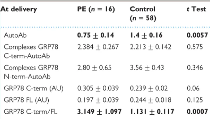
Table IV Various delivery markers of PE (mean +++++ SD)
At delivery PE (n 5 16) Control (n 5 58) t Test AutoAb 0.75 +++++ 0.14 1.4 +++++ 0.16 0.0057 Complexes GRP78 C-term-AutoAb 2.384 + 0.267 2.213 + 0.142 0.575 Complexes GRP78 N-term-AutoAb 2.80 + 0.65 3.56 + 0.43 0.346 GRP78 C-term (AU) 0.305 + 0.039 0.239 + 0.02 0.06 GRP78 FL (AU) 0.197 + 0.039 0.244 + 0.018 0.125 GRP78 C-term/FL 3.149 +++++ 1.097 1.131 +++++ 0.117 0.0007
Ab: antibodies; C/N-term: C/N-terminal; FL: full length; PE: pre-eclampsia; AU: arbitrary unit.
...
Table II Characteristics of patients recruited at first trimester of pregnancy Pre-eclamptics (n 5 10) Controls (n 5 26) P Nullipara (%) 87 61 0.14 Gestational age at the
time of sampling (weeks)
13.8 + 0.9 12.3 + 0.1 0.17 Maternal age (years) 27.4 + 1.3 29.3 + 0.8 0.21
Figure 1 ROC curves analysis. (A) First trimester. (B) Time of delivery.
Figure 2 Membrane localization of GRP78.
(A) FACS analysis of membrane GRP78. (a) cells stained with control antibody, (b) cells stained with GL-19 antibody, (c) cells stained with N-20 antibody. Lefthand plot shows FSC y axis and SSC x axis. Indicated percentage in righthand plot represents percentage of cells in R2 area (cells which are positive for GRP78 antibody). (B) Immunocytochemistry of purified CTB in permeabilized conditions (a, b, d, f) or non-permeabilized con-ditions (c, e). (a) cells stained with control goat antibody; (b) cells stained with control rabbit antibody; (c, d) cells stained with GL-19 antibody; (e, f) cells stained with N-20 antibody. (C) Cell surface localization of GRP78 was analysed by CELISA. (a) GL-19 antibody, and KDEL antibodies were used to detect membrane GRP78. PAb1620 antibody is directed against N-terminus of p53, and used here as a negative control. (b) Relative proportion of membrane GRP78 (estimated by non-permeabilized CELISA) over total GRP78 (estimated by permeabilized CELISA) detected with GL-19 and N-20 antibodies. mb ¼ membrane.
compared with healthy pregnant women that appears to allow or accel-erate degradation of circulating GRP78 in pre-eclamptic women at term. The biological significance of this observation is still unknown but interestingly some studies suggest a higher incidence of cancer in women who developed PE (Paltiel et al., 2007), although this association is debated (Terry et al., 2007). However, it would be noted that the delivery time is significantly lower in pre-eclamptic patients com-pared with healthy patient and could also explain this observation at delivery.
In conclusion, we demonstrate that the ratio of circulating C-terminal GRP78 over FL GRP78 is lower in plasma of women who will later be diagnosed with PE. This could be used as an early biologi-cal marker of PE and might allow novel strategies early in pregnancy to improve placentation before the consequences of defective tropho-blastic invasion become irreversible. Our preliminary assessment of circulating GRP78 constitutes a proof of principle rather than a defini-tive clinical study. Clearly the potential diagnostic potency of GRP78 in asymptomatic women prone to develop PE remains to be established and compared with known diagnostic parameters such as placental protein 13 (Chafetz et al., 2007; Gonen et al., 2008; Huppertz et al., 2008) for which ROC curves demonstrate an area of 0.91, a 90% specificity with 79% of sensitivity (Chafetz et al., 2007).
Acknowledgements
We wish to thank Antonina Chilin for collecting blood samples, enrolling patients.
Funding
The SNF and the ‘Hoˆpital Universitaire de Gene`ve’ for financial support.
References
Bischof P, Martelli M, Campana A, Itoh Y, Ogata Y, Nagase H. Importance of matrix metalloproteinases in human trophoblast invasion. Early Pregnancy 1995;1:263 – 269.
Castelli A, Delpino M. The 78 kDa glucose-regulated protein (GRP78/BIP) is expressed on the cell membrane, is released into cell culture medium and is also present in human peripheral circulation. Biosci Rep 2002; 22:407 – 420.
Chafetz I, Kuhnreich I, Sammar M, Tal Y, Gibor Y, Meiri H, Cuckle H, Wolf M. First-trimester placental protein 13 screening for preeclampsia and intrauterine growth restriction. Am J Obstet Gynecol 2007;197:35.e1 – 7.
Davidson DJ, Haskell C, Majest S et al. Kringle 5 of human plasminogen induces apoptosis of endothelial and tumor cells through surface-expressed glucose-regulated protein 78. Cancer Res 2005;65:4663– 4672.
Dong D et al. Spontaneous and controllable activation of suicide gene expression driven by the stress-inducible grp78 promoter resulting in eradication of sizable human tumors. Hum Gene Ther 2004;15:553 – 661. Fu Y, Lee AS. Glucose regulated proteins in cancer progression, drug
resistance and immunotherapy. Cancer Biol Ther 2006;5:741 – 744. Gerretsen G, Huisjes HJ, Elema JD. Morphological changes of the spiral
arteries in the placental bed in relation to pre-eclampsia and fetal growth retardation. Br J Obstet Gynaecol 1981;88:876 – 881.
Gonen R, Shahar R, Grimpel YI, Chefetz I, Sammar M, Meiri H, Gibor Y. Placental protein 13 as an early marker for pre-eclampsia: a prospective longitudinal study. BJOG 2008;115:1465 – 1472.
Huppertz B, Sammar M, Chefetz I, Neumaier-Wagner P, Bartz C, Meiri H. Longitudinal determination of serum placental protein 13 during development of preeclampsia. Fetal Diagn Ther 2008;24:230 – 236. Lee AS. The glucose-regulated proteins: stress induction and clinical
applications. Trends Biochem Sci 2001;26:504 – 510.
Lee AS. GRP78 induction in cancer: therapeutic and prognostic implications. Cancer Res 2007;67:3496 – 3499.
Li J, Lee AS. Stress induction of GRP78/BiP and its role in cancer. Curr Mol Med 2006;6:45 – 54.
Meisser A, Cohen M, Bischof P. Concentrations of circulating gelatinases (matrix metalloproteinase-2 and -9) are dependent on the conditions of blood collection. Clin Chem 2005;51:274 – 276.
Mintz PJ, Kin J, Do KA et al. Fingerprinting the circulating repertoire of antibodies from cancer patients. Nat Biotechnol 2003;21:57 – 63. Myers JE, Merchant SJ, Macleod M, Mires GJ, Baker PN, Davidge ST.
MMP-2 levels are elevated in the plasma of women who subsequently develop preeclampsia. Hypertens Pregnancy 2005;24:103 – 115. Narumiya H, Zhang Y, Fernandez-Patron C, Guilbert LJ, Davidge ST.
Matrix metalloproteinase-2 is elevated in the plasma of women with preeclampsia. Hypertens Pregnancy 2001;20:185 – 194.
Paltiel O, Fridlander Y, Tiram E, Barchana M, Xue S, Harlap S. Cancer after pre-eclampsia: follow-up of the Jerusalem perinatal study cohort. BMJ 2007;328:919.
Pang ZJ, Xing FQ. Expression profile of trophoblast invasion-associated genes in the pre-eclamptic placenta. Br J Biomed Sci 2003;60:97 – 101. Robertson WB, Brosens I, Dixon HG. The pathological response of the
vessels of the placental bed to hypertensive pregnancy. J Pathol Bacteriol 1967;93:581 – 592.
Shin BK, Wang H, Yim AM, Le Naour F, Brichory F, Jang JH, Zhao R, Puravs E, Tra J, Michael CW, Misek DE, Hanash SM. Global profiling of the cell surface proteome of cancer cells uncovers an abundance of proteins with chaperone function. J Biol Chem 2003;278:7607 – 7616. Terry MB, Perrin M, Salafia CM, Zhang F, Neugut AI, Teitelbaum S L,
Britton J, Gammon MD. Preeclampsia, Pregnancy-related Hypertension, and Breast Cancer Risk. Am J Epidemiol 2007; 165:1007 – 1014.
Zhang J, Jiang Y, Jia Z, Li Q, Gong W, Wang L, Wei D, Yao J, Fang S, Xie K. Association of elevated GRP78 expression with increased lymph node metastasis and poor prognosis in patients with gastric cancer. Clin Exp Metastasis 2006;23:401 – 410.
Submitted on November 19, 2008; resubmitted on May 12, 2009; accepted on May 23, 2009