Pulmonary artery banding: long-term telemetric adjustment
q
Antonio F. Corno
a,*, Nicole Sekarski
b, Marc-Andre´ Bernath
c, Maurice Payot
b,
Piergiorgio Tozzi
a, Ludwig K. von Segesser
aaCardiovascular Surgery, Centre Hospitalier Universitaire Vaudois (CHUV), 46 rue du Bugnon, CH-1011, Lausanne, Switzerland bPediatric Cardiology, Centre Hospitalier Universitaire Vaudois (CHUV), Lausanne, Switzerland
cAnaesthesia, Centre Hospitalier Universitaire Vaudois (CHUV), Lausanne, Switzerland
Received 10 September 2002; received in revised form 25 November 2002; accepted 27 November 2002
Abstract
Objective: Adjustment of pulmonary blood flow is difficult in pulmonary artery banding for complex congenital heart defects. A new
wireless, battery free, telemetrically controlled, implantable device (FloWatche, EndoArt, S.A., Lausanne, Switzerland) allowing for
progressive occlusion/reopening of the device through a remote control at the wanted percentage of occlusion (adjustable pulmonary artery
banding) underwent experimental evaluation. Methods: Eleven mini-pigs underwent FloWatche implantation around the main pulmonary
artery through left thoracotomy. The first group ðn ¼ 4Þ, mean age 18.2 ^ 0.1 weeks, mean body weight 12.0 ^ 0.1 kg, underwent
FloWatche implantation as device tolerance test. The second group ðn ¼ 7Þ, mean age 8.6 ^ 3.4 weeks, mean body weight 5.1 ^ 1.5 kg,
underwent functional evaluation: at implantation, 1, 3, 5, 8 and 10 weeks after implantation, the device was progressively occluded and
reopened, with Doppler evaluation of the developed pressure gradient. Results: The four mini-pigs of first group were sacrificed at mean age
of 42.3 ^ 0.1 weeks, mean body weight 25.1 ^ 3.2 kg (mean interval of 24 weeks after implantation); the device was still functioning and
histology revealed almost normal morphology of the pulmonary artery. In all seven mini-pigs of second group the possibility of narrowing/
releasing the pulmonary artery was confirmed at implantation and during follow-up: at last control their mean age was 20.5 ^ 2.8 weeks and
the body weight 12.7 ^ 3.7 kg. Conclusions: Complete adjustment of pulmonary blood flow is now possible with an implantable device
allowing for pulmonary artery banding with early and late telemetric flow control.
q
2002 Elsevier Science B.V. All rights reserved.
Keywords: Adjustable device; Congenital heart defects; Congenital heart surgery; Palliative procedure; Pulmonary artery banding; Pulmonary hypertension
1. Introduction
Indication for pulmonary artery banding is currently
limited by several factors:
(a) difficulty in determining the optimal perimeter of the
band,
(b) influence of several clinical variables with mutual
interference, including general anesthesia with positive
pressure ventilation, chest opening particularly with
thor-acotomy [1], heart rate and contractility, arterial PO
2and
PCO
2, acid–base status, hematocrit, systemic and
pulmonary vascular resistance [2]. Substantial changes
occur to all these variables, particularly within the first
few hours or days after the operation [2],
(c) variability of ventricular adaptive response,
particu-larly in ‘functionally’ univentricular hearts [3],
transposi-tion of the great arteries where a left ventricular training
is required in view of arterial switch operation [4], and
where simultaneous associated surgical procedures are
required,
(d) repeated operations frequently required to adjust the
band perimeter,
(e) long periods with intensive respiratory or
pharmaco-logical interventions to control the pulmonary blood flow
[4],
(f) frequent need for a reconstruction of the pulmonary
artery at the moment of the conventional de-banding for
surgical repair.
To overcome these difficulties, several attempts have
been made to find an adjustable pulmonary artery banding,
allowing for external regulation [5–19]. A MedLine
research for ‘adjustable pulmonary artery banding’ revealed
16 different techniques reported within the last 10 years, the
1010-7940/03/$ - see front matter q 2002 Elsevier Science B.V. All rights reserved. doi:10.1016/S 1 0 1 0 - 7 9 4 0 ( 0 2 ) 0 0 8 3 2 - 1
www.elsevier.com/locate/ejcts
q
Presented at the 16th Annual Meeting of the European Association for Cardio-thoracic Surgery, Monte Carlo, Monaco, September 22–25, 2002.
* Corresponding author. Tel.: 141-21-314.2280; fax: 141-21-314.2278. E-mail address: antonio.corno@chuv.hospvd.ch (A.F. Corno).
majority of which, however, did not result in a device
allow-ing for a precise, long-term, non-invasive, adjustment of
pulmonary blood flow in both ways, with repeated
narrow-ing and releasnarrow-ing of the pulmonary artery.
We report on the experimental evaluation of a new
wire-less, battery free, implantable device (FloWatche),
devel-oped by EndoArt, S.A. (Lausanne, Switzerland) [20]
allowing for repeated progressive occlusion and reopening
of the device through a remote control, at the wanted
percentage of occlusion (adjustable pulmonary artery
band-ing).
2. Materials and methods
2.1. Technical characteristics of the device
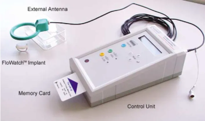
The FloWatche system comprises the implant and the
external control unit with an antenna (Fig. 1). The device is
placed with a surgical procedure around the main
pulmon-ary artery, with a minimal surgical dissection, enough to
allow for the passage of the clip of FloWatche around the
pulmonary artery, in a fashion similar to the conventional
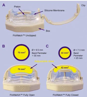
pulmonary artery banding (Fig. 2). With the device in
clipped position, the dimensions are: 26 mm (length) £ 18
mm (width) £ 18 mm (height), therefore due to the size
there are no chances of unwanted migration of the device
after implantation and pericardial closure. The change in the
adjustable area is obtained by means of a piston driven by an
incorporated electrical micro-motor. The concave form of
the adjustable area has been chosen so that during
compres-sion the area changes but the perimeter of the pulmonary
artery remains unchanged, which is optimal for long-term
use (i.e. reopening after several weeks of pulmonary artery
compression). The adjustable area in fully open position
correspond to a pulmonary artery banding with a perimeter
of 30 mm, and with fully closed position to a pulmonary
artery banding with a perimeter of 23 mm (Fig. 3).
Accord-ing to the Trusler’s rule [21], theoretically the device should
theoretically be suitable for pulmonary artery banding in
patients from 3 to 10 kg of body weight. The adjustment
is done via an external control unit delivering to the
implanted device, via the antenna, the energy as well as
the commands to drive the micro-engine. The device does
not contain any battery. The telemetric system is designed
such that the implant sends back to the control unit
informa-tion about its funcinforma-tioning, which allows for control of the
regulation by the treating doctor.
2.2. Experimental studies
A feasibility test performed with acute ðn ¼ 2Þ and
chronic ðn ¼ 2Þ experimental studies on mini-pigs (acute
study: 18 and 24 weeks of age, 12 and 17 kg of body weight;
chronic study: 8 and 10 weeks of age, 7 and 8 kg of body
weight), confirmed: (a) the applicability of the device
around the pulmonary artery; (b) the possibility of
progres-sively narrowing and releasing the pulmonary artery with
telemetric control after chest closure; (c) the functioning of
the device up to 6 weeks after implantation, with repeated
adjustments of pulmonary artery banding.
At this point a device tolerance test and a functional
evaluation test were performed on eleven mini-pigs.
After induction of general anaesthesia, tracheal
intuba-tion and mechanical ventilaintuba-tion, trough a left lateral
thora-cotomy the pericardium has been longitudinally opened
with an antephrenic incision, and a short segment of the
main pulmonary artery has been dissected free.
The first group ðn ¼ 4Þ, mean age 18.2 ^ 0.1 weeks,
mean body weight 12.0 ^ 0.1 kg, underwent FloWatche
implantation as device tolerance test, with a period of 24
weeks of observation. At the end of the 24 weeks the
mini-pigs underwent general anaesthesia, tracheal intubation and
mechanical ventilation. The chest has been reopened
through median sternotomy, followed by longitudinal
peri-Fig. 1. The FloWatche system comprises the implant and the external control unit with an antenna.
Fig. 2. The device is placed with a surgical procedure around the main pulmonary artery, in a fashion similar to the conventional pulmonary artery banding. The device is unclipped, with the clip already around the inferior aspect of the main pulmonary artery.
cardiotomy, as in any conventional surgical procedure for
repair of a congenital heart defect with previous pulmonary
artery banding. The device was progressively occluded and
reopened, with Doppler evaluation of the developed
pres-sure gradient, and with invasive prespres-sure meapres-surement by
insertion of a pressure catheter into the right ventricle and
another one into the distal pulmonary artery for
simulta-neous pressure monitoring. Then the device has been
explanted, and the animal sacrificed. Both the main
pulmon-ary artery and the tissues surrounding the device have been
sent for histological evaluation.
The second group ðn ¼ 7Þ, mean age 8.6 ^ 3.4 weeks,
mean body weight 5.1 ^ 1.5 kg (range 3.4–6.9 kg),
under-went functional evaluation test: at implantation, 1, 3, 5, 8
and 10 weeks after implantation, the device was
progres-sively occluded and reopened, with Doppler evaluation of
the developed pressure gradient. At the end of the
observa-tion period, the mini-pigs followed the same protocol of the
device tolerance test, with invasive pressure measurements
and device explantation through median sternotomy,
followed by sacrifice and harvesting of main pulmonary
artery and surrounding tissues for histological examination.
All animals received human care in compliance with the
‘Principles of Laboratory Animals’ formulated by the
National Society of Medical Research and the ‘Guide for
the Care and Use of Laboratory Animals’ prepared by the
Institute of Laboratory Animal Resources and published by
the National Institutes of Health (NIH publication 85 - 23,
revised 1985). The protocol was approved by the
institu-tional Committee on Animal Research.
3. Results
In all cases the device implantation was a fast track
surgi-cal procedure, with all animals extubated on at the end of the
procedure, and sent back to the farm within few hours,
with-out medications other than oral pain killers.
The four mini-pigs of the device tolerance test were
sacri-ficed at mean age of 42.3 ^ 0.1 weeks, mean body weight
25.1 ^ 3.2 kg (mean interval of 24 weeks after
implanta-tion); in all of them the device was still functioning.
Through median sternotomy and longitudinal
pericardiot-omy, the device has been easily identified in the same
posi-tion of implantaposi-tion, without any migraposi-tion of the device.
Macroscopic examination revealed a fibrous capsule
surrounding the device, very similar to the tissue generally
found around a pace-maker battery at the moment of battery
replacement. Removal of the device has been very easily
performed in all cases, without any lesion of the pulmonary
artery, any bleeding or other hemodynamic deterioration as
well. Macroscopic examination of the pulmonary artery
after sacrifice revealed a very pliable vessel wall, without
any sign of fibrosis externally as well as internally.
Histol-ogy revealed in all animals almost normal morpholHistol-ogy, with
limited thickening of the intima and some sign of
degenera-tion of the media of the pulmonary artery in correspondence
with the narrowing induced by the device (Fig. 4).
In all seven mini-pigs of the functional evaluation test the
possibility of repeatedly narrowing and releasing the
pulmonary artery was confirmed at implantation and during
the entire follow-up: 1, 3, 5, 8 and 10 weeks after
implanta-tion. At last control before sacrifice their mean age was
20.5 ^ 2.8 weeks and the body weight 12.7 ^ 3.7 kg.
Results of all Doppler evaluations (mean ^ SD), shown
in Table 1, showed a very good correlation between
percen-tage of occlusion and trans-banding pressure gradient, for
the narrowing as well as for the releasing of the device, even
weeks after the implantation of the device. In all cases
Fig. 4. Normal histology of the main pulmonary artery in correspondence of the narrowing induced by the device, 10 weeks after implantation. Fig. 3. Device in unclipped position (A) and in clipped position with the
progressive occlusion of the device was followed by
progressive increase of the pressure gradient, as well as
progressive re-opening of the device has been immediately
followed by reduction of the pressure gradient.
In all animals there was a great variability of the
ventri-cular response to the same percentage of occlusion of the
device ( ¼ narrowing of the pulmonary artery banding),
with the pressure gradient generally increasing with the
time elapsed from the day of implantation (example of a
single animal is represented in Fig. 5).
Macroscopic and histological examination in this group
of mini-pigs, after 10 weeks of implantation, showed the
same results as in the previous group (device tolerance
test) exposed to 6 months of device implantation.
4. Discussion
The clinical need for an adjustable pulmonary artery
banding has been confirmed by several attempts in
devel-oping such a technique reported in the literature ([5–19]). A
MedLine research confirmed the persistent unavailability of
a reliable device, capable of repeated narrowing and
releas-ing the pulmonary artery with effective remote control of the
pulmonary blood flow. Mostly because of the known
diffi-culties to control the pulmonary blood flow once the
conventional pulmonary artery banding is in place, the
indi-cation to pulmonary artery banding is currently limited to
the clinical situations without another suitable alternative
surgical option.
With our experimental research we tested a wireless,
battery free, telemetric controlled device (FloWatche) for
adjustable pulmonary artery banding [20]. Our experimental
studies demonstrated that the device is easy to implant and
to use, with proven evidence of repeated progressive
occlu-sion and reopening of the device through a remote control,
at the wanted percentage of occlusion. Furthermore, at the
time of explantation the procedure was absolutely smooth
and the pulmonary artery returned to the normal
morphol-ogy, without the localised narrowing in correspondence of
the banding and the fibrosis generally observed in the
pulmonary arteries with the band in place for some time;
therefore, another advantage of this device should be the
possibility of avoiding the need for a plastic repair after a
conventional banding.
In conclusion, our experimental studies demonstrated that
the FloWatche allows for unlimited external telemetric
adjustments of pulmonary blood flow, as expected and
required from a really adjustable pulmonary artery banding.
The potential clinical benefits of this device allowing for
unlimited external telemetric adjustments of pulmonary
artery banding are the following:
† fast track surgical procedure,
† effective control of pulmonary blood flow,
† no need for banding-related re-operations,
† simplified post-operative course,
† reduction of mortality and morbidity.
If the clinical studies with FloWatche implantation will
confirm our experimental results, and the potential clinical
benefits will be demonstrated, this device will represent a
major change in the management of children requiring for a
pulmonary artery banding, particularly for the most
complex pathophysiological situations, like univentricular
hearts or transposition of the great arteries requiring for a
left ventricular retraining because of a late referral. The size
of the currently available device will limit the clinical
appli-cation to children up to 15–20 kg of body weight, and
there-fore a larger size will need to be developed it be utilised in
older children and adolescents.
Finally, the indications for pulmonary artery banding
could be expanded in order to adapt the therapeutic
strate-gies to this promising technology.
References
[1] Corno AF, Carta MG, Giannico S. Pulmonary artery banding through median sternotomy. Clin Res 1989;37:91A.
[2] Corno AF. Revised pulmonary artery banding. Ann Thorac Surg 2000;69:1295–1296.
Fig. 5. Graphic showing the progression with the time from the day of implantation of the trans-banding pressure gradient for the same percentage of occlusion of the device.
Table 1
Correlation of second degree polynomial fit between % occlusion and Doppler gradient
Implantation 1 week 3 weeks 5 weeks 8 weeks 10 weeks
[3] Tchervenkov CI, Shum-Tim D, Beland MJ, Jutras L, Platt R. Single ventricle with systemic obstruction in early life: comparison of initial pulmonary artery banding versus the Norwood operation. Eur Cardi-othorac Surg 2001;19:671–677.
[4] Wernovsky G, Giglia TM, Jonas RA, Mone SM, Colan SD, Wessel DL. Course in the intensive care unit after ‘preparatory’ pulmonary artery banding and aortopulmonary shunt placement for transposition of the great arteries with low left ventricular pressure. Circulation 1992;86:II-133–II-139.
[5] Ahmadi A, Rein J, Hellberg K, Bastanier C. Percutaneously adjusta-ble pulmonary artery band. Ann Thorac Surg 1995;60:S520–S522. [6] Assad RS, Cardarelli M, Abduch MC, Aiello VD, Maizato M,
Barbero-Marcial M, Jatene A. Reversible pulmonary trunk banding with a balloon catheter: assessment of rapid pulmonary ventricular hyperthrophy. J Thorac Cardiovasc Surg 2000;120:66–72.
[7] Bonnet D, Sidi D, Vouhe´ PR. Absorbable pulmonary artery banding in tricuspid atresia. Ann Thorac Surg 2001;71:360–361.
[8] Dajee H, Benson L, Laks H. An improved method of pulmonary artery banding. Ann Thorac Surg 1984;37:254–257.
[9] Dabritz SH, Sachweh JS, Tiete AR, Engelhardt W, von Bernuth G, Messmer BJ. Experience with an adjustable pulmonary artery banding device in two cases: initial success-midterm failure. Thorac Cardio-vasc Surg 1999;47:51–52.
[10] Higashidate M, Beppu T, Imai Y, Kurosawa H. Percutaneously adjus-table pulmonary artery band. An experimental study. J Thorac Cardi-ovasc Surg 1989;97:864–869.
[11] Le Bret E, Bonhhoeffer P, Folliguet TA, Sidi D, Laborde F, de Leval MR, Vouhe´ P. A new percutaneously adjustable, thoracoscopically implantable, pulmonary artery banding: an experimental study. Ann Thorac Surg 2001;72:1358–1361.
[12] Muraoka R, Yokota M, Aoshima M, Nomoto S, Kyotu I, Kobayashi A, Nakano H, Ueda K, Saito A. Extrathoracically adjustable pulmon-ary banding. J Thorac Cardiovasc Surg 1983;86:582–586.
[13] Nahs R, Mundth ED, Ross B, Austen WG. Adjustable instrument for pulmonary artery banding. Description of instrument and technique of application. J Thorac Cardiovasc Surg 1972;63:732–734.
[14] Park SC, Griffith BP, Siewers RD, Hardesty RL, Ladowski J, Zoltun RA, Neches WH, Zuberbuhler JR. A percutaneously adjustable device for banding of the pulmonary trunk. Int J Cardiol 1985;9:477–484.
[15] Peek GJ, Arsiwala SS, Chan KC, Hickey MS. Absorbable pulmonary artery band. Ann Thorac Surg 1997;64:539–541.
[16] Schlensak C, Sarai K, Gildein HP, Beyersdorf F. Pulmonary artery banding with a novel percutaneously, bi-directional adjustable device. Eur J Cardiothorac Surg 1997;12:931–933.
[17] Solis E, Heck CF, Seward JB, Kaye MP. Percutaneously adjustable pulmonary artery band. Ann Thorac Surg 1986;41:65–69.
[18] Vince DJ, LeBlanc JG, Culham JAG, Taylor GP. A dilatable pros-thesis for banding the main pulmonary artery: human clinical trials. Int J Card Imaging 1996;12:205–212.
[19] Warren ET, Heath BJ, Brand WW. A staged expanding pulmonary artery band. Ann Thorac Surg 1992;54:240–242.
[20] Fridez P, Jordan A, Montavon JC, Stergiopulos N. FloWatch: an implantable device for telemetric control of flow after pulmonary artery banding. Cardiovasc Eng 2002;7:51.
[21] Trusler GA, Mustard WT. A method of banding the pulmonary artery for large isolated ventricular septal defect with and without transposi-tion of the great arteries. Ann Thorac Surg 1972;13:351–355.
Appendix A. Conference discussion
Dr G. Stellin (Padova, Italy): It is a very nice device and its implantation looks very appealing. However, I must say that most of the pulmonary artery banding that we need to place usually are in the neonatal period or in early infancy. I wonder, how can you get around the problem of
compres-sing the coronary arteries or comprescompres-sing the pulmonary artery branches with such a rigid body inside the chest?
Dr Corno: First of all, the size of the device is 26 mm length by 18 mm by 18 mm. Of course, it has to be rigid to sustain the increased pulmonary artery pressure when we do a banding. The size of the clip is exactly 4 mm, as the band that we used before having this device available. And I can tell you that in the experimental study, the smallest piglet with device implan-tation was 3.2 kilos. And we had never encountered a problem of compres-sion of coronary arteries, of pulmonary artery distortion, or left bronchial compression in the observed period. And I can even add that after this experimental study, we started a multicenter clinical trial, and the device has been already implanted down to 3.5 kilos in human beings without negative consequences.
Dr R. DiDonato (Rome, Italy): I don’t know all the methods of telemetric banding of the pulmonary artery, but I think this is the best one I have ever seen. And I would like to suggest one application in particular, namely for the cases of retraining of the left ventricle in corrected transposition that we were discussing earlier. Interestingly, for these cases the concept is coming out that the retraining has to be a gradual process, often with a need to be adjusted on a daily basis, maybe even an hourly basis, like the athlete that needs to train and build up muscle mass. So I think this is an ideal tool for this kind of application, i.e. for gradual banding and for retraining of the left ventricle.
Dr Corno: You are absolutely right. The main reason why we started this study is because, I’m sure you know, we had a complex group of patients who came with simple TGA, very late referral, up to 25 months of age, and we had to perform the left ventricular retraining. So we went for banding, shunt, plus/minus ASD and so forth. And I can tell you that it was a night-mare. We were successful, but in 50% of the patients we had to go back with a reoperation to adjust the banding, to further narrow the banding or to reopen the banding. And in one patient we had to go back three times. That’s why we started this study. So it is a perfect indication for left ventricular retraining.
For the left ventricular retraining after failing Mustard or Senning or in congenitally corrected transposition, the problem at the moment is the size, because generally these are patients who are later on in life, with a body weight of 30, 40, 50 kilos. And at the moment this available device is too small. But we are already starting to develop a larger size device. I’m very pleased with your comment, because this will stimulate the company to accelerate their study with a larger size device.
Dr V. Alexi-Meskishuili (Berlin, Germany): I just want to share with you a very limited experience of stage banding method, which we used in German Heart Institute. 3 years ago, I operated 2 patients sent to us for evaluation as inoperable with mitral atresia, restrictive foramen ovale and systolic pulmonary artery pressure of 80 mm Hg. In both patients I resected the atrial septum and tried to put a pulmonary artery band, but the patients cannot tolerate that. So I left the sternum open and tightened gradually the band during 4 days after the operation with the titan-clips, until mean pulmonary artery pressure fell to 18 mmHg. Now, 3 years after gradual banding both patients underwent very successful extracardiac Fontan operation. I think that in some cases such as ours it is a cheap and effective method of staged banding. This method may be useful also in older children and some adult patients for left ventricular retraining. In small children, we do banding very seldom, only in complex cases, it’s a very safe operation nowadays.
Dr Corno: I fully agree with you. I was trained in a research lab at UCLA where I was able to use adjustable banding very cheap with 2-0 polypro-pylene and a rubber tourniquet. The only problem is that with this kind of adjustable device you are almost sure then you can go back and narrow the pulmonary artery furthermore, but you still have to reopen the skin for 2-3 cm. But then, if you have few weeks after the banding, you are almost never able to release the pulmonary artery. So when you have a grown up child and you need to release the pulmonary artery, with our device you can do it. With the other one, we have never been able to do it.
Dr J. Amato ( Chicago, Illinois): Very, very ingenious device. And being an old-timer, I still think there’s need for bands. Two complications that I can foresee, and I wonder how you can possibly prevent them. One is
erosion. As you know, even the common pulmonary band will erode poster-iorly. I don’t know how soft this silastic material is and secondly is the possibility of clot formation. I noticed that as you compress the pulmonary artery, it’s not a circular compression but rather an elliptical compression, and I wonder whether you might anticoagulate, or do you anticoagulate these piglets and then children? May I also suggest that it might be safer to elevate the main pulmonary artery with a cord tape or vessel loop prior to inserting the device. It seems that the edges of the device are blunt and may injure the pulmonary artery tissues by inserting the device directly?
Dr Corno: Well, ingenious was the engineer who invented the device. Anyway, for your questions, we didn’t see any lesion in the pulmonary artery in the experimental study and that’s why at the moment of explanta-tion we harvested the entire block of the pulmonary artery. And the histol-ogy showed in all the cases almost normal pulmonary artery with very pliable tissue. And this is most probably due, but is a speculation, to the shape of the pulmonary artery, which doesn’t have a circular narrowing. With the conventional banding you always found a fibrosis. And if you go back after a few months, you have to reopen the pulmonary artery with a plasty. In all these animals, I can tell you, we explanted the last one 2 weeks ago, after 14 months, and we did need to reopen the pulmonary artery: simply amazing, the pulmonary artery opens up spontaneously.
We never gave any antiplatelets or anticoagulant. All the piglets were free of medication and we had no thrombus.
Dr K. Samir (Marseille, France): Firstly, I would like to thank you for the comments we had on line when we discussed the article before.
Secondly, when I calculated the effective diameters of your apparatus, I found it making bands between 28 and 32 to 33 cm. I think it is too loose bands to be applied for neonates. What do you think?
Dr Corno: First of all, the given circumference is 23 to 30 millimeters, not centimeters.
Before this device was available, I have done the banding following the Trusler rule. And the Trusler rule, you know better than I do probably, says that you should have a perimeter of the band of 20 mm plus 1 mm/kg. If you consider a neonate of 3 kilos, 23 mm should be the perfect size.
So far we have a relatively small group of patients, but we were never able to completely narrow the device even in a 3.5 kilogram patient. That means we never needed to reach the 100 percent occlusion, and therefore I speculate that this size should be enough even in neonates.
Now we have started to run a clinical trial, and we are not allowed to use the device in any child less than 3.5 kilos.
Dr A. Urban (Augustin, Germany): Which incision would you use to put in your device, median sternotomy or lateral thoracotomy, or what would you do in a human patient?
Dr Corno: In the experimental study all the devices have been implanted through a left thoracotomy and explanted through median sternotomy, like in a normal repair. But in the multicenter clinical trial we are performing, both in Switzerland and Paris, I can tell you the device has been implanted through both thoracotomy and median sternotomy, with associated proce-dure or without associated proceproce-dure, and even with proceproce-dure on cardio-pulmonary bypass.