Nephrol Dial Transplant (1993) 8: 1393-1394
Case Report
IMephrology
Dialysis
Transplantation
'Transient multiple myeloma' after intense immunosuppression in a renal
transplant patient
J. Passweg
1, H. A. Bock
1, A. Tichelli
2and G. Thiel
1Division of 'Nephrology and 2Hematology, Department of Internal Medicine, Kantonsspital Basel, Switzerland
Key words: monoclonal gammopathy; multiple
myeloma; renal transplantation
Introduction
B-cell lymphoproliferative disease, especially large cell lymphoma, is a well-recognized complication of immunosuppression early after organ transplantation, whereas the incidence of multiple myeloma does not appear to be increased [1]. We wish to report a patient who was diagnosed as having multiple myeloma during intense immunosuppression after renal transplantation. All abnormalities, however, later disappeared after reduction of immunosuppression.
Subjects and methods
The patient, a 51-year-old woman, had suffered from chronic glomerulonephritis since 1958. End-stage renal failure had been reached in 1976 and maintenance haemodialysis was begun. A first cadaveric renal transplant was rejected under conventional immunosuppression with azathioprine and prednisone within 6 weeks in 1978. Since she developed high titres of panel-reactive antibodies, she could not be retrans-planted for several years thereafter.
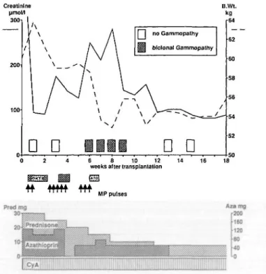
In October 1990 she finally received a second cadaveric kidney graft. Immunoelectrophoresis before transplantation was normal. Because of her history of rejection and lymph-ocytotoxic antibodies, an intensified immunosuppressive regi-men was administered, consisting of 5 mg OK.T3 for 14 days, cyclosporin 400 mg daily from day zero, azathioprine 2 mg/kg and methylprednisolone pulses for 3 days followed by 0.5 mg/kg of oral prednisone thereafter (Figure 1).
Although the graft functioned well from the first day, biopsy-proven vascular rejection on day 23 required therapy with seven daily doses of 5 mg OKT3, and seven pulses of methylprednisolone. The retreatement was well tolerated except for diarrhoea and slight pancytopenia. The patient had to be readmitted on day 40 because of an increase in
serum creatinine, and subfebrile temperature, dyspnoea, and cough.
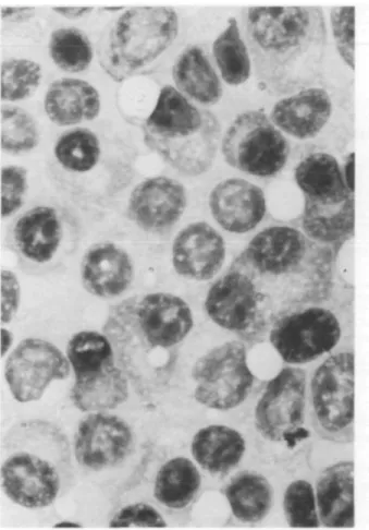
Admission serum protein electrophoresis revealed biclonal gammopathy, IgG kappa and lambda. Bronchoscopy speci-mens stained positive for Pneumocystis carinii, which prompted a therapy with co-trimoxazole. She was treated for suspected rejection with four additional steroid pulses and 7 days of rabbit anti-thymocyte globulin (ATG Fresenius) 2 mg/kg body weight. Serum IgG increased from 8.1 g/1 at transplantation to 33.1 g/1 by day 57. Urine electrophoresis was trace positive for the IgG kappa mono-clonal component. Bone marrow examination revealed plasma cell infiltrates of 30% (focally up to over 50%). These cells were polymorphic with atypical nuclei, some with blastic nuclear structure (Figure 2). A radiological work-up for osteolysis or organ infiltration was negative.
A diagnosis of multiple myeloma was made and removal of the renal transplant was therefore considered. However, since graft function improved in response to the rejection
no Gammopathy biclonal Gammopathy
0 2 4
11 I I I
8 10 12 weeks after transplantation
E3
LA ^ MP pulses
Correspondence and offprint requests to: Professor G. Thiel, Leiter Abteilung Nephrologie, Departement Innere Medizin, Kantonsspital Basel, CH-4031 Basel, Switzerland.
Fig. 1. Course of the patient's disease (top) and administered immunosuppression (bottom). B.Wt. = body weight.
1394
Fig. 2. Bone marrow smear with dense infiltrate of plasma cells.
treatment, immunosuppression could be reduced (see
Figure 1) at week 13. At day 90 the clonal gammopathy had
disappeared. A repeat bone marrow at that time showed less
than 5% plasma cells. Today, 18 months after
transplanta-tion, her graft functions satisfactorily with a serum creatinine
of 109 umol/1. Immunosuppression is maintained with 300 mg
cyclosporin and 7.5 mg prednisone daily. Serum protein
electrophoresis still shows no sign of clonal gammopathy.
Discussion
In summary, this patient developed clonal
gammopa-thy after intense immunosuppression with OKT3 (total
dose 105 mg) and MP pulses in addition to the baseline
medication with prednisone, azathioprine and
cyclosporin. Bone marrow examination disclosed
polymorphic infiltrates of plasma cells. By commonly
recognized criteria the combination of > 10% plasma
cell infiltrate and clonal gammopathy in serum or urine
allows the diagnosis of multiple myeloma [2]. However,
reduction of immunosuppression was followed by
com-plete disappearance of all abnormalities.
J. Passweg et al
Multiple myeloma has only rarely been reported
after renal transplantation [3], but clonal
gammo-pathies are frequently encountered in transplant
patients [4]. Their reported incidence ranges between
4 and 68%, depending on the organ transplantated,
the timing of sampling, and the sensitivity of the
methods used to detect clonality [5]. Not unexpectedly
the incidence appears to be greatest after bone marrow
transplantation [6] and may be least after renal
trans-plantation [7]. Clonal gammopathy after
transplanta-tion is often transient, and the quantity of clonal
immunoglobulin is generally small [4]. The cause of
pathological clonal expansion is believed to be
impaired control of B-cell clones by suppressor T cells,
most probably due to immunosuppression [8].
Spontaneous remissions have to our knowledge not
been reported in multiple myeloma. Since there is no
sharp distinction between monoclonal gammopathy
and multiple myeloma, there may be a zone of
dia-gnostic overlap. B-cell lymphomas after organ
trans-plantation are well known and their disappearance
after dose reduction of immunosuppressives has been
reported.
The lesson to be learned from the present case is
that a transient plasma cell dyscrasia may present like
multiple myeloma in the setting of heavy
immunosup-pression after organ transplantation. The alarming
findings suggesting myeloma after OKT3 therapy may
completely resolve simply by reducing
immunosuppres-sion without sacrificing the transplant.
References
1. Penn I. The changing pattern of posttransplant malignancies.
Transplant Proc 1991; 23: 1101-1103.
2. Kyle RA. Plasma cell proliferative disorders. In: Hoffman R, Benz EJ, Schattil SJ, Furie B, Cohen HJ. Hematology Basic
Principles and Practice. Churchill Livingstone, 1991,
pp.1021-1038.
3. Howard AD, Moore J, Tomaszewsky MM. Occurrence of mul-tiple myeloma three years after successful renal transplantation.
Am J Kidney Dis 1987; 10: 147-150.
4. Radl J, Valentijn RM, Haaijman JJ, Paul LC. Monoclonal gammopathies in patients undergoing immunosuppressive treat-ment after renal transplantation. Clin Immunol Immunopathol
1985; 37: 98-102.
5. Peest D, Schaper B, Nashan B et al. High incidence of monoclonal immunoglobulins in patients after liver or heart transplantation.
Transplantation 1988; 46: 389-393.
6. Fischer AM, Simon F, Le Deist F, Blanche S, Griscelli C, Fischer A. Prospective study of the occurrence of monoclonal gammo-pathies following bone marrow transplantation in young children.
Transplantation 1990; 49: 731-735.
7. Pollock CA, Mahony JF, Ibels LS et al. Immunoglobulin abnor-malities in renal transplant recipients. Transplantation 1989; 47: 952-956.
8. Stanko CK, Jeffery JR, Rush DN. Monoclonal and multiclonal gammopathies after renal transplantation. Transplant Proc 1989; 21: 3330-3332.
Received for publication 24.3.93 Accepted 24.6.93