Visual Neuroscience (1990), 4, 399-412. Printed in the USA.
Copyright © 1990 Cambridge University Press 0952-5238/90 $5.00 + .00
Neural elements in the pineal complex of the frog,
Rana esculenta, II: GABA-immunoreactive neurons
and FMRFamide-immunoreactive efferent axons
P. EKSTROM,1 T. OSTHOLM,1 H. MEISSL,2 A. BRUUN,3 J.G. RICHARDS4, AND H. MOHLER4 'Laboratory of Molecular Neuroanatomy, Department of Zoology, University of Lund, Lund, Sweden.
2
Max-Planck-Institute for Physiological and Clinical Research, W.G. Kerckhoff Institute, Bad Nauheim, FRG. 3
Department of Ophthalmology, University of Lund, Lund, Sweden.
4Pharmaceutical Research Department, F. Hoffmann-LaRoche & Co., Ltd., Basel, Switzerland.
(RECEIVED September 22, 1988; ACCEPTED February 19, 1990)
Abstract :
The photosensory pineal complex of anurans comprises an extracranial part, the frontal organ, and an intracranial part, the pineal organ proper. Although the pineal organ functions mainly as a luminosity detector, the frontal organ may monitor the relative proportions of short and intermediate/long wavelengths in the ambient illumination. The major pathway of information processing in the pineal and frontal organs is the photoreceptor to ganglion cell synapse. It is not known whether interneurons form part of the neural circuitry. In the present study, we demonstrate GABA-immunoreactive (GABA-IR) neurons in the pineal and frontal organs of the frog, Rana esculenta. No GABA-IR axons were observed in the pineal nerve between the frontal and pineal organs, or in the pineal tract that connects the pineal complex with the brain. The GABA-IR neurons differed in morphology from centrally projecting neurons visualized by retrograde labeling with horseradish peroxidase. Thus, we suggest that the GABA-IR neurons in the pineal and frontal organs represent local interneurons.
Axons of central origin, immunoreactive with a sensitive antiserum against the tetrapeptide Phe-Met-Phe-Arg-NH2 (FMRFamide), were observed in the intracranial portion of the photosensory pineal organ. The immunoreactive axons enter the caudal pole of the pineal organ via the posterior
commissure. The largest density of axons was observed in the caudal part, while fewer axons were detected in the rostral portion. The uneven distribution of the FMRFamide-immunoreactive axons may be related to the distribution of different types of intrapineal neurons. FMRFamide-immunoreactive varicose axons were observed in the extracranial frontal organ. A central innervation of the pineal organ, previously known exclusively from amniotes, is probably not per se linked with the evolutionary transition of the pineal organ from a directly photosensory organ to a neuroendocrine organ. It could rather represent a centrifugal input to a sensory system which has been retained when the directly sensory functions have changed, during phylogeny, to neuroendocrine functions.
Keywords: Pineal complex, •y-aminobutyric acid, GABAA/benzodiazepine receptor, FMRFamide, Rana esculenta (Amphibia)
Introduction
In evolution, the pineal organ has been transformed from a di-rectly photosensory organ, analogous to the lateral eye retinae, to a neuroendocrine organ (Studnicka, 1905; Bargmann, 1943; Collin, 1971; Oksche, 1971; Korf & Oksche, 1986). This trans-formation has been accompanied by a change in innervation pattern: the afferent fibers (pinealofugal fibers) of the pho-tosensory pineal organ, conveying neural signals related to am-bient illumination (Dodt, 1973), have gradually been lost and Reprint requests to: Peter Ekstrom, Laboratory of Molecular Neu-roanatomy, Department of Zoology, University of Lund, Helgonava-gen 3, S-223 62 Lund, Sweden.
efferent fibers (pinealopetal fibers) have become the dominant nervous connection with the brain. The sympathetic innervation of the mammalian pineal organ is the efferent system that has been the most extensively studied. Its main function appears to be the regulation of indoleamine synthesis, synchronizing it with the daily dark-light cycle (Wurtman et al., 1964; Klein et al., 1971). However, evidence has accumulated that the pineal or-gan of mammals also receives innervation from other sources, both peripheral (Shiotani et al., 1986) and central (Korf & Moller, 1984). The discovery of axons, immunoreactive with an antiserum against the neuropeptide FMRFamide, that innervate the photosensory pineal organ in teleost fish (Ekstrom et al., 1988) raised the possibility that the presence of a centrifugal in-399
nervation of the pineal organ may represent a pattern common to all vertebrate groups. To explore this possibility further, we investigated the possible presence of FMRFamide-immunore-active axons in the pineal complex of the frog, Rana esculenta.
Another question that is largely unresolved is whether inter-neurons form an integral part of the neural circuitry in the pho-tosensory pineal organ of anamniotes. The neural signals conveyed to the brain by projection neurons, that are analogous to retinal ganglion cells, carries simple information about the level of ambient illumination (Dodt, 1973; MeissI, 1986). These signals could conceivably be elaborated by a simple sign-conserving photoreceptor-to-neuron synapse (MeissI & Dodt, 1981). However, neuroanatomical data indicate the presence of intrapineal neurons that have no axonal connections with the brain (e.g. Paul et al., 1971; Wake, 1973; Wake et al., 1974; Korf, 1974; Ekstrom & Korf I986a,b; Ekstrom & MeissI, 1988). Here, we present evidence for putative GABAergic inter-neurons in the extracranial and intracranial portions of the pi-neal complex of the frog, Rana esculenta. We also report that although putative GABA receptor sites may be visualized in the retina and certain brain areas with monoclonal antibod-ies against GABAA/benzodiazepine receptor subunits (Haring et al., 1985; Richards et al., 1986), no such sites were observed in the pineal complex.
Material and methods
Fixation
Eyes, brains, pineal organs, and frontal organs from frogs
{Rana esculenta) were fixed either by immersion or by
perfu-sion followed by immerperfu-sion with one of the following fixa-tives: (1) 2% paraformaldehyde (w/v) + 0.1% glutaraldehyde (v/v) in phosphate-buffered saline (PBS), (2) 2% paraformal-dehyde + 0.1% glutaralparaformal-dehyde in 0.1 M phosphate buffer (PB), (3) 4% paraformaldehyde + 0.1% glutaraldehyde in PB, (4) 4% paraformaldehyde + 0.25% glutaraldehyde + 0.25% picric acid (v. saturated sol./v) in PB (only for GABA immu-nocytochemistry), or (5) 4% paraformaldehyde and 0.25% picric acid in 0.1 M PB (only for FMRFamide immunocyto-chemistry). All fixatives were held at pH 7.4. Total fixation time was 24 h for all fixatives used. Fixations (1), (2), and (3) gave essentially identical results with the monoclonal antibodies.
Tissue processing and immunocytochemistry
Tissue fixed in solutions 1, 2, 3, or 4, that was intended for cryostat sectioning, was thoroughly rinsed in Tyrode buffer, and infiltrated with Tyrode buffer containing 25% sucrose and 0.1% (w/v) sodium azide (Sigma Chemicals, St. Louis, MO). The brains were placed in Tissue Tek embedding medium and frozen on a metal plate cooled with liquid nitrogen. They were then mounted on metal chucks for sectioning in a Reichert-Jung cryostat. Serial 20-25 /xm sections were cut in the frontal and sagittal planes, thaw-mounted on chrome alum-gelatin-cov-ered slides, and allowed to air dry.
Pineal organs and frontal organs fixed in solution 4 and frontal organs fixed in solution 5 were washed in Tyrode buffer, dehydrated in a graded alcohol series, and embedded in soft Araldite (Durcupan, Fluka Chemie AG, Buchs, Switzerland) with propylene oxide as intermediate medium. After polymer-ization, serial 3-fim sections were cut and mounted on chrome alum-gelatinized slides.
Before incubation with the primary antiserum, the resin was
removed from the sections (Maxwell, 1978). Briefly, 40 g KOH was slowly dissolved in a mixture of 200 ml absolute methanol and 100 ml propylene oxide, and filtered. The resin was re-moved by immersing the slides in the filtered solution for 6 min. It is important that the mixture is cooled on ice throughout the procedure. After removal of the resin, the sections were rinsed in absolute methanol, and transferred to rinse buffer (PBS con-taining 0.25% Triton-X 100).
GABA immunocytochemistry
The slides were thoroughly rinsed in rinse buffer (see above). They were then processed for GABA immunocytochemistry, es-sentially as previously described (Ekstrom et al., 1987). Briefly, the sections were covered with rabbit anti-GABA, diluted 1:1000 in rinse buffer containing 1% bovine serum albumin. In-cubations at room temperature for 18 h and at 8°C for 72 h gave identical results. Following 2 x 10 min in rinse buffer, swine anti-rabbit IgG (Dakopatts, Copenhagen) diluted 1:50 was applied for 30 min at room temperature. After 2 x 10 min in rinse buffer, rabbit PAP-complex (Dakopatts, Copenhagen) diluted 1:50 was applied for 30 min at room temperature. The slides were then rinsed for 10 min in rinse buffer.
For visualization of the immunoreaction product, the slides were transferred to Tris buffer (Tris-HCl, 0.05 M, pH 7.6) for 10 min. They were then incubated in a solution of 0.05% (w/v) 3,3'-diaminobenzidine tetrahydrochloride (DAB; Sigma Chem-icals, St. Louis, MO; grade VI) and 0.025% (v/v) H2O2 in Tris buffer for 6 min at room temperature. After thorough rinsing in Tris buffer, the sections were dehydrated in a graded alco-hol series, cleared in xylene, and coverslipped with Permount mounting medium.
GABAA /benzodiazepine receptor immunocytochemistry
Frozen sections of tissue fixed in solutions 1, 2, or 3 were thoroughly rinsed in rinse buffer. They were then incubated overnight at room temperature with either of the hybridoma su-pernatants bdl7 (against the a-subunit of the receptor) or bd24 (against the /3-subunit) (Haring et al., 1985). Both supernatant bdl7 and bd24 were used undiluted, or diluted 1 + 1 with phosphate-buffered saline (pH 7.2) containing 0.25% Triton
X-100 and 1% bovine serum albumin. Following 2 x 1 0 min in rinse buffer, the sections were incubated in rabbit anti-mouse IgG (Dakopatts, Copenhagen) diluted 1:50 for 30 min, rinsed 2 x 10 min in rinse buffer, and incubated in mouse PAP-complex (Dakopatts, Copenhagen) diluted 1:50 for 30 min. After 10 min in rinse buffer, the sections were transferred to Tris buffer (see above) whereupon peroxidatic activity indicat-ing immunoreactive sites was visualized by reactindicat-ing with DAB (0.25 mg/ml), ammonium nickel sulphate (3 mg/ml), and H2O2 (final concentration 0.0075%) in Tris buffer for 6 min at room temperature.
FMRFamide immunocytochemistry
After thorough rinses in rinse buffer (see above), the slides were incubated overnight at room temperature with rabbit anti-FMRFamide antiserum (Incstar Corp., Stillwater, OK, USA) diluted 1:1500 in rinse buffer containing 1% bovine serum al-bumin. Following 2 x 1 0 min rinses in rinse buffer, swine anti-rabbit IgG (Dakopatts, Copenhagen) diluted 1:50 was applied for 30 min at room temperature. After 2 x 10 min in rinse buffer, rabbit PAP-complex (Dakopatts, Copenhagen) diluted 1:50 was applied for 30 min at room temperature. The slides were then rinsed for 10 min in rinse buffer.
GABA and FMRFamide in frog pineal 401 For visualization of the immunoreaction product, the slides
were transferred to Tris buffer and reacted with DAB as for GABA immunocytochemistry (see above). They were then de-hydrated, cleared in xylene, and mounted in Permount.
Controls
To control the method specificity (van Leeuwen, 1986), the entire incubation protocol was performed after liquid-phase ab-sorption of the antiserum: FMRFamide or GABA (Sigma Chemicals, St. Louis, MO) was added in excess to the FMRFa-mide or GABA antisera, respectively.
In the case of the monoclonal antibodies bdl7 and bd24, parallel sections were incubated with culture medium in which monoclonal antibody-producing cells had not been grown. In addition, C3H pigmented mouse retina (generously supplied by Dr. S. Sanyal, Department of Biochemistry, University of Rot-terdam), fixed in 4% paraformaldehyde in PB, was used as con-trol tissue.
Neuronal counting and measurements of somatal sizes
Calculation of the numbers of GABA-immunoreactive neurons in the pineal and frontal organs, as well as measurements of the sizes of GABA-immunoreactive somata, were performed as de-scribed (Ekstrom & Meissl, 1990, pp. 389-397 in this issue).
Results
GABA-immunoreactive elements in the pineal organ and frontal organ
Incubation with the GABA antibody resulted in a distinct label-ing of different types of neurons in four of the five pineal or-gans, and in all six frontal organs investigated. The numbers of GABA-immunoreactive (GABA-IR) neurons varied between in-dividual organs (Table 1). It could be assessed from reconstruc-tions of camera lucida drawings of serial thin secreconstruc-tions that most GABA-IR neurons in the pineal organ are unipolar or multipo-lar, although bipolar neurons were also observed in smaller numbers. For all types, the primary dendrites are coarse and branch abruptly into numerous thin processes ending with rather large varicosities (Figs. 1 and 2). The neurons do not seem to possess any centrally projecting axons; no GABA-IR axons were traced to the brain. However, several dorsally
situ-ated, smaller neurons extend neurites towards the unlabeled pi-neal tract (Fig. 3a-c). Such neurites exhibit swellings that encircle unlabeled intrapineal neurons (Fig. 3c and 3e) or the unlabeled pineal tract (Fig. 3d and 3f). Sometimes, small swellings were observed within the pineal tract (Fig. 3f). It could not be ascer-tained whether these short neurites constitute dendrites or short branching axons.
In the rostral part of the pineal organ, a few neurons were observed extending thin neurites towards the habenular com-missure. The habenular commissure contained numerous GABA-IR axons that formed a bundle consisting of crossing fibers. The GABA-IR neurites that emerged from the pineal or-gan did not appear to join the GABA-IR axons in the habenular commissure. Rather, they terminated with small swellings in the vicinity of the axon bundle.
Most GABA-IR neurons in the pineal organ were situated in the dorsal wall. They often occurred in small groups, where neurites from one neuron often could be seen to form swellings in close apposition to the soma of another (Figs, lc, le, If and 2b). Also, adjacent neurons often possessed closely intertwined neurites with numerous swellings (Fig. Id and If). Furthermore, neuronal somata were often in close apposition to each other (Fig. If).
GABA-IR neurons in the frontal organ were similar in morphology to those of the pineal organ. They encompassed two basic types: multipolar neurons with large dendritic trees (Figs. 4a-d, 5a and 5b) and unipolar neurons with extensively branching, although small, dendritic trees emerging from one primary dendritic trunk located in close apposition to the pineal nerve (Figs. 4e, 4f and 5c). No GABA-IR axons were observed in the pineal nerve.
As in the intracranial pineal organ, GABA-IR neurons in the frontal organ were situated close together, and neurites from adjacent neurons were often entwined, apparently sharing den-dritic fields (Fig. 4f).
GABA-IR somata in the frontal organ were somewhat larger than those in the pineal organ (Table 2). The difference was, however, not statistically significant.
GABAA/benzodiazepine receptor-immunoreactive elements
In all parts of the brain, except for the cerebellum (see below), immunolabeling with antibodies against the GABAA/benzodi-azepine receptor was very weak, and could only be discerned above background staining when using heavy metal
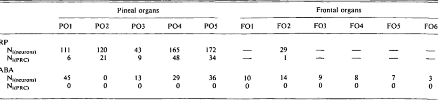
amplifica-Table 1. Comparison of the numbers of HRP-backfilled projection neurons and photoreceptor-like bipolar cells (PRC)
with the numbers of GABA-immunoreactive neurons in pineal and frontal organs of the frog Rana esculenta"
PO1
Pineal organs Frontal organs
PO2 PO3 PO4 PO5 FO1 FO2 FO3 FO4 FO5 FO6
HRP Ni(neurons) Ni(PRC) GABA Ni(neurons) 111 6 45 0 120 21 0 0 43 9 13 0 165 48 29 0 172 34 36 0 10 0 29 1 14 0
"Note that HRP-backfilling and GABA-immunocytochemistry was performed in different pineal organs. Numbers of neurons are given as numbers of counted profiles corrected according to Abercrombie's (1946) formula.
Fig. 1. Pineal organ. Reconstructions of GABA-immunoreactive neurons from camera lucida drawings of serial 3-/xm-thick frontal sections, [a] Unipolar neuron. The primary dendrite (arrow) gives off three main branches. Cf. Fig. 3a. [bj Unipolar neuron exhibiting neurites with numerous varicosities (small arrows), [c] Bipolar neuron with varicose neurites, one of which exhibits terminal-like swellings on another GABA-IR cell body (arrow), [d] Two dorsally situated GABA-IR neurons with closely intertwined neurites (arrows), [e] The primary dendrite of a unipolar neuron (black curved arrow) is in close apposition to the terminal-like swellings (small arrows) of a bipolar neuron (open curved arrow), [f] Dorsally situated GABA-IR neurons have neurites in close apposition (small arrows). The dotted lines denote the border of the central lumen of the pineal organ, b: bipolar neuron; m: multipolar neuron; and u; unipolar neuron. Scale bar = 50 pm.
tion of the diaminobenzidine-peroxidase reaction. In the pi-neal organ, only a few cells with a weak labeling (only slightly more intense than the background; Fig. 6) were observed after incubation with b d l 7 . The staining in the pineal organ lacked the somewhat granular appearance associated with the spe-cific immunoreactivities observed in the retina and in the brain (Figs. 6-10) and was thus considered to be unspecific back-ground staining. No labeling was observed in the frontal organ with b d l 7 , and no labeling was observed in either the pineal or frontal organs with bd24.
No attempt was made to map the total distribution of bdl7 immunoreactivity in the frog brain. Instead a few areas, known to receive input from the pineal organ and/or the retina, will be briefly described (nomenclature according to Neary & North-cutt, 1983).
A diffuse bdl7 immunoreactivity was observed in the cen-tral neuropil of the dorsal habenular nucleus (Fig. 7a). Its dis-tribution coincided with that of GABA-IR terminals (Fig. 7b). Caudally, the strongest labeling was confined to the lateral part of the medial subdivision (cf. Kemali & Lazar, 1985). In the
GABA and FMRFamide in frog pineal 403
Fig. 2. Pineal organ. Reconstructions of GABA-immunoreactive neu-rons from camera lucida drawings of 3-/mi-thick horizontal serial sec-tions, [a] A unipolar neuron with short dendritic branches, [b] Some GABA-IR neurons extend their neurites in the horizontal plane. One neurite of a multipolar neuron is seen in close contact (arrows) with the cell body of a unipolar neuron, m: multipolar neuron; and u: unipo-lar neuron. Scale bar = 50 jim.
ventral habenular nucleus, a relatively distinct band of immu-noreactivity was observed only caudally, in the lateral part (Fig. 6). The immunoreactivity was consistently strongest in the right habenular nuclei. In the anterior thalamic nucleus very weakly immunoreactive cell somata were observed in the ante-rior part. Posteante-riorly, diffuse non-perikaryal labeling extended into the neuropil of the central thalamic nucleus. The corpus geniculatum thalamicum contained diffuse immunoreactivity. Strong immunoreactions with bdl7 were observed in the granular layer of the cerebellum (Fig. 8a). Only weak immuno-reaction was observed in the molecular layer, whereas the Pur-kinje cells were immunonegative. No immunoreaction was observed with bd24. The granular layer and some of the cell bodies in the molecular layer were GABA-IR (Fig. 8b).
Of the monoclonal antibodies against the two GABAA/ benzodiazepine receptor (BZR) subunits, only bdl7 labeled neu-rons in the frog retina. Cell somata in the inner nuclear layer that were immunoreactive with bdl7 were confined to the mid-dle sublayer (Figs. 9 and 10). Their immunoreactive processes penetrated the inner portion to reach the inner plexiform layer. Here, immunoreaction was sometimes confined to three bands, of which the two innermost merged in most parts of the retina (Fig. 10). A diffuse immunoreactivity was associated with the
outer plexiform layer (Fig. 10). No immunoreactive cell bodies were observed in the ganglion cell layer.
In the mouse retina, cell somata immunoreactive with bdl7 were located in the inner nuclear layer. The largest numbers ap-peared to be situated approximately in the inner half of this layer. The somata were often very weakly labeled (Fig. lla). Weakly labeled cell bodies were situated in the ganglion cell layer (Fig. lib). Immunoreactivity was distributed throughout the inner plexiform layer, with a somewhat heavier labeling ap-parently associated with sublayers 2, 3, and 5 (Fig. 11).
FMRFamide-immunoreactive elements
FMRFamide-like immunoreactive (FMRF-IR) axons were ob-served in large numbers throughout the brain. FMRF-IR cell bodies were located in the periventricular hypothalamus: rela-tively large neurons (15-20 ^m diameter) were located close to the ependyma of the third ventricle (Fig. 12). Incubation with liquid phase-absorbed antiserum greatly decreased the number of immunoreactive structures in the brain and pineal organ, but did not abolish all immunoreactivity. Since it is known that FMRFamide antisera may have affinities for pancreatic poly-peptide-like substances, as well as other RFamides (see Discus-sion), we can only conclude that the structures recognized by the FMRFamide antibody contain one or more substances re-lated to, but possibly different from, FMRFamide.
FMRFamide-IR axons could be followed from the habenular commissure into the ventral wall of the pineal organ (Fig. 13a). However, the largest number of axons appeared to enter the pi-neal organ at its caudal pole from the posterior commissure. Here, some axons branched and apparently terminated in the pineal parenchyma (Fig. 13b and 13c), while others only seemed to make a detour through the pineal parenchyma on their way to the contralateral side of the brain via the posterior commis-sure. Although a small number of FMRFamide-IR axons could be followed rostrally in the pineal organ for some distance, most axons were confined to the caudal (proximal) portion of the pineal organ.
FMRFamide-IR axons were mostly located close to the basal lamina of the pineal parenchyma. It was, however, not possi-ble to ascertain whether they were associated with any special cell type. No FMRFamide-IR cell bodies were observed in the pineal organ.
Only very weak FMRF-immunoreactivity was observed in the frontal organ. Immunoreactive punctae, indicative of var-icose fibers, were sparsely distributed throughout the parenchyma.
Discussion
Are the GABAergic neurons interneurons?
We have demonstrated, by means of immunocytochemistry, the presence of putatively GABAergic neurons in the photosensory pineal complex of the frog. The GABA-IR neurons were of similar appearance in the extracranial frontal organ and in the intracranial pineal organ proper. Comparison of numbers, mor-phology, and location of GABA-IR neurons with retrogradely HRP (horseradish peroxidase)-labeled neurons led to the con-clusion that at least the large majority of GABA-IR neurons constitute interneurons. This interpretation was supported by the absence of GABA-IR axons in the pineal tract.
l\
a
rfl*
Fig. 3. Pineal organ. GABA-immunoreactive neurons. Frontal sections (3 nm). [a] The primary dendrite of a dorsally situated unipolar neuron (curved arrow) emits three branches (arrows) into the pineal parenchyma. This neuron is the one in Fig. la. [b] Dorsomedially situated neurons. One neuron (black curved arrow) extends its primary dendrite (arrow) ventrally. This neuron is the unipolar in Fig. le, whereas the one marked with the open curved arrow is the bipolar one. [c] Thin GABA-IR neurites (arrow) course towards the pineal tract (asterisk). GABA-IR neurites end with terminal-like swellings (small curved arrows) ad-jacent to an immunonegative cell body (black star; higher magnification in Fig. 3e). [d] The thin neurites from Fig. 3c end with terminal-like swellings around (arrows) and within (arrowhead) the pineal tract (asterisk; higher magnification in Fig. 3f). The neuron marked with a curved arrow is the one identically labeled in Fig. If. [e] Higher magnification of Fig. 3c. [f ] Higher magnification of Fig. 3d. Central lumen of the pineal organ, stars; pineal tract, asterisk. Scale bar is 50 ^m for Fig. a-d, and 10 fim for Fig. e-f.
GAB A and FMRFamide in frog pineal 405
Fig. 4. Frontal organ. Reconstructions of GABA-immunoreactive neurons from camera lucida drawings of 3-/xm-thick horizontal
serial sections, [a-d] Different morphological varieties of multipolar neurons (m). All have irregularly branching varicose neurites. [e] Unipolar neuron (u). [f] A unipolar neuron (u) and a multipolar neuron (m) with closely intertwined neurites (arrows). Scale bar = 50 jtm.
Pineal ganglion cells (projection neurons), as visualized when backfilled with HRP (Ekstrom & Meissl, 1990), tend to be located directly below the basal lamina of the pineal epithe-lium with dendrites forming a superficial plexus. GABA-IR neurons have also peripherally located cell bodies, but do not contribute neurites to a superficial plexus. Also, typical bipo-lar (photoreceptor-like) cells with a short dendritic protrusion towards the pineal lumen appear to constitute a class of pineal ganglion cells. No GABA-IR bipolar, photoreceptor-like, cells were observed in the pineal organ. It is worth noting that some of the so-called amacrine-like neurons visualized in the frog pi-neal organ, with supravital methylene blue-staining (Paul et al., 1971; Fig. 4g and 4h), have a morphology more similar with the
GABA-IR neurons than neurons with axonal projections into the pineal tract.
The absence of GABA immunoreactivity in the pineal nerve and tract is not per se evidence that all GABA-IR neurons are interneurons. Nevertheless, it is a strong indication, since the antibody employed is a powerful marker of GABAergic neurites in the retina (Agardh et al., 1987; Ostholm et al., 1988), tele-ost pineal organ (Ekstrom et al., 1987; Ostholm et al., 1988), and brain (Ekstrom, in preparation).
In the pineal organ, the numbers of GABA-IR neurons are much smaller than the numbers of HRP-backfilled ganglion cells (Table 1). Taken together with the morphological differences, it appears that at least the majority of the GABA-IR neurons
' : . ; - < & • '
Fig. 5. Frontal organ. GABA-immunorcactive neurons. Horizontal sec-tions (3 ^m). [a,b] Multipolar neurons with large dendritic trees (ar-rows), [c] Neurons with cell bodies (arrowheads) and dendrites (arrows) in close apposition to the exit of the pineal nerve (asterisk). Scale bar = 50 iim.
constitute a population different from the ganglion cells. If any GABA-IR neurons in the pineal organ are projection neurons, the most likely candidate would be the dorsally situated neurons that emit neurites toward the pineal tract.
Also in the frontal organ, the numbers of GABA-IR neurons are smaller than the number of HRP-backfilled neurons (al-though more difficult to compare with only one frontal organ sufficiently well backfilled; Table 1). However, when compar-ing the morphology of centrally projectcompar-ing neurons (Eldred & Nolte, 1981; see part Ekstrom & Meissl, part I, in this issue) with the GABA-IR neurons, it appears that most projection neurons are ventrally located with dorsally and laterally extend-ing dendrites. Cell somata and dendrites would then probably
not appear in the same (semithin) horizontal section. However, in semithin horizontal sections through the frontal organ, GABA-IR cell somata and dendrites can be observed in the same section. Also, GABA-IR neurons seem to be relatively evenly distributed along the dorsoventral axis. Taken together, these data suggest that also the GABA-IR neurons of the fron-tal organ are interneurons.
GABA-IR neurons in the frontal organ appeared to be of approximately the same size (Table 2), and to possess dendrites with a branching pattern closely similar to those of the pineal organ. Thus, morphological differences reflecting differences in the physiological responses to light stimulation should prob-ably not be sought in neuronal shapes and/or dendritic arbo-rizations but at the subcellular level, e.g. by electron-microscopic analysis of the synaptic organization of identified neural elements.
The close apposition of neurites from neighboring GABA-IR neurons indicate communication between these cells, perhaps reciprocal inhibitory synapses. However, this interpretation must await confirmation by immunocytochemical studies at the electron-microscopic level.
A direct way to prove that the GABA-IR neurons and the projection neurons constitute different neuronal populations would be to combine retrograde tracing with immunocytochem-istry in order to visualize both types of neurons in the same sec-tion. So far, this has not been successful in the pineal organ of the frog (unpublished observations). The reason for this is prob-ably that the conditions for optimal HRP backfilling is not compatible with optimal GABA immunocytochemistry in the pineal organ.
Comparison with earlier studies of GABA in the pineal complex
With the same GABA antiserum as in the present study, Ekstrom et al. (1987) demonstrated two populations of GABA-IR neu-rons in the pineal organ of the rainbow trout: large (20-30 /*m diameter) multipolar interneurons and small (ca. 10 ^m diam-eter) centrally projecting neurons. The numbers of neurons ob-served were small and varied greatly between specimens. In addition, a pronounced GABA immunoreaction was observed in glial elements (Ekstrom et al., 1987). In the present study, we observed no GABA-IR glial elements, although background labeling was somewhat higher in the pineal and frontal organs than in surrounding tissue (e.g. skin, paraphyseal complex), perhaps indicating diffusion of GABA during tissue processing. Further support for the presence of GABAergic neurons in pi-neal complexes may be gained from the study of the parietal eye of a lizard by Engbretson and Battelle (1985). Here it was shown that lizard parietal eyes may synthesize GABA from its precursor glutamic acid, and that [3H]-GABA is taken up by neurons (and lens cells and glial elements) in the parietal eye.
Physiologic action of GABA in the pineal organ
The pineal ganglion cells —the centrally projecting neurons — show a spontaneous firing of action potentials that is highest in the dark and is suppressed by light (Dodt & Jacobson, 1963). Iontophoretic application of GABA also suppresses the spon-taneous firing in the dark. However, the suppressive effects of GABA and light are not additive. Rather, a brief light
stimu-GAB A and FMRFamide in frog pineal 407
Table 2. Comparison of the mean sizes of neuronal somata revealed in the pineal and frontal organs of the frog, Rana esculenta, by means of retrograde filling with HRP or GABA-immunocytochemistry. HRP-backfilled
somata are divided in two classes, neurons and photoreceptor-like bipolar cells (PRC)a
HRP Neurons s S.D. I S.D. PRC s S.D. / S.D. GABA Neurons s S.D. / S.D. PO1 n = 21 10.29 (2.4) 13.88 (3.86) n= 1 7.05 — 17.62 — n = 30 10.88 (2.43) 15.98 (3.11) PO2 n = 33 11.54 (2.23) 14.95 (3.1) 7! = 5 7.77 (1.29) 24.44 (2.10) n = 0 Pineal PO3 n = 1 9.4 (2.35) 13.09 (4.04) n = 5 9.4 (3.71) 28.2 (4.7) n = 13 11.75 (1.66) 16.08 (2.85) organs PO4 n = 13 10.48 (2.06) 16.09 (3.82) n = l 8.39 (1.84) 21.82 (1.78) n= 18 11.35 (1.20) 13.84 (2.12) PO5 n= 15 9.24 (1.65) 13.63 (3.10) n = 6 7.44 (1.77) 31.33 (6.07) n = 28 11.16 (1.98) 14.93 (2.32) EPO n = 89 10.53 (2.30) 14.5 (3.51) n = 24 8.23 (1.7) 25.9 (5.55) n = 89 11.2 (1.97) 15.24 (2.75) FO1 n = 22 11.54 (1.61) 18.37 (3.53) FO2 n= 19 12.12 (2.11) 15.21 (2.52) n = 1 7.05 — 23.5 — n = 22 14.1 (1.45) 17.84 (2.48) Frontal organs FO3 n = 17 16.73 (3.51) 22.67 (5.51) FO4 71 = 10 13.87 (1.33) 17.16 (2.49) FO5 n = 8 14.98 (1.22) 17.62 (1.26) FO6 n = 4 15.28 (1.36) 18.21 (1.18) EFO TI = 83 14.07 (2.71) 18.88 (3.93)
Measures are given as mean lengths in microns for the short (s) and long (I) axes of the cells (see also the text and Ekstrom & MeissI, 1990). n = the number of measured profiles; this may thus be larger than the actual (corrected) number of neurons (cf. Table 1). Standard deviations (S.D.) of the samples are given in parentheses.
lus interferes with the suppressive effect of GABA, eliciting an OFF response and generally shortening the duration of inhibi-tion by GABA (MeissI & George, 1985).
The suppressive effect of light on the spontaneous firing of ganglion cells may be explained by taking the major pathway of information processing in the pineal organ as the photore-ceptor to ganglion cell synapse. Similar to retinal photorecep-tor cells, pineal phophotorecep-torecepphotorecep-tors appear to release excitaphotorecep-tory amino acid(s) in the dark (MeissI & George, 1984) that stimu-late the postsynaptic ganglion cells. Light hyperpolarizes the photoreceptor cell membrane, the release is reduced, and the ex-citatory input to the ganglion cells is reduced.
To explain the interactive effects of light and GABA on gan-glion cell activity, we have to introduce a mini-circuit where the excitatory feedforward pathway from photoreceptor to gan-glion cell is balanced by an inhibitory feedforward pathway via a GABAergic neuron that itself is postsynaptic to the photore-ceptor cell. In order to achieve this balance, the GABAergic neuron can be assigned several possible identities: GABAergic interneurons that are postsynaptic to photoreceptor cells and make (1) feedforward inhibitory contact on ganglion cells or (2) feedback inhibitory contacts on photoreceptor cells, GABAer-gic interneurons that receive input from excitatory ganglion cell axon collaterals and make feedback inhibitory contacts with (3) ganglion cells or (4) photoreceptor cells, or GABAergic gan-glion cells that are postsynaptic to photoreceptors and inhibit (5) ganglion cells or (6) photoreceptor cells by axon collaterals. Our results indicate that at least the large majority of GABA-IR neurons in the pineal organ are interneurons, and that at least some of the GABA-IR neurons are presynaptic to other neurons (Fig. 3e).
Assigning to the GABA-IR neurons the role of inhibitory in-terneurons that are presynaptic to ganglion cells and
postsyn-aptic to photoreceptor cells [(1) above], the interactive effects of light and GABA may be explained as follows. Ganglion cells are activated in the dark by direct excitatory inputs from pho-toreceptor cells, but this activation is balanced by the inhibitory input from GABAergic interneurons that are activated by input from photoreceptor cells. A light flash would then reduce the direct excitatory input to ganglion cells from photoreceptor cells, but would also reduce the excitation of GABAergic inter-neurons, thus reducing their inhibition of ganglion cell activity. In this way, the mini-circuit possesses two pathways for infor-mation transfer from photoreceptor cells to ganglion cells, first, the direct feedforward excitatory pathway from photoreceptor to ganglion cells, and second, the indirect feedforward inhibi-tory pathway to ganglion cells via GABAergic interneurons.
If the GABAergic interneurons make inhibitory feedback on photoreceptor cells [(2) above] that are presynaptic to ganglion cells, the same net result would ensue: a light flash would de-crease the activation of the ganglion cells, but also the tonic feedback inhibition of photoreceptors in the dark, thus attenu-ating the net decrease in release of excitatory transmitter. We cannot, on the basis of our immunocytochemical results, ex-clude the possibility that GABA-IR neurons form feedback in-hibitory synapses on photoreceptors.
If the GABAergic neurons are interneurons that are acti-vated by input from axon collaterals of excitatory ganglion cells, and make feedback inhibitory synapses on the ganglion cells [(3) above] or photoreceptor cells [(4) above], the light-in-duced decrease in direct activation of ganglion cells by photo-receptors would be balanced by a decreased inhibition by the GABAergic elements.
Also, if the GABAergic neurons are ganglion cells with recurrent axon collaterals that make inhibitory synapses on themselves or on other ganglion cells [(5) above] or on
photo-&
>7a
V .-6
• MS$$%?%:
9 a
9b
/
?- \f r »•.10
l l a
l i b
Figs. 6-11. bdl7-immunoreactive (bdl7-IR) and GABA-immunoreactive elements in the CNS and retina of frog and mouse. [6] The frog pineal organ (p) contains no labeling above background, whereas a weak bdl7 immunoreactivity is observed in the dorsal habenular nucleus (open arrow) and the ventral habenular nucleus (small arrows), d: dorsal habenular nucleus; /: central lumen of the pineal organ; and v: ventral habenular nucleus. [7] Dorsal habenular nucleus, frog. Adjacent sections show-ing that the distribution of bdl7-IR in the neuropil (a) corresponds well with that of GABA-IR (b). Note that the peripherally situated cell bodies (star) are unlabeled. [8] Cerebellum, frog. Adjacent sections showing that the granular layer (black star) is both bdl7-IR (a) and GABA-IR (b). Note that the cell bodies of the Purkinje cells (open stars) are surrounded by bdl7-IR pro-cesses, and that several cell bodies in the molecular layer are weakly GABA-IR (small arrows). [9] Frog retina, [a] bdl7-IR cell bodies (small arrows) are located in the middle of the inner nuclear layer (INL), whereas the inner plexiform layer (between ar-rows) is interspersed with bdl7-IR neurites, that in some locations can be seen to occur in higher densities in sublaminae 1, 3, and 5. [b] Control section: culture medium in which no monoclonal antibody-producing cells were grown was substituted for bdl7. [10] Frog retina. Higher magnification of Fig. 9a. Note the diffuse immunoreactivity in the outer plexiform layer (ar-rowheads). [lla,b] Mouse retina. bdl7-IR cell bodies occur primarily in the inner half of the INL, where strongly immunoreactive cells are located close to the IPL (small arrows) and weakly immunoreactive cells are farther away from it (curved arrow). Weakly bdI7-IR cell bodies are located in the ganglion cell layer (curved arrows). Scale bar is 50 ^m for Figs. 6, 8, and 9; 75 fim for Fig. 7; and 25 fim for Figs. 10 and 11.
CABA and FMRFamide in frog pineal 409
13c
Figs. 12-13. FMRFamide-immunoreactivity (FMRF-IR) in the frog brain and pineal organ. [12] FMRF-IR neurons located close to the ependyma lining the third ventricle. Note the large number of varicose processes. [13] Pineal organ, [a] At its rostral pole, close to the habenular commissure, a varicose FMRF-IR axon (arrow) enters the pineal parenchyma. Numerous FMRF-IR pro-cesses (arrowheads) course in the diencephalic roof. [b,c] Numerous FMRF-IR axons (arrows) enter the pineal organ from its caudal pole, close to the posterior commissure. Dark structures are erythrocytes (e) and pigment cells (star). Scale bar is 50 ^im for Fig. 12, and 28 /im for Fig. 13.
receptors [(6) above], the net result of an interaction between light and GABA would be the same.
Here, a word of caution is appropriate. The interaction of GABA and light was observed when exogenous GABA was in-troduced to the pineal neural circuitry (Meissl & George, 1985). Thus, the effects of the exogenous GABA must be expected to have been superimposed on the effects of endogenous GABA release, and to have saturated the GABA response. Although picrotoxin and bicuculline were able to block the inhibition by GABA of ganglion cell activity in the dark, these substances did not appear to affect the ganglion cell activity by themselves. Unfortunately, their effect on the interactive effects of GABA and light was not investigated. To explain the attenuation by light of GABA-induced inhibition, we probably have to include non-GABAergic inhibitory neurons that are connected to pho-toreceptor cells and ganglion cells in the same way(s) as neurons (l)-(6) above.
Unfortunately, we have no information about the actions of GABA in the frontal organ. Although the intracranial pineal organ mainly functions as a luminance meter, the extracranial frontal organ could be a chromaticity detector, at least within a moderate range of light intensities (Dodt & Heerd, 1962; Dodt & Jacobson, 1963; Eldred & Nolte, 1978; Meissl & Dodt, 1981). The different actions of short and midspectral wavelengths on the projection neurons of the frontal organ might be mediated by inhibitory interneurons (Meissl & Dodt, 1981), although al-ternative mechanisms have been proposed. Hamasaki (1970) hy-pothesized that there may be two receptor populations with different photopigments that make synaptic contacts with a common ganglion cell. To date, we have no direct physiologi-cal recordings from pineal photoreceptors with different spec-tral sensitivities that may support this theory, although three types of photoreceptor cells may be distinguished on the basis of ultrastructure and opsin immunoreactivity (Vigh & Vigh-Teichmann, 1986; cf. Eldred & Nolte, 1981). Dodt (1963) and later Eldred and Nolte (1978) suggested that the chromaticity re-sponse may be due to photointerconversion of two states of a single visual pigment in a single type of photoreceptor. For this, again, we have no direct evidence.
GABAA/benzodiazepine receptors
A direct way to investigate whether pineal projection neurons are postsynaptic to GABAergic interneurons would be to dem-onstrate GABA receptors in their plasma membranes. Histolog-ical analysis of the distribution of GABA receptors is usually performed by exposing unfixed tissue to radioactively labeled GABA or GABA ligands, whereupon the binding sites are visualized by autoradiography (Richards et al., 1986). This ap-proach gives a poor resolution and would not allow identifica-tion of cell types in the minute pineal organ. Instead, we have taken advantage of the recent development of monoclonal an-tibodies against the a and 0 subunits of the GABAA/benzodi-azepine receptor complex (GABAA/BZR; Haring et al., 1985; Richards et al., 1986). Immunocytochemical detection of this receptor complex yields a resolution at the subcellular level (Richards et al., 1986).
GABAA/BZR-IR cell bodies and neurites were readily de-tectable with bdl7 in control tissue, the mouse retina, with a distribution similar to that reported from the rat retina (Richards et al., 1986). In frog brain and retina, bdl7 im-munoreactivity was distinct but much weaker. The
concentra-tion of GABAA/BZR-IR to the inner plexiform layer of the frog retina agrees with radiohistochemical studies of other ver-tebrates, using tritiated benzodiazepines (Yazulla, 1986). In light of this, the absence of immunoreactivity in the pineal organ is somewhat surprising, since Meissl and George (1985) showed that the GABAA receptor antagonists bicuculline and picro-toxin blocked the effect of GABA on pineal projection neurons. On the basis of the present results, we can only conclude that even if GABA receptors, with a structural similarity to the GABAA/BZR of mammals, are present in the central nervous system (CNS) of the frog, their concentration in the frog pineal and frontal organs are too low to allow immunocytochemical detection by use of the monoclonal antibodies bdl7 or bd24.
FMRFamide-like immunoreactive efferent axons
Although it is of great importance for the understanding of the functions of the photosensory pineal organ to identify the neu-roactive substance of the FMRF-IR axons*, is it of less impor-tance for the problem addressed in the present investigation: is there a central innervation of the pineal complex in the frog, the most extensively studied anamniote pineal organ? Earlier stud-ies have not been unequivocal on this point (see below), and we have explored the possibility that axons innervating the pineal complex of the frog may be immunoreactive with a commercial FMRFamide antiserum, as in teleosts (Ekstrom et al., 1988).
One difficulty connected with the interpretation that the FMRF-IR axons are efferent to the pineal and frontal organs lies in the possibility that absence of immunoreactivity in pineal nerve cell bodies only reflects lower levels of antigen than that present in the axons. This difference relates to the process of packaging of a propeptide in secretory granules, in which the propeptide molecules undergo posttranslational modifications during transport down the axon. Thus, an antibody directed against the final signal peptide may label only axons and termi-nals, unless (1) secretory granules containing processed signal peptide are accumulated in the cell body, or (2) the signal pep-tide epitope recognized by the antibody is present and accessi-ble also in the propeptide before packaging in secretory granules. Accumulation of secretory granules in' the cell bodies may be achieved by interruption of the axonal transport systems, e.g. by local application of colchicine. Unfortunately, the minute size of the frog pineal organ makes this approach impractical. Also, analysis of the synthesis ipathways of peptides likely to cross-react with the FMRF antibodies gives no further clue to the origins of the intrapineal FMRF-IR axons: pancreatic poly-peptides that cross-react with FMRFamide antisera (Triepel & Grimmelikhuijzen, 1984) are C-amidated in the secretory gran-ules along their passage down the axon (Boel et al., 1984), and could give rise to a similar staining of axons but not cell
bod-*The question whether the FMRF-IR axons in the intracranial, di-rectly photosensory, pineal organ of the frog (Rana esculenta) really contain FMRFamide cannot be answered by simple absorption tests; such tests only indicate the degree of recognition of similar epitopes on related molecules, i.e. the specificity of the antiserum. Even laborious solid-phase pre-absorptions with all substances that might conceivably be recognized by the polyclonal antiserum do not exclude the possibility that the purified antiserum still cross-reacts with unknown substances with epitope(s) very similar to the antigen. However, it is important to bear in mind that FMRFamide antisera may recognize pancreatic poly-peptides, and some neuropeptides that have the carboxyterminus-RFamide (Triepel & Grimmelikhuijzen, 1984; Ebberink et al., 1987).
GAB A and FMRFamide in frog pineal 411 ies. Instead, we have to look for correlative evidence from
mor-phological studies of frog pineal organs.
Neuroanatomical evidence for the presence of axons of cen-tral origin innervating the frontal organ has, until now, been somewhat contradictory. After applying HRP to the frontal or-gan, Eldred et al. (1980) found no retrogradely filled perikarya in the brain, and claimed that Zilles and Nickeleit (1979) had misinterpreted neuromelanin granules for HRP reaction prod-uct when describing widespread cell groups innervating the frontal organ. Later Kemali and De Santis (1983), using cobal-tic lysine tracing, described neurons in the pineal organ, hypo-thalamus, and retina that would provide an innervation of the frontal organ. The location of these neurons did not coincide with those described by Zilles and Nickeleit (1979).
In a comparative study of different types of nerve fibers in the pineal organ, Vigh-Teichmann et al. (1973) described nerve terminals containing large granular vesicles apparently terminat-ing on the basal parts of pinealocytes (photoreceptor cells) in the pineal organ of an urodele amphibian, Pleurodeles walt/ii. These axons, which may well represent a peptidergic innerva-tion of the pineal parenchyma, were similar to axons of hypo-thalamic neurons (Vigh-Teichmann et al., 1973).
Observations of axonal degeneration after lesions of the pi-neal nerve (i.e. the nerve connecting the intracranial pipi-neal or-gan with the frontal oror-gan) led Bottger and Bottger (1973) to the conclusion that axons, both afferent and efferent to the frontal organ, course in this nerve. This conclusion supported the physiological experiments of Morita (1971), who interpreted changes in the photic responses of the frontal organ to electri-cal stimulation of the pineal nerve as evidence for the presence of central axons innervating the projection neurons ("ganglion cells") of the frontal organ. The efferent impulses seemed to po-tentiate the light responses of the ganglion cells in the frontal organ (Morita, 1971). These results may be compared with the observations by Engbretson and Lent (1976) who were able to elicit efferent impulses in the parietal nerve of a lizard by expos-ing the intracranial pineal organ to norepinephrine and seroto-nin. Notably, these substances had differential effects on the light responses of the parietal eye, and the effects depended on whether the experiments were conducted during the animals' natural photophase or scotophase.
Because of the large numbers of FMRF-IR axons in the brain, it was not possible to follow the FMRF-IR axons from the pineal organ to their cell bodies of origin. Thus, our results do not allow the identification of the cell group(s) that supply the FMRF-IR innervation of the pineal complex in the frog. A recent study of the pineal organ in teleost fish demonstrated an innervation of FMRF-IR axons that seemed to originate, at least partly, in the telencephalic nucleus of the terminal nerve or nucleus olfactoretinalis (Ekstrom et al., 1988). No similar distribution of terminal nerve fibers is known from amphibians (Wirsig & Getchell, 1986). It seems probable that the FMRF-IR axons in the pineal complex of the frog originate elsewhere in the brain.
A central innervation is apparently not a feature directly linked with the evolution of the pineal organ from a photosen-sory to a photoneuroendocrine structure, but may represent a phylogenetically older trait. It is probably more related to the general pattern of centrifugal feedback innervations of sensory structures. However, there is so far no evidence for homologies either with the innervation of the pineal organ from the hypo-thalamus and/or habenular complex, as known from amniotes
(Korf & Wagner, 1980, 1981; Korf et al., 1982), or the innerva-tion by the nervus terminalis as known from teleosts (Ekstrom et al., 1988).
Conclusions
We have demonstrated the presence of GABA-immunoreactive neurons and FMRFamide-like immunoreactive axons in both the intracranial and extracranial portions of the pineal complex of the frog Rana esculenta. On the basis of neuronal morphol-ogy, it appears likely that at least the majority of the GABA-IR neurons represent a neuronal population different from the centrally projecting neurons. Thus, they appear to constitute a population of inhibitory interneurons. The FMRF-IR axons may represent a central innervation of this photosensory organ, although the location of their neuronal somata was not identi-fied in the present study.
Acknowledgments
The technical assistance of Ms. C. Rasmussen and Ms. R. Wallen, and photographical assistance of Ms. I. Norling are gratefully acknowl-edged. The authors would like to thank the two anonymous reviewers for a number of very useful suggestions to improve the manuscript. This study was supported by a Research grant from the Swedish Natural Science Research Council (Bl 8554-103) to P. Ekstrom.
References
AGARDH, E., BRUUN, A., EHINGER, B., EKSTROM, P., VAN VEEN, T H .
& Wu, J.-Y. (1987). Gamma-aminobutyric acid- and glutamic acid decarboxylase-immunoreactive neurons in the retina of different ver-tebrates. Journal of Comparative Neurology 258, 622-630. BARGMANN, W. (1943). Die Epiphysis cerebri. In Lehrbuch der
Ver-gleichenden Mikroskopischen Anatomie des Menschen, Vol. Vl/4, ed. VON MOLLENDORF, W., pp. 309-502. Berlin: Springer-Verlag.
BOEL, E., SCHWARTZ, T.W., NORRIS, K.E. & Fm., N.P. (1984). A cDNA
encoding a small common precursor for human pancreatic polypep-tide and pancreatic icosapeppolypep-tide. EMBO Journal 3, 909-912. BOTTGER, W.V. & BQTTGER, E.-M. (1973). Degenerationsstudien am
Nervus pinealis von Rana esculenta L. nach stirnorgannaher und -ferner Durchtrennung. Zeitschrift fur Zellforschung 136, 365-391. COLON, J.-P. (1971). Differentiation and regression of the cells of the
sensory line in the epiphysis cerebri. In The Pineal Gland, ed. WOL-STENHOLME, G.E.W. & KNIGHT, J. pp. 79-120. Edinburgh, England: Churchill Livingstone.
DODT, E. (1963). Photosensitivity of the pineal organ in the teleost, Salmo irideus (Gibbons). Experientia 19, 642-643.
DODT, E. (1973). The parietal eye (pineal and parietal organs) of lower vertebrates. In Handbook of Sensory Physiology, Vol. VII/3B,ed. JUNG, R., pp. 113-140. New York, Berlin, Heidelberg: Springer-Verlag.
DODT, E. & HEERD, E. (1962). Mode of action of pineal nerve fibers in frogs. Journal of Neurophysiology 25, 405-429. ' DODT, E. & JACOBSON, M. (1963). Photosensitivity of a localized region
of the frog diencephalon. Journal of Neurophysiology 26, 752-758.
EBBERINK, R.H.M., PRICE, D.A., VAN LOENHOUT, H., DOBLE, K.E.,
RIEHM, J.P., GERAERTS, W.P.M. & GREENBERG, M.J. (1987). The
brain of Lymnaea contains a family of FMRFamide-like peptides. PeptidesS, 515-522.
EKSTROM, P. & KORF, H.W. (1986a). Putative cholinergic elements in the photosensory pineal organ and retina of a teleost, Phoxinus phoxinus L. (Cyprinidae). Distribution of choline acetyltransferase
immunoreactivity, acetylcholinesterase-positive elements, and pi-nealofugally projecting neurons. Cell and Tissue Research 246, 321-329.
EKSTROM, P. & KORF, H.W. (1986ft). Substance P-like-immunoreactive neurons in the photosensory pineal organ of the rainbow trout, Salmo gairdneri Richardson (Teleostei). Cell and Tissue Research 246, 359-364.
EKSTROM, P. & MEISSL, H. (1988). Intracellular staining of physiolog-ically identified photoreceptor cells and hyperpolarizing interneurons in the teleost pineal organ. Neuroscience 25, 1061-1070. EKSTROM, P. & MEISSL, H. (1990). Neural elements in the pineal
com-plex of the frog, Rana esculenta, I: Centrally projecting neurons. Vi-sual Neuroscience 4, 389-397.
EKSTROM, P., VAN VEEN, T H . , BRUUN, A. & EHINGER, B. (1987).
GABA-immunoreactive neurons in the photosensory pineal organ of the rainbow trout form two distinct neuronal populations. Cell and Tissue Research 250, 87-92.
EKSTROM, P., HONKANEN, T. & EBBESSON, S.O.E. (1988). FMRFamide-like immunoreactive neurons of the nervus terminalis of teleosts in-nervate both retina and pineal organ. Brain Research 460, 68-75. ELDRED, W.D. & NOLTE, J. (1978). Pineal photoreceptors: evidence for a vertebrate visual pigment with two physiologically active states.
Vision Research 18, 29-32.
ELDRED, W.D. & NOLTE, J. (1981). Multiple classes of photoreceptors and neurons in the frontal organ of Rana pipiens. Journal of Com-parative Neurology 203, 269-295.
ELDRED, W.D., FINGER, T.E. & NOLTE, J. (1980). Central projections of the frontal organ of Rana pipiens, as demonstrated by the antero-grade transport of horseradish peroxidase. Cell and Tissue Research 211, 215-222.
ENGBRETSON, G.A. & BATTELLE, B.A. (1985). Identification of putative neurotransmitters in the lizard parietal eye. Investigative Ophthal-mology and Visual Science 26, 670-678.
ENGBRETSON, G.A. & LENT, CM. (1976). Parietal eye of the lizard: neu-ronal photoresponses and feedback from the pineal gland. Proceed-ings of the National Academy of Sciences of the U.S.A. 73, 654-657.
HAMASAKI, D.I. (1970). Interaction of excitation and inhibition in the Stirnorgan of the frog. Vision Research 10, 307-316.
HARING, P., STAHLI, C , SCHOCH, P., TAKACS, B., STAEHELIN, T. &
MOHLER, H. (1985). Monoclonal antibodies reveal structural homo-geneity of •y-aminobutyric acid/benzodiazepine receptors in different brain areas. Proceedings of the National Academy of Sciences of the U.S.A. 82, 4837-4841.
KEMALI, M. & DE SANTIS, A. (1983). The extracranial portion of the pi-neal complex of the frog (frontal organ) is connected to the pipi-neal, the hypothalamus, the brain stem, and the retina. Experimental Brain Research 53, 193-196.
KEMALI, M. & LAZAR, G. (1985). Cobalt injected into the right and left fasciculi retroflexes clarifies the organization of this pathway. Jour-nal of Comparative Neurology 2 3 3 , 1 - 1 1 .
KLEIN, D., WELLER, J. & MOORE, R. (1971). Melatonin metabolism: neural regulation of pineal serotonin: acetyl coenzyme A N-acetyl transferase activity. Proceedings of the National Academy of Sci-ences of the U.S.A. 68, 3107-3110.
KORF, H.W. (1974). Acetylcholinesterase-positive neurons in the pineal and parapineal organs of the rainbow trout, Salmo gairdneri (with special reference to the pineal tract). Cell and Tissue Research 155, 475-489.
KORF, H.-W. & MOLLER, M. (1984). The innervation of the mammalian pineal gland with special reference to central pinealopetal projec-tions. Pineal Research Reviews 2, 41-86.
KORF, H.-W. & OKSCHE, A. (1986). In Vertebrate Endocrinology: Fun-damentals and Biomedical Implications, ed. PANG, P.K.T. & SCHREIBMAN, M.P., pp. 105-145. Orlando, Florida: Academic Press. KORF, H.-W. & WAGNER, U. (1980). Evidence for a nervous connection between the brain and the pineal organ in the guinea pig. Cell and Tissue Research 209, 505-510.
KORF, H.-W. & WAGNER, U. (1981). Nervous connections of the pari-etal eye in adult Lacerta s. sicula Rafinesque as demonstrated by an-terograde and retrograde transport of horseradish peroxidase. Cell and Tissue Research 219, 567-583.
KORF, H.-W., ZIMMERMAN, N.H. & OKSCHE, A. (1982). Intrinsic neu-rons and neural connections of the pineal organ of the house spar-row, Passer domesticus, as revealed by anterograde and retrograde transport of horseradish peroxidase. Cell and Tissue Research 222, 243-260.
MAXWELL, M.H. (1978). Two rapid and simple methods used for the removal of resins from 1.0-^m-thick epoxy sections. Journal of Mi-croscopy 112, 253-255.
MEISSL, H. (1986). Photoneurophysiology of pinealocytes. In Pineal and
Retinal Relationships, ed. KLEIN, D.C. & O'BRIEN, P.J., pp.
33-45. New York: Academic Press.
MEISSL, H. & DODT, E. (1981). Comparative physiology of pineal pho-toreceptor organs. In The Pineal Organ: Photobiology —
Biochro-nometry—Endocrinology, ed. OKSCHE, A. & PEVET, P., pp. 61-80.
Amsterdam: Elsevier/North-Holland Biomedical Press.
MEISSL, H. & GEORGE, S. (1984). Electrophysiological studies on neu-ronal transmission in the frog's photosensory pineal organ. The ef-fect of amino acids and biogenic amines. Vision Research 24, 1727-1734.
MEISSL, H. & GEORGE, S. (1985). Effect of GABA and its antagonists, bicuculline and picrotoxin, on nerve cell discharges of the photosen-sory pineal organ of the frog, Rana esculenta. Brain Research 332, 39-46.
MORITA, Y. (1971). Post-tetanic activity changes of the frog's neurosen-sory pineal end vesicle (Stirnorgan). Pfliigers Archiv 328, 135-144. NEARY, T.J. & NORTHCUTT, R.G. (1983). Nuclear organization of the bullfrog diencephalon. Journal of Comparative Neurology 213, 262-278. •
OKSCHE, A. (1971). Sensory and grandular elements of the pineal or-gan. In The Pineal Gland, ed. WOLSTENHOLME, G.E.W. & KNIGHT, J. pp. 127-146. Edinburgh, England: Churchill Livingstone.
OSTHOLM, T., EKSTROM, P., BRUUN, A. & VAN VEEN, Th. (1988).
Tem-poral disparity in pineal and retinal ontogeny. Developmental Brain Research*!, 1-13.
PAUL, E., HARTWIG, H.-G. & OKSCHE, A. (1971). Neurone und zentral-nervose Verbindungen des Pinealorgans der Anuren. Zeitschrift fur Zellforschung 112, 466-493.
RICHARDS, G., MOHLER, H. & HAEFELY, W. (1986). Mapping benzodi-azepine receptors in the CNS by radiohistochemistry and immuno-histochemistry. In Neurohistochemistry: Modern Methods and Applications, pp. 629-677, New York: Alan R. Liss, Inc.
SHIOTANI, Y., YAMANO, M., SHJOSAKA, S., EMSON, P.C., HILLYARD, C.J.,
GIRGIS, S. & MACINTYRE, I. (1986). Distribution and origins of sub-stance P (SP)-, calcitonin gene-related peptide (CGRP)-, vasoactive intestinal polypeptide (VIP)-, and neuropeptide Y (NPY)-contain-ing nerve fibers in the pineal gland of gerbils. Neuroscience Letters 70, 187-192.
STUDNICKA, A.K. (1905). Die Parietalorgane. In Lehrbuch der ver-gleichende Anatomie der Wirbeltiere, Vol. 5, ed. OPPEL, A., pp. 1-254. Jena: Springer-Verlag.
TRIEPEL, J. & GRIMMELIKHUUZEN, C.J.P. (1984). A critical examination of the occurrence of FMRFamide immunoreactivity in the brain of guinea pig and rat. Histochemislry 80, 63-71.
VAN LEEUWEN, F. (1986). Pitfalls in immunocytochemistry with special reference to the specificity problems in the localization of neuropep-tides. American Journal of Anatomy 175, 363-377.
VIGH, B. & VIGH-TEICHMANN, I. (1986). Three types of photoreceptors in the pineal and frontal organs of frogs: ultrastructure and opsin immunoreactivity. Archivum Histologicum Japonica 49, 495-518.
VIGH-TEICHMANN, 1., VIGH, B. & AROS, B. (1973). CSF contacting
ax-ons and synapses in the lumen of the pineal organ. Zeitschrift fur Zellforschung 144, 139-152.
WAKE, K. (1973). Acetylcholinesterase-containing nerve cells and their distribution in the pineal organ of the goldfish, Carassius auratus. Zeitschrift fur Zellforschung 145, 287-298.
WAKE, K., UECK, M. & OKSCHE, A. (1974).
Acetylcholinesterase-con-taining nerve cells in the pineal complex and subcommissural area of the frogs, Rana ridibunda and Rana esculenta. Cell and Tissue Research 154, 423-442.
WIRSIG, C.R. & GETCHELL, T.V. (1986). Amphibian terminal nerve: dis-tribution revealed by LHRH and AChE markers. Brain Research 385, 10-21.
WURTMAN, R.J., AXELROD, J. & FISCHER, J.E. (1964). Melatonin
syn-thesis in the pineal gland: effect of light mediated by the sympathetic nervous system. Science 143, 1328-1330.
YAZULLA, S. (1986). GABAergic mechanisms in the retina. Progress in Retinal Research 5, 1-52.
ZnxES, K. & NICKELEIT, V. (1979). Efferent connections from the brain to the frontal organ in Rana temporaria demonstrated by labeling with horseradish peroxidase. Cell and Tissue Research 196, 189-192.
![Fig. 1. Pineal organ. Reconstructions of GABA-immunoreactive neurons from camera lucida drawings of serial 3-/xm-thick frontal sections, [a] Unipolar neuron](https://thumb-eu.123doks.com/thumbv2/123doknet/14929591.664643/4.939.184.749.122.793/pineal-reconstructions-immunoreactive-neurons-drawings-frontal-sections-unipolar.webp)
![Fig. 2. Pineal organ. Reconstructions of GABA-immunoreactive neu- neu-rons from camera lucida drawings of 3-/mi-thick horizontal serial sec-tions, [a] A unipolar neuron with short dendritic branches, [b] Some GABA-IR neurons extend their neurites in the h](https://thumb-eu.123doks.com/thumbv2/123doknet/14929591.664643/5.939.95.462.127.584/reconstructions-immunoreactive-drawings-horizontal-unipolar-dendritic-branches-neurites.webp)
![Fig. 3. Pineal organ. GABA-immunoreactive neurons. Frontal sections (3 nm). [a] The primary dendrite of a dorsally situated unipolar neuron (curved arrow) emits three branches (arrows) into the pineal parenchyma](https://thumb-eu.123doks.com/thumbv2/123doknet/14929591.664643/6.939.186.762.121.974/immunoreactive-frontal-sections-dendrite-dorsally-situated-unipolar-parenchyma.webp)
![Fig. 4. Frontal organ. Reconstructions of GABA-immunoreactive neurons from camera lucida drawings of 3-/xm-thick horizontal serial sections, [a-d] Different morphological varieties of multipolar neurons (m)](https://thumb-eu.123doks.com/thumbv2/123doknet/14929591.664643/7.939.208.737.128.799/frontal-reconstructions-immunoreactive-horizontal-different-morphological-varieties-multipolar.webp)
![Fig. 5. Frontal organ. GABA-immunorcactive neurons. Horizontal sec- sec-tions (3 ^m). [a,b] Multipolar neurons with large dendritic trees (ar-rows), [c] Neurons with cell bodies (arrowheads) and dendrites (arrows) in close apposition to the exit of the pi](https://thumb-eu.123doks.com/thumbv2/123doknet/14929591.664643/8.939.108.457.134.791/frontal-immunorcactive-horizontal-multipolar-dendritic-arrowheads-dendrites-apposition.webp)
