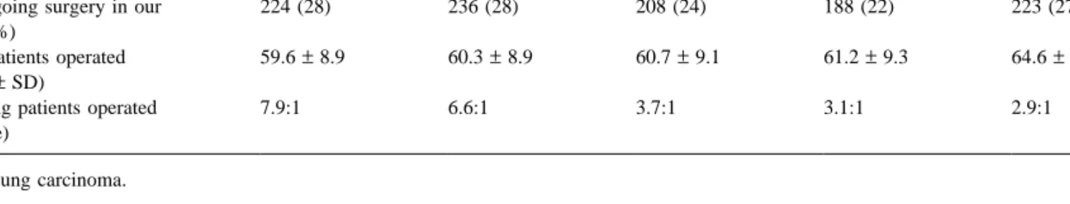
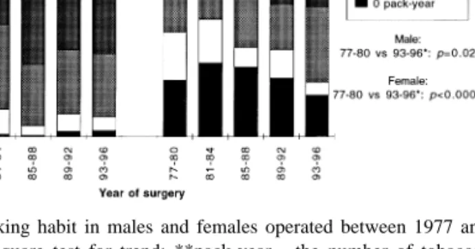
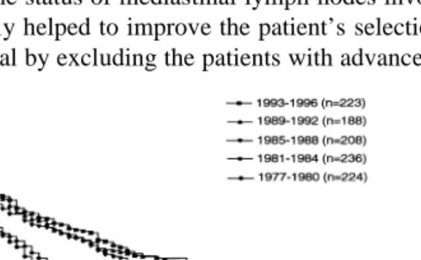
Time trend in the surgical management of patients with lung carcinoma
5
0
0
Texte intégral
Figure
Documents relatifs