© The Author 2012. Published by Oxford University Press on behalf of the British Occupational Hygiene Society doi:10.1093/annhyg/mes087
456
Evaluation of Decontamination Efficacy of Cleaning
Solutions on Stainless Steel and Glass Surfaces
Contaminated by 10 Antineoplastic Agents
ThomAS QuEruAu LAmEriE,
1SuSAnnE nuSSbAumEr,
2,5bErTrAnD DéCAuDin,
1,3*
SAnDrinE FLEury-SouvErAin,
2JEAn-FrAnçoiS GooSSEnS,
4PASCAL bonnAbry
2,5and PASCAL oDou
1,31Biopharmacy, Galenic and Hospital Pharmacy Department (EA 4481, IFR114), UFR Pharmacie,
Université Lille Nord de France, F-59000 Lille, France; 2Pharmacy, Geneva University Hospitals,
Gabrielle-Perret-Gentil 4, 1211 Geneva 14, Switzerland; 3Pharmacy, Lille University Hospital,
F-59000 Lille, France; 4Analytical Chemistry Department (EA 4481, IFR114), UFR Pharmacie,
Université Lille Nord de France, F-59000 Lille, France; 5School of pharmaceutical sciences,
University of Geneva, University of Lausanne, Switzerland
Received 21 March 2012; in final form 9 October 2012; Advance Access publication 7 December 2012
objectives: The handling of antineoplastic agents results in chronic surface contamination that must be minimized and eliminated. This study was designed to assess the potential of several chemical solutions to decontaminate two types of work surfaces that were intentionally contaminated with antineoplastic drugs.
methods: A range of solutions with variable physicochemical properties such as their hydrophilic/hydrophobic balance, oxidizing power, desorption, and solubilization were tested: ultrapure water, isopropyl alcohol, acetone, sodium hypochlorite, and surfactants such as dishwashing liquid (DWL), sodium dodecyl sulfate (SDS), Tween 40, and Span 80. These solu-tions were tested on 10 antineoplastic drugs: cytarabine, gemcitabine, methotrexate, etoposide phosphate, irinotecan, cyclophosphamide, ifosfamide, doxorubicin, epirubicin, and vincris-tine. To simulate contaminated surfaces, these molecules (200 ng) were deliberately spread onto two types of work surfaces: stainless steel and glass. recovered by wiping with a specific aqueous solvent (acetonitrile/hCooh; 20/0.1%) and an absorbent wipe (Whatman 903®), the residual contamination was quantified using high-performance liquid chromatography (hPLC) coupled to mass spectrometry. To compare all tested cleaning solutions, a perfor-mance value of effectiveness was determined from contamination residues of the 10 drugs.
results: Sodium hypochlorite showed the highest overall effectiveness with 98% contamina-tion removed. ultrapure water, isopropyl alcohol/water, and acetone were less effective with effectiveness values of 76.8, 80.7, and 40.4%, respectively. ultrapure water was effective on most hydrophilic molecules (97.1% for cytarabine), while on the other hand, isopropyl alcohol/ water (70/30, vol/vol) was effective on the least hydrophilic ones (85.2% for doxorubicin and 87.8% for epirubicin). Acetone had little effect, whatever the type of molecule. Among products containing surfactants, DWL was found effective (91.5%), but its formulation was unknown. Formulations with single surfactant non-ionics (tween 40 and span 80) or anionic (SDS) were also tested. Finally, solutions containing 10–2 m anionic surfactants and 20% isopropyl
alco-hol had the highest global effectiveness at around 90%. more precisely, their efficacy was the highest (94.8%) for the most hydrophilic compounds such as cytarabine and around 80.0% for anthracyclines. Finally, the addition of isopropyl alcohol to surfactant solutions enhanced
*Author to whom correspondence should be addressed. Tel: +33320964029; Fax: +33320959009. e-mail: bertrand. [email protected]
their decontamination efficiency on the least hydrophilic molecules. measured values from the stainless steel surface were similar to those from the glass one.
Conclusion: This study demonstrates that all decontamination agents reduce antineoplastic contamination on work surfaces, but none removes it totally. Although very effective, sodium hypochlorite cannot be used routinely on stainless steel surfaces. Solutions containing anionic surfactant such as SDS, with a high efficiency/safety ratio, proved most promising in terms of surface decontamination.
Keywords: decontamination/methods; detergents; equipment contamination/prevention and control; hazardous
substances/analysis; occupational exposure/analysis
inTroDuCTion
Nowadays, antineoplastic drugs are widely used in cancer therapies. Given their high toxicity, these sub-stances represent a potential risk for professionals at each step of the healthcare process. The National Institute for Occupational Safety and Health has esti-mated that around 5.5 million healthcare workers are potentially exposed to hazardous drugs in the USA. Despite publications of guidelines describing han-dling protocols and the use of biology safety cabinets (BSCs) or barrier isolators, surface contamination still exists in hospital pharmacy units (Acampora
et al., 2005; Crauste-Manciet et al., 2005; ISOPP, 2007). Environmental monitoring has indicated that all surfaces could be potentially contaminated (Turci
et al., 2003; Bussières et al., 2006; Heinemann et al., 2008; Käslin et al., 2010). Biological monitoring has proved that genotoxic effects can be detected by the Ames test or SOS chromo tests (urine muta-genicity, chromosomal aberrations, sister chromatid exchanges, and micronuclei) in the urine of nurses and pharmacy technicians (Poyen et al., 1988;
Sessink et al., 1994; Cavallo et al., 2005; Quillardet and Hofnung, 2009). Physical effects such as skin rashes, adverse reproductive effects (abortions, still-births, and congenital malformations), leukaemia, or cancers can occur (Skov et al., 1990; Connor and
McDiarmid, 2006). Traces of contamination have
been described in patients’ rooms and hospital efflu-ents, among operating theatre personnel, pharmacy technicians, and pharmacists (Mahnik et al., 2004, 2006; Sottani et al., 2010, 2011). Several papers have reported antineoplastic drug contamination on vials, surfaces, floors, countertops, carts, storage bins, waste containers, tabletops, chairs, and linen and in the atmosphere of pharmacy units (Mason, 2003; Connor and McDiarmid, 2006; Touzin et al., 2008). The main exposure routes have been by der-mal contact with contaminated surfaces and by inha-lation of particles (Kromhout et al., 2000; Fransman
et al., 2005; Connor and McDiarmid, 2006). To
confront this challenge, the pharmacist strategy is first to confine contamination in specific phar-macy areas within closed working areas (biosafety cabinets and isolators) and secondly to reduce the risk of contamination on pharmacists and on phar-macy technicians by using specific devices such as containment safety devices rather than needles for example. However, there is still a risk of accumula-tion over time. Efficient decontaminaaccumula-tion of surfaces is therefore of the utmost importance.
Several studies are available on the impact of decon-tamination procedures to reduce chemical contami-nation by cytotoxic agents. Raghavan et al. studied a water rinsing method on cisplatin decontamina-tion using liquid chromatography (Raghavan et al., 2000). Chlorine-based agents reduced the mutagen-icity of methotrexate (MTX) by inactivating it (Wren
et al., 1993). Earlier studies described various other solutions for cytotoxic agents on different surfaces. Multiple compounds [carmustine (BCNU), lomus-tine (CCNU), chlorozotocin, N-[2-chloroethyl]-N’-[2,6-dioxo-3-piperidinyl]-N-nitrosourea (PCNU), 1-(2-Chloroethyl)-3-(4-methylcyclohexyl)-1-nitrosoure (Methyl-CCNU), mechlorethamine, mel-phalan, chlorambucil, cyclophosphamide, ifosfamide, uracil mustard, and spiromustine] were degraded using nickel aluminium in a potassium hydroxyde solution without any toxic degradation (Lunn et al., 1989). Barek et al. proposed two methods for sur-face decontamination: the first reported almost total degradation of melphalan based on its oxidation by potassium permanganate in a sodium hydroxide solu-tion, and the second degraded multiple compounds (amsacrine, azathioprine, asparaginase, and thiotepa) using sodium hypochlorite and a Fenton reagent [an oxidizing solution based on hydrogen peroxide oxi-dized by catalyst ferrous iron (II)] (Barek et al., 1987, 1998). Oxidizing agents had already been tested on antineoplastic agents (Hansel et al., 1996; Castegnaro
et al., 1997; Roberts et al., 2006) and assessments established on different antineoplastic classes: oxa-zophosphorine or anthracycline molecules, using
hydrogen peroxide and sodium hypochlorite, which proved to be effective. Vaporized hydrogen peroxide and detergents were also investigated with positive results on 5-Fluorouracil, doxorubicin (DOX), and cyclophosphamide. Despite all previous studies, to date, no clear, effective, and evidence-based cleaning recommendations for daily practice exist. The aim of this paper is to evaluate the surface decontamina-tion efficacy of different cleaning soludecontamina-tions through a step-by-step controlled study, to provide advice for cleaning steps in pharmacy units.
The first part of this experimental work was per-formed on stainless steel, where aqueous solutions, aqueous alcohol solutions, or organic solutions were first screened. An improvement was then made on selected solutions and finally the optimal volume required for decontamination was determined. The second part was performed on glass to test the effec-tiveness of selected solutions. The final objective is to provide an effective and clear review of cleaning solu-tions for the periodic decontamination of work areas.
ExPErimEnTAL
Chemicals and reagents
Antineoplastic agents. The study was performed with the following commercially available cytotoxic drugs (Table 1). Reconstitution of etoposide phos-phate (Etopophos®), gemcitabine (Gemcitabine Teva®), and ifosfamide (Holoxan®) was obtained
with water for injection (Bichsel Laboratories, Interlaken, Switzerland). 5% sterile glucose (Sintetica, Bioren SA, Couvet, Switzerland) was used for the reconstitution of Endoxan®.
Liquid chromatography–mass spectrometry/mass spectrometry. Lichrosolv® HPLC grade acetoni-trile (ACN) and ultrapure water were purchased from Merck (Darmstadt, Germany), and formic acid (FA) came from Biosolve (Valkenswaard, the Netherlands).
Wiping and desorption material. Filter paper (Protein Saver TM 903 Card) was from Whatman (Dassel, Germany), and 1.5 ml polyethylene (PE) safe-lock tubes were from Eppendorf AG (Hamburg, Germany). Texwipe 3210 cleaning wipers, used as received as desorption material, were from ITW Texwipe (Kernersville, USA).
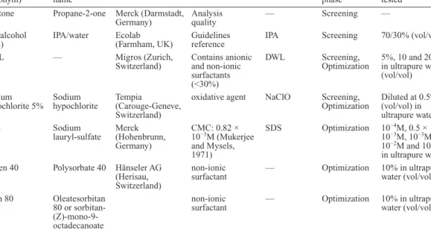
Cleaning solutions. Products used in cleaning solu-tion formulasolu-tions are summarized in Table 2. Simple solutions were tested as decontamination procedures. The choice of these solutions was based on current pharmaceutical practice and on scientific publica-tions. Two kinds of solutions were tested: “elimina-tion-type” solutions whose main action is to dissolve chemical products on the surface and “degradation-type” solutions that react with the chemical structure of compounds, leading to their degradation and the formation of expected non-cytotoxic compounds. Among “elimination-type” solutions, ultrapure Table 1. Commercially available cytotoxic drugs used in the study.
Molecules (acronym) Brand name Hydrophilic (H)/
Hydrophobic (h) Concentration Manufacturer Town, country Molecules in test
Irinotecan (IRI) Campto® h 20 mg ml−1 Pfizer AG Zürich, Switzerland
Cytarabine (CYT) Cytosar® H 20 mg ml−1
Gemcitabine (GEM) Gemcitabin Teva® H 20 mg ml−1 Teva Pharma AG Aesch, Switzerland
Vincristine (VI) Vincristine Teva® h 1 mg ml−1
Ifosfamide (IF) Holoxan® H 40 mg ml−1 Baxter AG Volketswil, Switzerland
Cyclophosphamide
(CP) Endoxan® H 20 mg ml
−1
Methotrexate
(MTX) Methotrexate Farmos® H 2.5 mg ml
−1 Orion Pharma Zug, Switzerland
Etoposide
phosphate (ETO) Etopophos® h 20 mg ml
−1 Bristol-Myers
Squibb SA Baar, Switzerland Doxorubicin (DOX) Doxorubine
Ebewe® h 2 mg ml
−1 Ebewe Pharma Cham, Switzerland
Epirubicin (EPI) Epirubicin Actavis
Solution® h 2 mg ml
−1 Actavis Regensdorf, Switzerland
Internal standard
water was tested single as a cleaning solution ref-erence and as solvent when mixed with surfactants such as dishwashing liquid (DWL), span 80, tween 40, and sodium dodecyl sulphate (SDS). Isopropyl alcohol (IPA) 70/30 was also studied because of rec-ommendations from guidelines for microbiological decontamination in chemotherapy production units (Le Garlantezec et al., 2011). Hydrophobic solvents such as acetone were used to determine its expected efficacy on the more hydrophobic compounds. Finally, among “degradation-type” solutions, a sodium hypochlorite solution, the most currently used solution to wash surfaces today, was also tested. Preparations of compound stock solutions,
calibration standards, and internal standard All solutions (i.e. drug reconstitutions and sample dilutions) were prepared in appropriate conditions (BSC, individual protection) for handling hazardous compounds such as cytotoxic agents. The preparation of solutions and standards was performed with brand drugs to avoid any direct contact of the operator with cytotoxic powder and to minimize contamination risk during the preparation of solutions. Aliquots of the internal standard (IS; 250 µg.ml−1) were pre-pared with a mixture of ACN and water (75/25, vol/ vol) and stored at −22°C for 12 months with no sample degradation observed. Stock solutions of IS were diluted daily to 50 ng ml−1 in 20% ACN (vol/ vol) with 0.1% FA (vol/vol) and were kept stable for at least 2 weeks at 2–8°C. A main stock solution
containing the 10 cytotoxic drugs was prepared by diluting at 20 µg.ml−1 concentration each cytotoxic compound in water. This solution was further diluted to obtain five independent stock solutions at 20, 40, 200, 1 000, and 4 000 ng ml−1 in 20% ACN (vol/ vol) and 0.1% FA (vol/vol). For calibration standards (CS), stock solutions were diluted with the IS solu-tion at 50 ng ml−1 to obtain five CS at 1, 2, 10, 50, and 200 ng ml−1.
Equipment and liquid chromatography–mass spectrometry/mass spectrometry conditions
Analyses were carried out with the Accela liq-uid chromatography system from Thermo Fisher Scientific Inc. (Waltham, MA, USA) consisting of a quaternary pump equipped with an online degas-ser, an auto sampler and a solvent platform. The chromatographic system was coupled to a Quantum Discovery MS from Thermo Fisher Scientific Inc. equipped with Ion Max electrospray ionization (ESI) interface and a triple quadrupole. The liquid chroma-tography–mass spectrometry/mass spectrometry sys-tem was monitored with Xcalibur software (Thermo Fisher Scientific). Separations were obtained on a ZORBAX SB-C18 RR column with an inner diameter of 2.1 mm, a length of 10 cm, and a par-ticular diameter of 3.5 µm from Agilent Technologies (Waldbronn, Germany). The liquid chromatogra-phy–mass spectrometry/mass spectrometry condi-tions and method validation have been described in detail elsewhere (Nussbaumer et al., 2010).
Table 2. Products used in formulations of cleaning solutions tested. Products
(acronym) International name Manufacturer Commentaries Abbreviation Experimental phase Concentrations tested Acetone Propane-2-one Merck (Darmstadt,
Germany) Analysis quality — Screening — Kleralcohol
(IPA) IPA/water Ecolab (Farmham, UK) Guidelines reference IPA Screening 70/30% (vol/vol)
DWL — Migros (Zurich,
Switzerland) Contains anionic and non-ionic surfactants (<30%)
DWL Screening,
Optimization 5%, 10 and 20% in ultrapure water (vol/vol) Sodium
hypochlorite 5% Sodium hypochlorite Tempia (Carouge-Geneve, Switzerland)
oxidative agent NaClO Screening,
Optimization Diluted at 0.5% (vol/vol) in ultrapure water
SDS Sodium
lauryl-sulfate Merck (Hohenbrunn, Germany) CMC: 0.82 × 10⁻³M (Mukerjee and Mysels, 1971) SDS Optimization 10−4M, 0.5 × 10⁻³M, 10−3M, 10−2M and 10−1M in ultrapure water Tween 40 Polysorbate 40 Hänseler AG
(Herisau, Switzerland)
non-ionic
surfactant — Optimization 10% in ultrapure water (vol/vol) Span 80 Oleatesorbitan
80 or sorbitan- (Z)-mono-9-octadecanoate
non-ionic
Decontamination
All tests were performed under a laminar airflow hood. The surface to be investigated (10 × 10 cm) was contaminated with 50 µl of stock solution sprayed on surface (solution containing all 10 cytotoxic agents at 4000 ng ml−1) using an adjustable volume micropi-pette. This voluntary contamination was repeated 10 times for each cleaning solution. For the drying step, contaminated surfaces were protected from light in a laminar airflow hood for a period of 1 h. After drying, different cleaning solutions were applied. These were prepared extemporaneously and used directly. 300 µl of each cleaning solution was poured onto a 100 cm² Texwipe 3210 wipe. A single standard motion from top to bottom was adopted to clean each surface. Wiping and analytical procedure
The wiping step can recover the residual con-tamination present on the surface after the decon-tamination step. A validated wiping procedure was performed to reclaim remaining cytotoxic com-pounds (Nussbaumer et al., 2012). To do so, a 1-cm² blotting paper (Whatman 903®) was soaked with 100 µl of an aqueous desorbing solution [ACN: water, 20/80 (vol/vol) with 0.1% FA]. The contami-nated surface was then wiped for 30 s, turning the blotting paper regularly. Blotting papers were placed in PE safe-lock tubes, and 1 ml IS solution at 50 ng ml−1 was added. Then samples were ultrasonicated for 20 min and centrifuged at 4000 rd min−1 for 5 min. All samples were immediately placed in the LC auto sampler at 15°C and analysed within the day. Decontamination evaluation
Data extracted from the analytical procedure cor-respond to residual contamination (RCi,m) of each antineoplastic agent. For each molecule, an effi-ciency index was generated (Effi,m; Equation 1). Then, to be able to compare cleaning solutions with each other, an overall effectiveness index was calcu-lated (Effi). It was the average of the 10 efficiency indexes (Equation 2). So, this Effi corresponded to the overall effectiveness of a solution on the 10 antineoplastic agents, during a single attempt. To validate the overall effectiveness of a solution, each cleaning procedure was tested 10 times. As a con-clusion, in this paper, the median value of those 10 attempts (EPvalue or Efficiency performance value) was used to compare cleaning solutions (Equation 3). Results were presented as follow: median value [minimum value − maximum value].
Effi m, =100−RCi m, (%) (1)
Effi Effi m
i
n
=∑ , (2)
EPvalue=median value (Eff ) ( =10)i n n (3) Standard deviation (SD) per compound for each cleaning solution was also calculated on 10 attempts. It indicated the reproducibility of the cleaning solu-tion on each compound. Due to the numerous man-ual steps throughout the procedure, its acceptance threshold was arbitrarily set at 10%.
Sequence of experiments
In the first part of the study, tests were performed on stainless steel. The first “Screening” step involved screening solutions with various physicochemical characteristics. Working on a solubility procedure, tests were carried out with ultrapure water, aqueous-alcoholic solutions and organic solvents such as ACN. An oxidative solution was assessed using an aqueous solution of 0.5% sodium hypochlorite. At last, complex micellar formulations such as DWL diluted in ultrapure water were tested to focus on sur-factant molecules. In the “Optimization” step, other detergent solutions were also tested using single anionic and neutral surfactants. SDS was especially focused on to consider the impact of its concentra-tion on decontaminaconcentra-tion efficacy. Different formula-tions of aqueous-alcoholic soluformula-tions with stable SDS concentration were also tested to reduce surfactant deposit. Up to this point, tests were performed with normalized surfaces and volumes, non-representa-tive of current decontamination activity. So, in the “Practical” step, additional tests were carried out over a 0.2 m² surface area with different volumes of optimized solution to simulate current cleaning methods. Finally, the second part involved tests on glass to validate the effectiveness of our solution on the most commonly used materials in closed working areas. All data are summarized in Table 2.
Statistical analysis
Statistical analysis was performed by analysis of variance on ranks following the method of Conover and Iman (Conover and Iman, 1981). This method was used to compare the effectiveness performance value of cleaning solutions. When this analysis revealed a significant P value (P < 0.05), contrasts were established with the Tukey–Kramer test to detect significant differences between couples of cleaning solutions with a statistical threshold of 5%. Analysis was performed with XLSTAT® software (Addinsoft).
rESuLTS
Screening phase
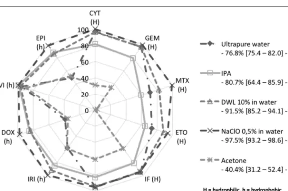
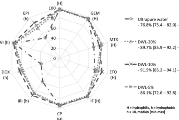
Considering physicochemical properties of the 10 antineoplastic agents, two groups of molecules can be distinguished: first one corresponding to the most hydrophilic substances with cytarabine (CYT), gemcitabine (GEM), MTX, etoposide phos-phate (ETO), cyclophosphamide (CP), and ifos-famide (IF) and second one to more hydrophobic compounds with irinotecan (IRI), DOX, vincristine (VI), and epirubicin (EPI). All data and statistical analyses performed on the stainless steel surface are summarized in Fig. 1 and Table 3.
Ultrapure water, aqueous alcohol, and organic sol-vents. Ultrapure water effectiveness was consid-ered to be insufficient (Fig. 1). It was effective to remove CYT, GEM, IF, CP, and VI, but for MTX, ETO, IRI, DOX, and EPI, Effi,m values were between 39 and 73%. Reproducibility was low on hydropho-bic molecules (e.g. DOX 15.5%), except for VI. For IPA/water 70/30 (vol/vol), EPvalue was slightly higher than that of ultrapure water (P = 0.041). Effi,m values for hydrophilic molecules (CYT, GEM, MTX, ETO, IF, and CP) were lower than for ultrapure water and inferior to 90.0%. On the other hand, efficacy on the
most hydrophobic molecules (IRI, DOX, and EPI) was superior to that obtained with ultrapure water (Fig. 1). Acetone EPvalue was significantly lower than ultrapure water and IPA (both with P < 0.0001). Sodium hypochlorite. The 0.5% sodium hypochlo-rite had the highest EPvalues (97.5%) and was signifi-cantly superior to all other solutions (all P < 0.0001). All removal values were superior to 90.0% Table 3. For CYT, GEM, MTX, IRI, and VI, Effi,m were even superior to 99.0%. For these compounds, SDs were inferior to 5%. The lowest Effi,m were found for ETO, IF, and CP.
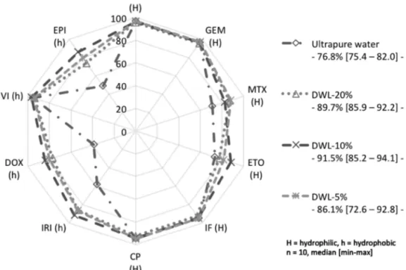
Surfactants. As shown in Fig. 1, 10% DWL obtained a 91.5% EPvalue. Results are reported in Fig. 2. Optimization phase: focus on surfactants molecules
Complex surfactants assessments (DWL). During screening phase, 10% DWL reached a promis-ing 91.50% EPvalue. Two more concentrations (5 and 20%) were also tested in order to observe the potential of DWL concentration on the antineoplas-tic removal. No significant difference was observed between the three DWL concentrations tested. 20% DWL obtained an EPvalue (89.7%) significantly
Fig. 1. Efficacy per compound and effectiveness performance of each cleaning solution on ten antineoplastic agents during tests
higher than ultrapure water (all P < 0.001), but not 5% DWL (86.1%, P = 0.001). Effi,m values for CYT, GEM, IF, CP, and VI were superior to 90.0% and SD values close to 10%, whatever the dilutions tested. On the other hand, for IRI, DOX, and EPI, the high-est Effi,m values were obtained using 10% DWL (Table 3).
Single surfactant assessments. Attempts realized on non-ionic surfactants (Tween 40 and Span 80):
Fig. 3 reports results obtained with surfactant solu-tions. Span 80 effectiveness was not significantly different from ultrapure water. It was significantly inferior to 10% DWL (P = 0.0001) and to 10−2M SDS (P > 0.0002). All of its Effi,m values were
inferior to the 90.0% threshold, whatever the polarity of the molecules. On the other hand, Tween 40 EPvalue was significantly superior to Span 80 (P = 0.018) but not significantly different from ultrapure water (P > 0.0610). Its Effi,m values were superior to 90.0% for CYT, GEM, and VI. Its lowest Effi,m values were obtained for DOX and EPI. 10−2M-SDS was sig-nificantly superior to Span 80 (P = 0.0002) but not to Tween 40 (P = 0.0610) and to 10% DWL (P = 0.9276; Fig. 3). Statistically as effective as 10−2 M-SDS, Tween 40 had nevertheless an SD superior to 10−2M-SDS values for CYT, GEM, MTX, ETO, IF, CP, and DOX. As a result, subsequent evaluations were made with SDS on a concentration range of 10−4–10−1M.
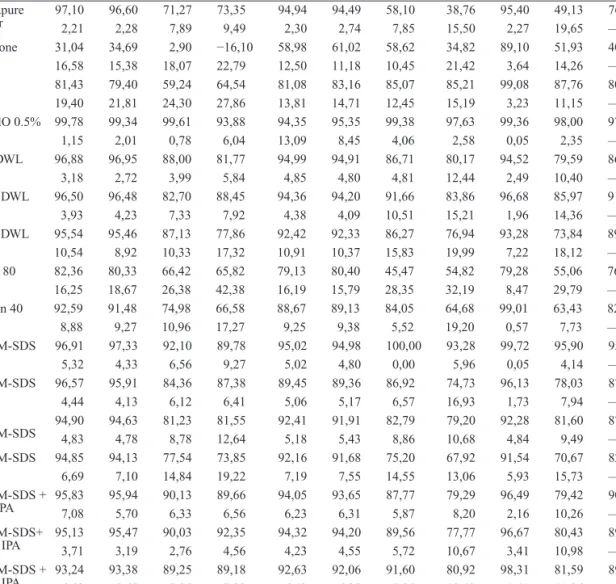
Table 3. Efficacy per compound of surfactant solutions on 10 antineoplastic agents and on stainless steel surface.
Modalities CYT GEM MTX ETO IF CP IRI DOX VI EPI EPvalue
Ultrapure water 97,102,21 96,602,28 71,277,89 73,359,49 94,942,30 94,492,74 58,107,85 15,5038,76 95,402,27 19,6549,13 76.8— Acetone 31,04 34,69 2,90 −16,10 58,98 61,02 58,62 34,82 89,10 51,93 40.4 16,58 15,38 18,07 22,79 12,50 11,18 10,45 21,42 3,64 14,26 — IPA 81,43 79,40 59,24 64,54 81,08 83,16 85,07 85,21 99,08 87,76 80.7 19,40 21,81 24,30 27,86 13,81 14,71 12,45 15,19 3,23 11,15 — NaClO 0.5% 99,78 99,34 99,61 93,88 94,35 95,35 99,38 97,63 99,36 98,00 97.5 1,15 2,01 0,78 6,04 13,09 8,45 4,06 2,58 0,05 2,35 — 5% DWL 96,88 96,95 88,00 81,77 94,99 94,91 86,71 80,17 94,52 79,59 86.1 3,18 2,72 3,99 5,84 4,85 4,80 4,81 12,44 2,49 10,40 — 10% DWL 96,50 96,48 82,70 88,45 94,36 94,20 91,66 83,86 96,68 85,97 91.5 3,93 4,23 7,33 7,92 4,38 4,09 10,51 15,21 1,96 14,36 — 20% DWL 95,54 95,46 87,13 77,86 92,42 92,33 86,27 76,94 93,28 73,84 89.7 10,54 8,92 10,33 17,32 10,91 10,37 15,83 19,99 7,22 18,12 — Span 80 82,36 80,33 66,42 65,82 79,13 80,40 45,47 54,82 79,28 55,06 76.8 16,25 18,67 26,38 42,38 16,19 15,79 28,35 32,19 8,47 29,79 — Tween 40 92,59 91,48 74,98 66,58 88,67 89,13 84,05 64,68 99,01 63,43 82.7 8,88 9,27 10,96 17,27 9,25 9,38 5,52 19,20 0,57 7,73 — 10−1M-SDS 96,91 97,33 92,10 89,78 95,02 94,98 100,00 93,28 99,72 95,90 95.4 5,32 4,33 6,56 9,27 5,02 4,80 0,00 5,96 0,05 4,14 — 10−2M-SDS 96,57 95,91 84,36 87,38 89,45 89,36 86,92 74,73 96,13 78,03 87.8 4,44 4,13 6,12 6,41 5,06 5,17 6,57 16,93 1,73 7,94 — 0.5 × 10⁻²M-SDS 94,90 94,63 81,23 81,55 92,41 91,91 82,79 79,20 92,28 81,60 87.5 4,83 4,78 8,78 12,64 5,18 5,43 8,86 10,68 4,84 9,49 — 10−3M-SDS 94,85 94,13 77,54 73,85 92,16 91,68 75,20 67,92 91,54 70,67 82.6 6,69 7,10 14,84 19,22 7,19 7,55 14,55 13,06 5,93 15,73 — 10−2M-SDS + 5% IPA 95,837,08 95,945,70 90,136,33 89,666,56 94,056,23 93,656,31 87,775,87 79,298,20 96,492,16 10,2679,42 90.3— 10−2M-SDS+ 20% IPA 95,133,71 95,473,19 90,032,76 92,354,56 94,324,23 94,204,55 89,565,72 10,6777,77 96,673,41 10,9880,43 89.6— 10−2M-SDS + 30% IPA 93,246,53 93,385,67 89,257,35 89,187,28 92,636,42 92,066,27 91,605,95 10,4380,92 98,311,41 11,3681,59 89.9— Notes: n = 10.
Attempts realized on anionic surfactant (SDS): Results obtained for decontamination solutions con-taining SDS at different concentrations are shown in
Fig. 4. The effectiveness of SDS increased propor-tionally to concentration. Indeed, the lowest EPvalue was obtained with 10−3M-SDS. These results were significantly lower than 10−1M-SDS (P < 0.0001) and 10−2M-SDS (P = 0.026) but not significantly different from 0.5 × 10⁻³M-SDS (P = 0.062). For both concentrations around the critical micellar con-centration (CMC) value (10−2M and 0.5 × 10⁻²M), no significant difference was found [87.8% (83.9 – 92.3) and 87.5% (83.9 – 92.3); P = 0.997]. For both concentrations, all Effi,m values were close to each other. For CYT, GEM, and VI, they were superior to 90.0% but slightly higher with “10−2M-SDS”. However, for DOX and EPI, efficacy was slightly lower with 10−2M-SDS. The highest effectiveness was obtained with 10−1M-SDS. Despite results sig-nificantly superior to 10−2M-SDS (P < 0.0001), this concentration presented a major drawback. Indeed, a thin surfactant film appeared from time to time on the surface after the cleaning step. Microbiological con-tamination could appear inside, making it necessary to reduce the risk of formation of the residual film. Attempts realized with improved anionic surfactant (SDS + IPA): To overcome the problem of surfactant
deposit and to increase solution evaporation, the for-mulation was tested with the addition of IPA. Despite containing as much as 20% IPA, a large deposit of surfactant still remained on the stainless steel sur-face when “10−1M-SDS + 20% IPA” was spread over it. Therefore, an SDS concentration of 10−2M was selected for further experiments. Results are reported in Fig. 5. IPA ranging from 5 to 30% was diluted in an aqueous solution and mixed with 10−2M-SDS. For all aqueous alcohol mixtures, EPvalues were significantly higher than those obtained for an ultrapure water solu-tion (P < 0.0001 except with “10−2M-SDS + 10%-IPA”, P = 0.031). However, no significant difference was found between 30%-IPA, 20%-IPA, 5%-IPA (and 10−2M-SDS without IPA; Fig. 5). EP
values around 90% were obtained in all cases. Nevertheless, the mixture containing 20%-IPA was the most suitable solution, thanks to SD values inferior to our threshold of 10% and lower than those of other mixtures (Table 3). As already mentioned, IPA improved the decontamina-tion efficacy of the most hydrophobic compounds, while SDS acted on hydrophilic molecules. More precisely, Effi,m obtained with “10−2M-SDS + 20%-IPA” were superior to SDS alone as far as the most hydrophobic molecules were concerned, but they were slightly lower for the two most hydrophilic mol-ecules, CYT and GEM (Table 3).
Fig. 2. Efficacy per compound and effectiveness performance of DWL dilutions on ten antineoplastic agents and on stainless
Fig. 3. Efficacy per compound and effectiveness performance of surfactant solutions on ten antineoplastic agents and on
stainless steel surface
Fig. 4. Efficacy per compound and effectiveness performance of SDS range concentration on ten antineoplastic agents and on
Practical phase: volume sprayed and decontamination effectiveness
In this test, the solution was sprayed directly on the contaminated surface (0.2 m²), and a new whole Texwipe 3210 was used for each trial. Results are presented in Table 4. Whatever the volume of mix-ture used (“10−2M-SDS + 20 %-IPA”), EP
value was superior to 90% and even to 93%. Effectiveness with “6 ml” was significantly higher than with “3 ml” [97.3% (96.5 – 98.1) versus 96.12% (92.77 – 97.27); P = 0.008] or with “1 ml” [97.3% (96.5 – 98.1) ver-sus 93.9% (78.4 – 94.4); P < 0.0001]. Moreover, SD was inferior to the 10% threshold (Table 4).
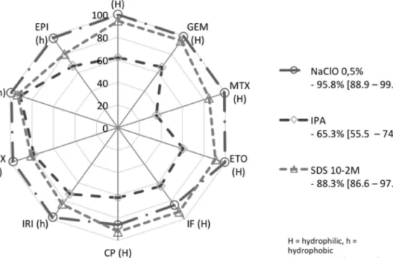
Decontamination procedure glass versus stainless steel surface
Results are presented in Fig. 6, and all data are compiled in Table 5. Similar to results found on stainless steel, IPA was the least effective on glass. With Effi,m values superior to the 90% threshold except for IF and CP, sodium hypochlorite was the most effective and was significantly different from IPA (P < 0.0001) on glass. Nevertheless, it was not statistically different from “10−2M-SDS”, unlike its use on stainless steel. With an EPvalue of 88.3%, “10−2M-SDS” had the same effectiveness on glass as
on stainless steel. The dispersion of its values was less important than those of sodium hypochlorite, which may explain the non-statistical difference between the two modalities.
DiSCuSSion
As the most widely used solvent in cleaning solu-tions, ultrapure water had to be evaluated. Its per-formance highlighted the minimal perper-formance required for all other aqueous cleaning solutions. According to our results, its effectiveness is not suf-ficient and optimization was required, especially for hydrophobic compounds. IPA/water 70/30 (vol/ vol) was expected to improve decontamination of the most hydrophobic compounds. In practice, an improvement was found on IRI and anthracyclin compounds, but at the same time, deterioration was measured on the most hydrophilic ones. Acetone was no more suitable at improving the decontamination process. Despite a lower polarity than IPA, hydro-phobic molecules were not more effectively removed than with IPA. Furthermore, as far as hydrophilic molecules are concerned, acetone was the least effec-tive solution tested. As a result, evaluating the solu-bility of single solvents did not seem to be the proper solution for decontamination procedure. Two other
Fig. 5. Efficacy per compound and effectiveness performance of concentration range of optimised solutions, on ten
hypotheses were considered: oxidative action and modification of solvents’ solubility by adjunction of surfactants. For sodium hypochlorite, results are in accordance with those obtained by Hansel et al. who reported degradation efficacy for CP and an IF superior to 98.0% (Hansel et al. 1996). Nevertheless, despite its high decontamination potential, the use of sodium hypochlorite solutions has major drawbacks. First of all, the possibility of cytotoxic agents to be degraded in mutagenic residues has already been mentioned (Barek et al., 1987, 1998). To avoid this phenomenon, a time gap after cleaning (minimum 1 h) should be respected, but this delay is not feasi-ble in everyday pharmacy routine (Castegnaro et al., 1997). It is necessary to clean the surface after use with a soaked wipe, otherwise corrosion phenomena appear on metals such as stainless steel. Nowadays, most barrier isolators and BSCs are made of stainless steel and manufacturers do not recommend the use of sodium hypochlorite. Finally, according to United States Pharmacopeia (USP) (797, Table 2), sodium hypochlorite can cause side effects such as skin, eye, and respiratory irritations or systemic toxicity. To overcome these inconveniences, another decontami-nation method as surfactant should be considered. Already available on the food market, DWL could be convenient. The poorer efficacy of 5% DWL can be explained by an insufficient concentration of sur-factants to remove hydrophobic compounds properly because of the lack of micelle structures. With 20% DWL, a residual film was observed on the stainless steel surface, which persisted after the wiping step. This was probably the reason for the higher residual contamination observed (Table 3). This film can be removed with a large volume of water spilled over the surface, but this solution is not suitable within BSCs. None of these limits were found with the inter-mediate dilution 10% DWL. Even if its results were less effective than those obtained with 0.5% sodium hypochlorite, the main advantage of 10% DWL was undoubtedly its safety not only for humans but also for work surfaces. These results confirmed a previ-ous work that studied cyclophosphamide chemical contamination on a glass vial surface (Touzin et al., 2008). Nevertheless, as already mentioned, the exact composition of DWL was unknown and depending
on the supplier tested. DWL formulations are usu-ally based on mixtures of anionic and non-ionic sur-factants. For a better understanding of DWL action and to simplify formulation of cleaning solutions, subsequent experiments were focused on a single surfactant. Span 80 did not appear to be efficient, so attempts were not pursued further. Tween 40 and SDS were both effective on stainless steel surface. Nevertheless, after a brief literature review, SDS appeared to be the most widely employed surfactant on decontamination products. An additional benefit of SDS is that it is commercially available in cer-tified laboratory quality powdered form. The use of a standardized formulation would allow users to guarantee the quality of the cleaning agent. Despite its high effectiveness, SDS 10−1M was not selected because a residual film was noticed after each decon-tamination procedure. This residual film was similar to the one observed with 20% DWL. CMC is the main characteristic to take into account when using surfactants. This is found in our results. With a con-centration 10 times inferior to CMC (SDS 10−3M), the effectiveness of the cleaning solution decreased. The highest ratio “effectiveness/residual film on surface” was found for concentrations around CMC (10−2M and 0.5 × 10⁻²M). To promote the formation of micelles, concentration has to be superior to CMC. So, SDS 10−2M was selected as the cleaning solution for further experiments. To further minimize the risk of residual film in everyday use, adjunction of IPA in SDS formulation was tested. Deposit of surfactants was less serious on a stainless steel surface, and its removal by evaporation was found to be especially fast with the 20%-IPA concentration. Moreover, no decrease of effectiveness (compared with SDS 10−2M) was noticed using the “10−2M-SDS + 20%-IPA” mixture. Finally, the “10−2M-SDS + 20%-IPA” mixture presented the best balance between decon-tamination profile and reduced deposit and so was selected for further trials.
In our research so far, effectiveness has been tested on 100 cm² surfaces with 300 µl of decon-tamination solution, which is not representative of current decontamination in an isolator or a laminar airflow hood. Simulations of practical decontamina-tion on larger surfaces with different volumes were Table 4. Efficacy per compound of “10−2M-SDS + 20% IPA” solutions on 0.2 m² stainless steel surface.
Modalities (ml) CYT GEM MTX ETO IF CP IRI DOX VI EPI EPvalue
1 94,68 94,82 94,57 90,52 93,79 93,58 94,47 91,51 96,64 91,76 93.9
3 97,47 97,77 97,35 93,72 97,35 97,19 96,44 93,83 97,11 93,96 96.1
6 97,46 97,65 97,91 95,96 97,69 97,47 99,00 95,22 98,36 95,23 97.3
needed to validate both the effectiveness of the solu-tion and to quantify the volume needed to clean a large surface properly. The best results were obtained when the largest volume was used. Even when using “6 ml”, no deposition of surfactant film was found after the wiping procedure. Consequently, a calcu-lation by proportionality between the standardized surface (0.2m²) and a theoretical surface of 1 m² can be evaluated. As a result, ratio of cleaning agent to surface area between 20 and 30 ml m−² should be recommended as an informal rule to clean a stainless steel surface properly.
The most frequently encountered materials in BSCs and isolators are stainless steel and glass. Consequently, further experiments were performed on a glass surface with only three selected aqueous
mixtures. IPA/water 70/30 (vol/vol) is recommended by guidelines and has microbiological decontamina-tion effectiveness. During our study, 0.5% sodium hypochlorite, a “degradation-type” solution, pre-sented the best overall EPvalue, and finally, “10−2 M-SDS”, developed and tested by ourselves, presented the best ratio between performance and safety. IPA had the lowest EPvalue (65.3%) and presented no more advantage on glass than on stainless steel. NaOCl effectiveness (95.8%) was equivalent on both stain-less steel and glass surfaces. The variations observed between the two surfaces can be accounted for by their physicochemical characteristics. Glass as a more hydrophilic surface has high wettability, and stainless steel as metal has higher hydrophobicity and lower wettability. Finally, 10−2M-SDS with an
Fig. 6. Efficacy per compound and effectiveness performance of hypochlorite, IPA and SDS 10-2M on glass surface
Table 5. Efficacy per compound of cleaning solutions on glass surface.
Modalities CYT GEM MTX ETO IF CP IRI DOX VI EPI EPvalue
0.5% NaClO 99,748,33 99,028,41 99,235,83 99,536,66 84,9119,08 17,6186,34 98,227,81 97,588,60 99,420,68 97,356,88 95.8— 10−2M- SDS 94,115,74 94,694,82 84,5810,12 90,846,51 92,367,33 92,217,71 10,4983,76 16,0780,68 93,465,62 18,5880,14 88.3— IPA 62,11 65,85 35,70 60,67 64,09 62,08 72,52 78,11 91,15 67,36 65.3 16,44 18,08 23,97 15,91 19,02 27,45 11,38 10,36 0,96 9,47 — Notes: n = 6.
88.3 EPvalue was as effective on glass as on stainless steel surfaces.
ConCLuSion
The “degradation-type” solution represented by sodium hypochlorite was very attractive because of oxidation. However, stainless steel as a build-ing material in isolators and BSC prohibits its use. Moreover, this recommendation conveyed by manufacturers themselves is reinforced by the risk of producing agents with unknown chemical struc-tures and cytotoxic potential. The “elimination-type” solutions demonstrated promising results. The use of surfactants such as DWL proves to be efficient and reproducible. Previous studies had already proved its efficiency on glass surface, but those studies were performed with a single antineoplastic agent, the cyclophosphamide or the carboplatin (Lê
et al., 2012). In our study, the DWL efficiency was again found on 10 antineoplastic agents. Approved on both hydrophilic and hydrophobic agents, DWL had nevertheless a major drawback. Indeed, many manufacturers are on the household cleaning market with their own unknown formulation. Nevertheless, DWLs were very convenient and practical prod-ucts, and further tests will be performed to evaluate the relevance of their use in daily practical condi-tions. During our study, surfactants used alone have proved to be effective especially the SDS. Moreover, they have the advantage of being available with the pharmaceutical certification, which eases their use and approval in pharmacy units. SDS allows the desorption of antineoplastic agents and reinforces their solubility. Their effectiveness was successfully proved on both stainless steel and glass surfaces. However, the appearance of residual film at high concentrations can be a potential source of cross and bio contamination. To overcome this problem, IPA was added into the formulation. With a quantity of 20–30 ml m−², the “10−2M-SDS + 20% IPA” formu-lation was efficient when sprayed on both stainless steel and glass surfaces with an efficacy superior to 97% on a single run. This effectiveness confirms its suitable use in current practice. Nevertheless, this study was performed on standardized surfaces that cannot be substituted for production units used daily by healthcare workers, where additional parameters have to be taken into account. The best rated decon-tamination solvents will have to be tested further in real environments, as well as on other materials (transparent thermoplastic such as poly-methyl-methacrylate and polycarbonate) and on molecules (platine derivatives and 5-fluorouracil) for which no
data is available. The decontamination procedure could also be tested on various supports such as cyto-toxic packaging which is known to be contaminated in its industrial area.
FunDinG
Pharmacy of Geneva University Hospital (Geneva, Switzerland).
Acknowledgements— The authors kindly thank the Proteomics Core Facility at the Faculty of Medicine, University of Geneva (Switzerland) for the loan of the LC-MS/MS instrument. The authors wish to acknowledge the help and advice given on the development of the LC-MS/MS method by Dr L. Geiser, Swiss Centre for Applied Human Toxicology, Geneva, Switzerland.
rEFErEnCES
Acampora A, Castiglia L, Miraglia N et al. (2005) A case study: surface contamination of cyclophosphamide due to working practices and cleaning procedures in two Italian hospitals. Ann Occup Hyg; 49: 611–8.
Barek J, Castegnaro M, Malaveille C et al. (1987). A method for the efficient degradation of melphalan into nonmuta-genic products. Microchem J; 36: 192–7.
Barek J, Cvacka J, de Méo M et al. (1998) Chemical degra-dation of wastes of antineoplastic agents amsacrine, aza-thioprine, asparaginase and thiotepa. Ann Occup Hyg; 42: 259–66.
Bussières J-F, Sessink PJM, Prot-Labarthe S et al. (2006). Évaluation de l’exposition professionnelle aux antinéopla-siques dans une unité de pharmacie hospitalière. Archives Des Maladies Professionnelles Et De l’Environnement; 67: 880–8.
Castegnaro M, De Méo M, Laget M et al. (1997) Chemical degradation of wastes of antineoplastic agents. 2: six anthra-cyclines: idarubicin, doxorubicin, epirubicin, pirarubicin, aclarubicin, and daunorubicin. Int Arch Occup Environ Health; 70: 378–84.
Cavallo D, Ursini CL, Perniconi B et al. (2005) Evaluation of genotoxic effects induced by exposure to antineoplastic drugs in lymphocytes and exfoliated buccal cells of oncol-ogy nurses and pharmacy employees. Mutat Res; 587: 45–51.
Connor TH, McDiarmid MA. (2006) Preventing occupational exposures to antineoplastic drugs in health care settings. CA Cancer J Clin; 56: 354–65.
Conover WJ, Iman RL. (1981). Rank transformations as a bridge between parametric and nonparametric statistics. Am Stat; 35: 124–9.
Crauste-Manciet S, Sessink PJ, Ferrari S et al. (2005) Environmental contamination with cytotoxic drugs in healthcare using positive air pressure isolators. Ann Occup Hyg; 49: 619–28.
Fransman W, Vermeulen R, Kromhout H. (2005) Dermal expo-sure to cyclophosphamide in hospitals during preparation, nursing and cleaning activities. Int Arch Occup Environ Health; 78: 403–12.
Le Garlantezec P, Rizzo-Padoin N, Lamand V et al. (2011). Manipulation des médicaments anticancéreux à l’hôpital : le point sur l’exposition et sur les mesures de
prévention. Archives Des Maladies Professionnelles Et De l’Environnement; 72: 24–35.
Hansel S, Castegnaro M, Sportouch MH et al. (1996) Chemical degradation of wastes of antineoplastic agents: cyclophosphamide, ifosfamide and melphalan. Int Arch Occup Environ Health; 69: 109–14.
Heinemann A, Kiffmeyer T, Stüter H et al. (2008). Monitoring Effekt-Studie für Wischproben in Apotheken. Hambourg: Berufsgenossenschaft für Gesundheitsdienst und Wohlfahrtspflege (BGW).
ISOPP. (2007). Section 13 cleaning procedures. J Oncol Pharm Pract; 13: 55–61.
Käslin E, Merz B, Rüegger M et al. (2010). Contamination de surface lors de la manipulation de médicaments cytostatiques dans les établissements de santé. Suva Med; 2010: 59–72. Kromhout H, Hoek F, Uitterhoeve R et al. (2000) Postulating
a dermal pathway for exposure to anti-neoplastic drugs among hospital workers. Ann Occup Hyg; 44: 551–60. Lê LM, Jolivot PA, Sadou Yaye H et al. (2012) Effectiveness
of cleaning of workplace cytotoxic surface. Int Arch Occup Environ Health, in press.
Lunn G, Sansone EB, Andrews AW et al. (1989) Degradation and disposal of some antineoplastic drugs. J Pharm Sci; 78: 652–9.
Mahnik SN, Rizovski B, Fuerhacker M et al. (2004) Determination of 5-fluorouracil in hospital effluents. Anal Bioanal Chem; 380: 31–5.
Mahnik SN, Rizovski B, Fuerhacker M et al. (2006) Development of an analytical method for the determination of anthracyclines in hospital effluents. Chemosphere; 65: 1419–25.
Mason HJ. (2003) Cytotoxic drug contamination on the out-side of vials delivered to a hospital pharmacy. Ann Occup Hyg; 47: 681–5.
Mukerjee P, Mysels KJ. (1971) Critical Micelle Concentrations of Aqueous Surfactant Systems. Washington, DC: US Department of Commerce, US Government Printing Office. Nussbaumer S, Fleury-Souverain S, Antinori P et al. (2010)
Simultaneous quantification of ten cytotoxic drugs by a vali-dated LC-ESI-MS/MS method. Anal Bioanal Chem; 398: 3033–42.
Nussbaumer S, Geiser L, Sadeghipour F et al. (2012) Wipe sampling procedure coupled to LC-MS/MS analysis for the
simultaneous determination of 10 cytotoxic drugs on differ-ent surfaces. Anal Bioanal Chem; 402: 2499–509. Poyen D, De Méo MP, Botta A et al. (1988) Handling of
cyto-static drugs and urine mutagenesis. Int Arch Occup Environ Health; 61: 183–8.
Quillardet P, Hofnung M. (2009). Le SOS chromotest : des cel-lules bactériennes pour détecter et caractériser produits et radiations génotoxiques. Radioprotection; 29: 539–56. Raghavan R, Burchett M, Loffredo D et al. (2000) Low-level
(PPB) determination of cisplatin in cleaning validation (rinse water) samples. II. A high-performance liquid chro-matographic method. Drug Dev Ind Pharm; 26: 429–40. Roberts S, Khammo N, McDonnell G et al. (2006) Studies on
the decontamination of surfaces exposed to cytotoxic drugs in chemotherapy workstations. J Oncol Pharm Pract; 12: 95–104.
Sessink PJ, Cerná M, Rössner P et al. (1994) Urinary cyclo-phosphamide excretion and chromosomal aberrations in peripheral blood lymphocytes after occupational exposure to antineoplastic agents. Mutat Res; 309: 193–9.
Skov T, Lynge E, Maarup B et al. (1990) Risks for physicians handling antineoplastic drugs. Lancet; 336: 1446.
Sottani C, Porro B, Comelli M et al. (2010) An analysis to study trends in occupational exposure to antineoplastic drugs among health care workers. J Chromatogr B Analyt Technol Biomed Life Sci; 878: 2593–605.
Sottani C, Porro B, Imbriani M et al. (2011) Occupational exposure to antineoplastic drugs in four Italian health care settings. Toxicol Lett; 213:107–15
Touzin K, Bussières JF, Langlois E et al. (2008) Cyclophosphamide contamination observed on the external surfaces of drug vials and the efficacy of cleaning on vial contamination. Ann Occup Hyg; 52: 765–71.
Turci R, Sottani C, Spagnoli G et al. (2003) Biological and environmental monitoring of hospital personnel exposed to antineoplastic agents: a review of analytical methods. J Chromatogr B Analyt Technol Biomed Life Sci; 789: 169–209.
Wren AE, Melia CD, Garner ST et al. (1993) Decontamination methods for cytotoxic drugs. 1. Use of a bioluminescent technique to monitor the inactivation of methotrexate with chlorine-based agents. J Clin Pharm Ther; 18: 133–7.