Inactivation in vitro of the Escherichia coli outer membrane
protein FhuA by a phage T5-encoded lipoprotein
Ivo Pedruzzi, Jurg P. Rosenbusch, Kaspar P. Locher *
Biozentrum, University of Basel, Klingelbergstrasse 70, CH-4056 Basel, Switzerland Received 31 August 1998; received in revised form 10 September 1998; accepted 12 September 1998Abstract
Bacteriophage T5-encoded lipoprotein, synthesized by infected Escherichia coli cells, prevents superinfection of the host cell by this virus. The molecular basis of its ability to inactivate the receptor of phage T5, the FhuA protein, was investigated in vitro. Fully competent T5 lipoprotein, with a His tag attached to the C-terminus, was purified in detergent solution. Co-reconstitution with homogeneous FhuA protein into liposomes revealed that the lipoprotein inhibited the irreversible inactivation of phage T5 by FhuA protein. This phenomenon correlated with the inhibition of phage DNA ejection determined by fluorescence monitoring. Addition of detergent abolished the interaction between T5 lipoprotein and FhuA protein. When the signal sequence and N-terminal cysteinyl residue of the lipoprotein were removed by genetic truncation, the soluble polypeptide could be refolded and purified from inclusion bodies. The truncated lipoprotein interfered with infection of E. coli by phage T5, but only at very high concentrations. Circular dichroism spectra of both forms of T5 lipoprotein exhibited predominantly L-structure. T5 lipoprotein is sufficient for inactivation of the FhuA protein, presumably by inserting the N-terminal acyl chains into the membrane, thus increasing its local concentration. An in vitro stoichiometry of 10:1 has been calculated for the phage-encoded T5 lipoprotein to FhuA protein complex. z 1998 Published by Elsevier Science B.V. All rights reserved.
Keywords: FhuA protein; Phage T5; Lytic conversion; Lipoprotein; Liposome
1. Introduction
In the early stage of infection of Escherichia coli by bacteriophage T5, a phage-encoded lipoprotein is expressed which not only prevents superinfection but
also protects progeny phages from being inactivated by the receptor present in envelope fragments of lysed host cells [1,2]. The T5 lipoprotein has been proposed to exert its function by binding to the phage T5 receptor, the FhuA protein, on the surface of E. coli [3]. The interaction with, and the inhibition of, the FhuA protein has been dubbed lytic conver-sion [2]. The FhuA protein [4^6], which is multifunc-tional, requires the energy-transducing TonB system for all functions except the binding of phage T5 [7^12]. This interaction causes the TonB-independent opening of a channel through the FhuA protein [13]
* Corresponding author. Tel.: +41 (61) 267-2114; Fax: +41 (61) 267-2118; E-mail: [email protected] Abbreviations: SDS-PAGE, sodium dodecyl sulfate polyacryl-amide gel electrophoresis; HEPES, 4-(2-hydroxyethyl)-piper-azine-1-ethane-sulfonic acid ; IPTG, isopropyl-L-D
-thiogalactopyranoside; CD, circular dichroism
and triggers T5 DNA ejection [14], invariably result-ing in the irreversible inactivation of the virus. This has allowed us to establish an in vitro assay for the FhuA-mediated translocation of both ferrichrome and phage DNA across E. coli outer membranes [15,16]. Our results provided the basis for the study of the mechanism of action by which phage T5-en-coded lipoprotein inhibits the FhuA protein.
We have now overexpressed the T5 lipoprotein in E. coli and puri¢ed it to homogeneity in detergent solution. We have also constructed a truncated lipo-protein lacking the N-terminal cysteine and conse-quently the covalently attached fatty acyl moiety. The polypeptide formed inclusion bodies and could be puri¢ed in aqueous solution. The in vitro assay system presented allows the function of the lipopro-tein to be examined and lytic conversion to be under-stood at a molecular level.
2. Materials and methods
2.1. Expression and puri¢cation of acylated, C-terminally His-tagged lipoprotein
Phage T5 was prepared as described [13]. The DNA encoding T5 lipoprotein (llp gene) was ampli-¢ed by PCR (Pwo DNA Polymerase, Boehringer Mannheim) using T5 DNA as template and the fol-lowing primers: llpXbaIF, GTATCTAGAAT-TAAATAGGGGAGAATGTAA-3P; llpXhoIR, 5P-T 5P-T 5P-TC5P-TCGAG GAAAAC5P-TCCC5P-TCGCA5P-TG5P-TA5P-T5P-T- GAAAACTCCCTCGCATGTATT-3P. The fragment obtained was cloned into expres-sion vector pET32a(+) (Novagen) by means of XbaI and XhoI sites. This resulted in plasmid pIP-1, with the llp sequence under the control of a T7 promoter [17] and fused to a His6 coding sequence at the
C-terminus of the protein. Sequences were con¢rmed using the T7 sequencing kit (Pharmacia Biotech). For expression, E. coli strain B834(DE3) [18] was transformed with pIP-1. Cells were grown at 30³C in 2YT medium, containing 100 Wg ml31 ampicillin,
until late stationary phase. For puri¢cation, 30 g of cells were broken in the French pressure cell, and membranes were prepared as described [19]. T5 lipo-protein was extracted from the membranes using 60 ml of 20 mM Tris, 50 mM imidazole, 0.1 M NaCl, 3% octyl-polyoxyethylene (octyl-POE, Alexis,
Laëu£ingen, Switzerland), ¢nal pH 8.0, and stirring at 4³C overnight. The protein was applied to an a¤nity column (3 ml chelating Sepharose, Pharmacia Biotech, saturated with Ni2) preequilibrated with
bu¡er A (20 mM Tris, 50 mM imidazole, 0.1 M NaCl, 1% octyl-POE, ¢nal pH 8.0). After washing, the lipoprotein was eluted by using the same bu¡er but containing 0.25 M imidazole. The protein was precipitated at 320³C with 10 vol. of ethanol (370³C) for 20 min, collected by centrifugation and resolubilized at 4³C in bu¡er A overnight. Af-¢nity chromatography was repeated once. The pro-tein was dialyzed overnight against bu¡er A without imidazole at 4³C. The ¢nal protein concentrations were estimated [20] and gave a yield of 1.8 mg. 2.2. Expression in E. coli, refolding from inclusion
bodies, and puri¢cation of lipoprotein truncated at the N-terminus
A lipoprotein mutant lacking the N-terminal cys-teine was constructed as described above but using as primers llpNdeIF, 5P-TCGCATATGTC-TACTTTTGGACCTAAAGAT-3P and llpBamHIR, 5P-GATGGATCCTAAGGTGGTTTTTACTTAGA-A-3P with pET32a(+) as expression vector. The pro-tein was overexpressed as above but at 37³C and 0.1 mM IPTG was added when the cultures reached an OD600 of 0.6. Inclusion bodies were isolated as
described [21] and dissolved in bu¡er B (20 mM Tris, 0.1 M NaCl, pH 7.6) containing 6 M urea. Refolding was performed by slow (18 h) dilution at room tem-perature in bu¡er B (¢nal urea concentration of 0.6 M), followed by 10-fold concentration and sub-sequent dialysis overnight against bu¡er B for com-plete removal of urea. After removing aggregates by centrifugation, gel ¢ltration chromatography using a Sephadex G-50 column in bu¡er B followed. The protein was soluble at concentrations 91 mg ml31.
2.3. Physical characterizations of lipoprotein
SDS-PAGE was performed at 4³C as described [22]. Gels were stained with Coomassie brilliant blue. Matrix-assisted laser desorption/ionization mass spectroscopy (Maldi-MS) was performed as de-scribed [23]. CD spectra of the protein were recorded on a Jasco J-720 spectropolarimeter in a range from
250 to 195 nm, with dynode voltages 6 0.6 kV. Prior to the measurement, lipoprotein was treated with 0.1 M EDTA for 3 h at room temperature, and EDTA was subsequently removed by gel ¢ltration (Sepha-dex G50) in 20 mM NaPi, 1% octyl-POE, pH 7.6.
2.4. Reconstitution of FhuA and lipoprotein
Homogeneous FhuA protein [19] and T5 lipopro-tein were co-reconstituted 14C-labeled liposomes as
described [15]. Before use, labeled proteoliposomes were washed by gel ¢ltration chromatography (9 ml Sephacryl S-200 HR, Pharmacia Biotech) in `Hepes bu¡er' (20 mM HEPES, 0. 15 M NaCl, pH 7.2), and the fractions counted for 14C in a Packard
scintilla-tion counter.
2.5. Phage inactivation assay
The procedure was adapted from Braun et al. [3]. FhuA protein (0.4 Wg) was mixed with lipoprotein in `Hepes bu¡er' containing 1 mM of both MgSO4and
CaCl2 to a ¢nal volume of 400 Wl. Di¡erent ratios
were used, and mixtures were either in detergent sol-ution with octyl-POE at di¡erent concentrations, or reconstituted. Phage T5 (10 Wl/5U104 pfu ml31) was
added, and the mixture incubated at 37³C for 15 min. Ice cold 0.15 M NaCl (200 Wl) was added, and 300 Wl of the solution combined with 100 Wl of freshly grown E. coli BE(tester strain) to OD
600W0.5
in 2YT medium, mixed with 3 ml of molten soft agar containing 1 mM both of MgSO4 and CaCl2 and
plated onto nutrient agar plates. Plates were incu-bated at 37³C for 12 h and single plaques counted. 2.6. Phage T5 DNA ejection
Ejection of phage T5 DNA into the medium, and injection into proteoliposomes, were measured using YO-PRO 1 (Molecular Probes) in 1.5 ml `Hepes bu¡er' (above) as described [14^16]. Measurements were performed at 37³C in a SLM 8000C £uorimeter (SLM-Aminco, Urbana, IL), using excitation and emission wavelengths of 491 nm and 509 nm, respec-tively. Data points were integrated over 0.9 s. At t = 30 s, co-reconstituted FhuA (7.5 pmol) and lip-oprotein (in variable amounts) were added to the cuvette. At t = 70 s, 5U106 phage T5 particles were
added. Where indicated, octyl-POE was added to a ¢nal concentration of 1% to solubilize liposomes, and 20 U DNase I was added to degrade DNA. 3. Results
T5 lipoprotein, expressed in E. coli strain B834(DE3) with a His tag at its C-terminus, ren-dered the cells as resistant to phage T5 as does the native protein, demonstrating that the construct pro-duced active lipoprotein. When cells were grown at 30³C without induction, a new band appeared on SDS-PAGE, with a mobility corresponding to a mass of 9 kDa (Fig. 1). Attempts to prepare large quantities of the protein by induction with IPTG or by incubating cultures at 37³C resulted in cell lysis, indicating a toxic e¡ect of overexpression.
Puri¢ca-Fig. 1. Purity of overexpressed and isolated T5 lipoprotein with a His tag. Discontinuous SDS-PAGE was performed, and gels were stained with Coomassie brilliant blue. Lanes 1 and 2, total cells of the expression strain E. coli B834(DE3), with and with-out transformation. The band corresponding to T5 lipoprotein is indicated by the arrow. Lane 3, homogeneous lipoprotein, with its acyl chains presumably inserted into the SDS micelles. The gel pattern was similar to that of truncated lipoprotein. Lane 4, marker proteins, with molecular masses indicated in kDa on the right.
tion from 30 g of intact cells a¡orded 1.8 mg pure lipoprotein, which migrated as a single band in gel electrophoresis (Fig. 1). Mass spectroscopy [23] yielded a single peak with a mass of 8988 þ 14 Da, consistent with acylation of the N-terminus by three fatty acids, analogous to the posttranscriptional modi¢cation of cellular lipoprotein of E. coli [24]. When the signal peptide and the ¢rst residue (Cys) of the mature T5 lipoprotein were removed by trun-cation, a fatty acyl-free, water-soluble polypeptide was expressed and puri¢ed from inclusion bodies. CD spectra of both the native and the truncated T5 lipoprotein revealed a high content of L-structure (Fig. 2).
Inactivation of phage T5 by puri¢ed FhuA protein (Table 1) was found to be inhibited in vitro by iso-lated T5 lipoprotein. When proteoliposomes contain-ing both FhuA and lipoprotein were incubated with phage T5, the integrity of the virions remained un-a¡ected. For quantitative inactivation of the FhuA protein, and hence full phage recovery, a molar ratio
of lipoprotein to FhuA protein of v10:1 is neces-sary. Addition of detergent (octyl-POE) solubilized the vesicles and restored the activity of FhuA pro-tein, resulting in the inactivation of all virus par-ticles. The truncated, soluble lipoprotein inhibited neither reconstituted nor detergent-solubilized FhuA protein (not shown). When added to tester strain, the soluble polypeptide inhibited infection of the cells by phage T5, with the inactivation of FhuA protein of the tester strain by one order of magni-tude higher in the presence of detergent. In order to exclude an unspeci¢c interaction of the polypeptide, we performed control experiments using albumin and horse myoglobin at similar concentrations, with the result that infection of the cells was not inhibited.
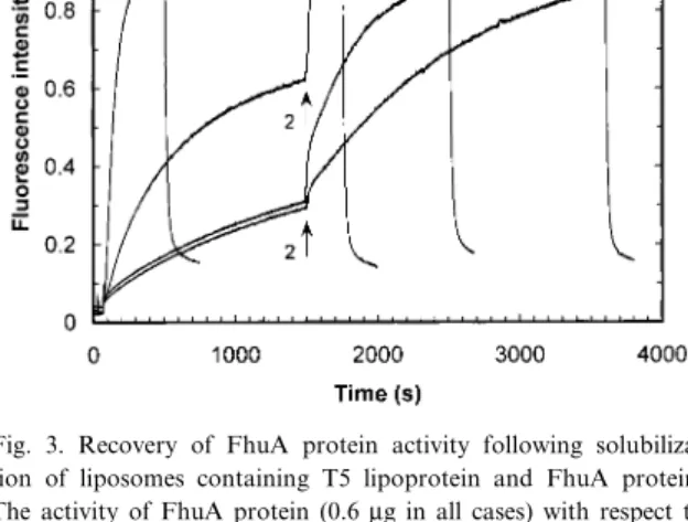
The results of the £uorescence studies (Fig. 3) are in excellent agreement with the inactivation studies described. Virus DNA ejection, which occurs upon interaction of phage T5 with FhuA protein, was in-hibited in the presence of T5 lipoprotein. A rise in
Table 1
Inhibition of FhuA protein by native and truncated lipoprotein
Vesicles FhuA protein T5 lipoprotein Truncated lipoprotein Detergent pfu
A 3 ^ ^ ^ ^ 256 þ 23 (n = 4) B 3 ^ ^ ^ 4 234 þ 16 (n = 2) C + ^ ^ ^ 1 239 þ 44 (n = 6) D 3 5 pmol ^ ^ 4 0 þ 0 (n = 3) E + 5 pmol ^ ^ 1 2 þ 2 (n = 10) F + 5 pmol ^ ^ 4 1 þ 1 (n = 5) G 3 5 pmol 50 pmol ^ 4 0 þ 0 (n = 4) H + 5 pmol 50 pmol ^ 1 245 þ 49 (n = 7) I + 5 pmol 50 pmol ^ 4 1 þ 1 (n = 2) K 3 ^ ^ 0.75 nM ^ 220 þ 4 (n = 2) L 3 ^ ^ 7.5 nM ^ 165 þ 5 (n = 2) M 3 ^ ^ 75 nM ^ 0 þ 0 (n = 2) N 3 ^ ^ 75 pM 0.3 109 þ 9 (n = 2) O 3 ^ ^ 0.75 nM 0.3 30 þ 8 (n = 2) P 3 ^ ^ 7.5 nM 0.3 2 þ 1 (n = 2)
The results of the phage T5 inactivation study are given with the deviations and the number of experiments (n) performed. Plaque forming units (pfu) of uniform aliquots of bacteriophage T5 were determined after incubation with mixtures of FhuA protein and T5 lipoprotein. Incubations were carried out for 15 min, and aliquots of the mixtures were added to the tester strain (E. coli BE), plated on agar plates, and
incubated at 37³C. The proteins were used either in detergent-solubilized form, or reconstituted into vesicles. Detergent concentrations are expressed as multiples of the critical micelle concentration (CMC, for octyl-POE 6.7 mM or 0.24%). At 1UCMC, the e¡ective concentration of detergent monomers is less than the CMC because of detergent binding to both protein and lipids, thus keeping the T5 lipoprotein in solution without solubilizing the liposomes. At concentrations of 4UCMC, vesicles were solubilized completely. Rows D^I: concentrations of proteins refer to the total volume of the incubation mixtures (400 Wl). The critical values are given in bold. Rows K^P: truncated lipoprotein was incubated with phage T5 in the absence of puri¢ed FhuA protein. Concentrations of protein and detergent refer to the total volume of top agar (3 ml) and reaction mixture added (400 Wl).
£uorescence showed that addition of detergent and the resulting solubilization of the proteoliposomes restored the activity of FhuA protein. The kinetics of recovery of activity varied with the molar ratio of T5 lipoprotein to FhuA protein used.
4. Discussion
The irreversible inactivation of bacteriophage T5 by puri¢ed FhuA protein is inhibited in vitro by a phage-encoded lipoprotein. The receptor and the vi-ral lipoprotein, co-reconstituted into liposomes, ap-pear to form a complex which is not recognized by the virus. For complete inhibition, a molar excess of 10:1 of T5 lipoprotein over FhuA protein is neces-sary. An independent con¢rmation of the stoichiom-etry was obtained by £uorescence studies which also revealed that the molar ratio of T5 lipoprotein to FhuA protein a¡ects the kinetics of recovery of FhuA protein activity upon detergent-mediated sol-ubilization. The value of 10 mol lipoprotein per mol
FhuA protein refers, of course, to the total concen-trations supplied and thus depends on whether re-ceptor and inhibitor are distributed evenly among liposomes, as has been observed with FhuA protein [16]. Moreover, it is signi¢cant to know whether T5 lipoprotein distributes randomly between inner and outer lea£ets of the liposomes, and on what side of the membrane the interaction with FhuA protein occurs.
Aiming to resolve these topological questions, we have constructed and puri¢ed a mutant T5 lipopro-tein lacking the N-terminally acylated cyslipopro-teine. The resulting polypeptide is water-soluble and exhibits a
Fig. 3. Recovery of FhuA protein activity following solubiliza-tion of liposomes containing T5 lipoprotein and FhuA protein. The activity of FhuA protein (0.6 Wg in all cases) with respect to triggering phage T5 DNA ejection was monitored by recording the time course of the £uorescence signal of YO-PRO 1 (1 WM), a dye interacting with double stranded DNA. At t = 70 s, phage T5 was added to a suspension of co-reconstituted T5 lipoprotein and FhuA protein (curves C and D). A gradual rise of the £uo-rescence was attributed to penetration of the dye into the phage capsid [19]. Addition of octyl-POE (1% ¢nal concentration, ar-row 2) caused an increase of the £uorescence signal, indicative of phage DNA ejected in the medium. The higher the molar ratio of T5 lipoprotein to FhuA protein, the slower the recovery of FhuA protein activity. A ratio of 10:1 is shown in curve C, one of 100:1 in curve D. Free DNA was degraded by DNase I (20 U, arrow 3), causing a sharp decrease of the £uorescence signal. As control experiments, phage T5 was added to detergent-solubi-lized FhuA protein (curve A) as well as to FhuA protein recon-stituted into lipid vesicles (curve B). In curve A, all the DNA is ejected into solution, whereas in curve B, a quenched signal is in-dicative of a signi¢cant amount of viral DNA injected into the liposomes [15]. Liposomes devoid of protein or containing only lipoprotein did not cause any phage DNA ejection.
Fig. 2. Circular dichroism spectrum of lipoprotein. CD spectra of isolated T5 lipoprotein were recorded at 20³C in NaPi bu¡er
containing 1% octyl-POE. The protein concentration was 0.8 mg/ ml, the path length of the cuvette 1 mm. When treatment of the protein with 100 mM EDTA was omitted (see Section 2), a peak with a maximum at 235 nm was observed (dotted line), possibly re£ecting binding of a divalent cation to the polypeptide [25]. Ac-tivity in vitro was not in£uenced with or without EDTA treat-ment. The spectrum obtained with the truncated, non-acylated mutant of the T5 lipoprotein in the absence of octyl-POE is in-distinguishable from that shown. The spectra obtained are indica-tive of L-sheet as the predominant secondary structure element.
CD spectrum indistinguishable from that obtained with wild-type T5 lipoprotein. When incubated with cells of the E. coli tester strain at a ratio of soluble lipoprotein to surface-exposed FhuA protein of 106:1, the bacteria became completely resistant to
the phage. This result re£ects the dilution of the soluble protein in the bulk solvent, and suggests the role of N-terminal acylation of wild-type T5 lipo-protein to be that of increasing the e¡ective local concentration in two dimensions. Indeed, truncation a¡ects lipoprotein function in a way comparable to the addition of detergent to full-size lipoprotein: in either case, its interaction with the FhuA protein is weakened, with the result that the receptor function of the FhuA protein is much less inhibited. With respect to topology, the result obtained suggests that soluble T5 lipoprotein may prevent infection by binding to the extracellular face of the FhuA protein located at the surface of the tester strain. This would represent an alternative mechanism to that proposed previously, which suggested the inhib-itory action of T5 lipoprotein to occur by insertion on the periplasmic side [3].
In conclusion, we have demonstrated that the presence of T5 lipoprotein is necessary and su¤cient for the inactivation of the FhuA protein in vitro, and that the interactions between the two proteins are speci¢c. Our results suggest that several lipoprotein molecules cluster around the FhuA protein in the lipid bilayer, thereby shielding, directly or indirectly, the receptor protein from recognition by the phage. The critical question to be addressed now is the in-teraction between receptor and lipoprotein at the molecular level. We are currently investigating the complex formation by studying the interaction of the integral FhuA protein and the phage T5 lipopro-tein by structural methods.
Acknowledgments
We thank Dr. K. Strupat, Institute for Medical Physics, University of Muënster, Germany, for carry-ing out the Maldi-MS analysis, and R. Koebnik (Biozentrum, University of Basel) for critical reading of the manuscript. This work was supported by a grant from the Swiss National Fund for Scienti¢c Research to J.P.R.
References
[1] McCorquodale, D.J. and Warner, H.R. (1988) In: The Bac-teriophages (Calendar, R., Ed.), pp. 439^475. Plenum Press, New York.
[2] Decker, K., Krauel, V., Meesmann, A. and Heller, K. (1994) Lytic conversion of Escherichia coli by bacteriophage T5: blocking of the FhuA receptor protein by a lipoprotein ex-pressed early during infection. Mol. Microbiol. 12, 321^332. [3] Braun, V., Killmann, H. and Herrmann, C. (1994)
Inactiva-tion of FhuA at the cell surface of Escherichia coli K-12 by a phage T5 lipoprotein at the periplasmic face of the outer membrane. J. Bacteriol. 176, 4710^4717.
[4] Braun, V., Hancock, R.E.W., Hantke, K. and Hartmann, A. (1976) Functional organization of the outer membrane of Es-cherichia coli: phage and colicin receptors as components of iron uptake systems. J. Supramol. Struct. 5, 37^58. [5] Fecker, L. and Braun, V. (1983) Cloning and expression of the
fhu genes involved in iron(III)hydroxamate uptake by E. coli. J. Bacteriol. 156, 1301^1314.
[6] Coulton, J.W., Mason, P. and DuBow, M.S. (1983) Molecular cloning of the ferrichrome-iron receptor of E. coli K12. J. Bacteriol. 156, 1315^1321.
[7] Neilands, J.B. (1981) Microbial iron compounds. Annu. Rev. Biochem. 50, 715^731.
[8] Braun, V. (1995) Energy-coupled transport and signal trans-duction through the Gram-negative outer membrane via TonB-ExbB-ExbD-dependent receptor proteins. FEMS Mi-crobiol. Rev. 16, 295^307.
[9] Kadner, R.J. (1990) Vitamin B12 transport in Escherichia coli: energy coupling between membranes. Mol. Microbiol. 4, 2027^2033.
[10] Klebba, P.E., Rutz, J.M., Liu, J. and Murphy, C.K. (1993) Mechanisms of TonB-catalyzed iron transport through the enteric bacterial cell envelope. J. Bioenerg. Biomembr. 25, 603^611.
[11] Postle, K. (1990) TonB and the Gram-negative dilemma. Mol. Microbiol. 4, 2019^2025.
[12] Braun, V., Hantke, K. and Koster, W. (1998) Bacterial iron transport: mechanism, genetics, and regulation. Metal Ions Biol. Syst. 35, 67^145.
[13] Bonhivers, M., Ghazi, A., Boulanger, P. and Letellier, L. (1996) FhuA, a transporter of the Escherichia coli outer mem-brane, is converted into a channel upon binding of bacterio-phage T5. EMBO J. 15, 1850^1856.
[14] Boulanger, P., Le Maire, M., Bonhivers, M., Dubois, S., Des-madril, M. and Letellier, L. (1996) Puri¢cation and structural and functional characterization of FhuA, a transporter of the Escherichia coli outer membrane. Biochemistry 35, 14216^ 14224.
[15] Locher, K.P. and Rosenbusch, J.P. (1997) Modeling ligand-gated receptor activity: FhuA-mediated ferrichrome e¥ux from lipid vesicles triggered by phage T5. J. Biol. Chem. 272, 1448^1451, 8836.
[16] Planc°on, L., Chami, M. and Letellier, L. (1997) Reconstitu-tion of FhuA, an Escherichia coli outer membrane protein, into liposomes. J. Biol. Chem. 272, 16868^16872.
[17] Studier, F.W., Rosenberg, A.H., Dunn, J.J. and Dubendor¡, J.W. (1990) Use of T7 RNA polymerase to direct expression of cloned genes. Methods Enzymol. 185, 60^89.
[18] Doherty, A.J., Ashford, S.R., Brannigan, J.A. and Wigley, D.B. (1995) A superior host strain for the over-expression of cloned genes using the T7 promoter based vectors. Nucleic Acids Res. 23, 2074^2075.
[19] Locher, K.P. and Rosenbusch, J.P. (1997) Oligomeric states and siderophore binding of the ligand-gated FhuA protein that forms channels across Escherichia coli outer membranes. Eur. J. Biochem. 247, 770^775.
[20] Bradford, M.M. (1976) A rapid and sensitive method for the quantitation of microgram quantities of protein utilizing the principle of protein-dye binding. Anal. Biochem. 72, 248^254. [21] Marston, F.A.O. (1986) The puri¢cation of eucaryotic poly-peptides synthesized in Escherichia coli. Biochem. J. 240, 1^12. [22] Schaëgger, H. and von Jagow, G. (1987) Tricine-sodium
do-decyl sulfate-polyacrylamide gel electrophoresis for the sepa-ration of proteins in the range from 1 to 100 kDa. Anal. Biochem. 166, 368^379.
[23] Rosinke, B., Strupat, K., Hillenkamp, F., Rosenbusch, J.P., Dencher, N., Kruëger, U. and Galla, H. (1995) Matrix-assisted laser desorption/ionization mass spectrometry (MALDI-MS) of membrane proteins and non-covalent complexes. J. Mass Spectrom. 30, 1462^1468.
[24] Braun, V. and Rehn, K. (1969) Chemical characterization, spatial distribution and function of a lipoprotein (murein-lipoprotein) of the E. coli cell wall. The speci¢c e¡ect of trypsin on the membrane structure. Eur. J. Biochem. 10, 426^438.
[25] Gri¤n, J.H., Rosenbusch, J.P., Blout, E.R. and Weber, K.K. (1973) Conformational changes in aspartate transcarbamylase. II. Circular dichroism evidence for the involvement of metal ions in allosteric interactions. J. Biol. Chem. 248, 5057^5062.