Fractional magnesium absorption is significantly lower in human
subjects from a meal served with an oxalate-rich vegetable,
spinach, as compared with a meal served with kale, a vegetable
with a low oxalate content
Torsten Bohn, Lena Davidsson*, Thomas Walczyk and Richard F. Hurrell
Laboratory for Human Nutrition, Institute of Food Science and Nutrition, Swiss Federal Institute of Technology, Zurich, Switzerland
(Received 27 May 2003 – Revised 7 November 2003 – Accepted 28 November 2003)
The aim of the present study was to evaluate Mg absorption from a test meal served with an oxalate-rich vegetable, spinach, as compared with a test meal served with a vegetable with a low oxalate content, kale. Mg absorption was measured by a stable-isotope technique based on extrinsic labelling of the test meals and faecal monitoring of the excreted isotope labels. Nine healthy adults participated in the study. The test meals were based on 100 g phytate-free white bread, served with 300 g spinach (6·6 mmol oxalate; 0·7 mmol25Mg label added, 5·0 mmol total Mg) or 300 g kale (0·1 mmol oxalate; 1·2 mmol26Mg label added, 4·8 mmol total Mg). The test meals were served on days
1 and 3, at breakfast and lunch, using a cross-over design. The results from the present study demonstrated that apparent Mg absorption was significantly lower from the meal served with spinach (26·7 (SD10·4) %) than the meal served with kale (36·5 (SD11·8) %) (P¼ 0·01). However, the lower fractional apparent Mg absorption from the test meal served with spinach can be assumed to be, at least partly, coun-terbalanced by the higher native Mg content of spinach as compared with kale. Although based on indirect evidence, i.e. not based on an evaluation of added (or removed) oxalic acid, the difference in Mg absorption observed in the present study is attributed to the difference in oxalic acid content between the two vegetables.
Magnesium absorption: Spinach: Oxalate: Stable isotopes: Faecal monitoring
Oxalic acid and its salts are ubiquitous in plant cells and relatively large amounts are found in leafy vegetables such as spinach (Tabekhia, 1980), and also in fruits, grains, nuts, tea, coffee, and cocoa (Zarembski & Hodgkinson, 1962; Souci et al. 1994). Oxalate intakes vary with dietary habits and have, for example, been reported to be in the range of 70 – 150 mg/d in the UK (Zarembski & Hodgkinson, 1962; Anderson et al. 1971; Hodgkinson 1977a).
Although oxalic acid forms insoluble complexes at phys-iological pH with divalent cations such as Ca2þ, Zn2þ, and Mg2þ(Weast, 1989), the influence of oxalate or foods rich in oxalate on mineral and trace element absorption in man has not been evaluated systematically. However, Ca absorption from a vegetable rich in oxalate, spinach, has been reported to be significantly lower than from kale (a vegetable with a low oxalate content) in adults (Heaney et al. 1988; Heaney & Weaver, 1990). In addition, Schwartz et al. (1984) reported significantly lower net (apparent) Mg absorption from bran muffins with added spinach as compared with added collard greens, a vegetable botanically similar to kale, but no difference as compared with added lettuce or turnip greens. However, these results are difficult to interpret as the quantity
of spinach and the content of oxalate in the test meal are not reported and the study only included four subjects. An inhibitory effect of oxalic acid-rich vegetables on Mg and Zn absorption in man is indicated by the observation that spinach added to the diet resulted in negative Mg and Zn balances (Kelsay & Prather, 1983). Furthermore, Mg has been shown to inhibit oxalate absorption in man (Berg et al. 1986; Hanson et al. 1989), suggesting that Mg forms insoluble, non-absorbable complexes with oxalic acid in the gastrointestinal tract.
The aim of the present study was to evaluate Mg absorp-tion from test meals served with vegetables with high or low oxalate content (spinach or kale) in healthy adults. Mg absorption was measured by a stable-isotope technique based on extrinsic labelling of the meals and faecal moni-toring of the excreted isotope label.
Subjects and methods Subjects
Apparently healthy, free-living, i.e. non-hospitalised, sub-jects (ten adult men and women) were recruited. Lactating
* Corresponding author: Dr Lena Davidsson, fax þ 41 1 7045710, email [email protected] qThe Authors 2004
and pregnant women were excluded from the study. No medication was allowed during the study except for oral con-traceptives. The intake of mineral and vitamin supplements was not allowed from 2 weeks before the start of the study and during the entire study. The participants were asked not to change their dietary habits or lifestyle during the study. Information about the aims and the procedures of the study was given orally and in writing. Written informed con-sent was obtained from all participants. The study protocol was reviewed and approved by the ethical committee at the Swiss Federal Institute of Technology, Zurich.
Isotopic labels
Highly enriched 25MgO (1·04 (SD 0·01) % 24Mg, 98·73 (SD0·01) %25Mg and 0·23 (SD0·01) %26Mg) and 26MgO (0·39 (SD0·01) %24Mg, 0·11 (SD 0·01) %25Mg and 99·50 (SD0·01) %26Mg) labels were purchased from Chemotrade (Dusseldorf, Germany). The enriched25Mg label (28 mmol as25MgO) and26Mg label (43 mmol as26MgO) were each dissolved in 4M-HCl (10 ml) and diluted to 100 ml with water. Solid NaHCO3 (Merck, Darmstadt, Germany) was
added to adjust to pH 6. The concentration of 25Mg and
26Mg isotope labels in solution was determined by
isotope-dilution MS against a commercial Mg standard of natural isotopic composition (Titrisol; Merck, Darmstadt, Germany). Unless otherwise specified, all chemicals were of analytical grade, and acids were further purified by surface distillation. Only 18 MV water (Milli Q water system; Millipore, Zurich, Switzerland) was used for laboratory work and test-meal preparation.
Test meals
Kale and spinach were purchased frozen at a local supermar-ket, then cooked and pure´ed to prepare one batch of each vegetable. Individual servings were weighed and frozen (2 258C) immediately after preparation. Weighed samples of the isotope-label solutions were added to the test meals about 15 min before administration to ensure the accurate monitoring of individual doses. Stable-isotope labels were mixed with the pure´ed vegetables. Higher doses of26Mg label than of 25Mg label were used in the present study due to differences in measurement precision (see p. 604).
Phytate-free wheat bread was prepared from white wheat flour (Migros, Zurich, Switzerland), water, salt, sugar and dry yeast. The dough was left to ferment for 5 h at room temperature. Bread rolls were baked for 15 min at 2008C, and stored frozen (2 258C) until served.
The rare-earth elements Yb and Eu were used as non-absorbable faecal markers to evaluate the completeness of faecal collections. Both compounds (in chloride form; Aldrich, Buchs, Switzerland) were added to water served as a drink together with the labelled test meals.
Study design
Before the start of the study, a faecal sample was collected to determine the baseline Mg isotope ratios. In addition, brilliant blue (100 mg; Warner Jenkinson Europe, King’s Lynn, UK), a dye used as a faecal marker, was administered
in a gelatine capsule to indicate the start of the faecal pool-ing. After an overnight fast, a venous blood sample (10 ml) was drawn into a heparinised glass tube (Vacutainer Sys-tems, Plymouth, UK) for the determination of Mg concen-tration in plasma. Plasma was separated by centrifugation (Omnifuge 2.0 RS; Heraeus, Zurich, Switzerland) at 208C and about 500 g (5 min), and stored in acid-washed plastic vials at 2 258C until analysed.
The study had a cross-over design, with each subject acting as his or her own control. Test meals A and B were randomly allocated to be served on day 1 or day 3. All test meals were based on 100 g phytate-free wheat bread served with either 300 g spinach (test meal A), to which the25Mg label was added, or with 300 g kale (test meal B), to which the26Mg label was added. Spinach and kale were heated in a microwave oven before serving. Water (18 MV water; 600 ml) with the added rare-earth elements was served as a drink with test meals A and B. The test meals were divided into two identical portions served at breakfast (07.30 – 08.30 hours) and at lunch (12.00 – 13.00 hours).
No food or drink was allowed between breakfast and lunch on days 1 and 3 and for 3 h after lunch. Standardised dinners (pizza and white wheat crisp bread) were provided on days 1 and 3. Drinking water (18 MV water; 2 litres) was provided on days 1 and 3. No additional foods or drinks were allowed on days 1 and 3. Diet was unrestricted at all other times.
Pre-weighed polypropylene containers (Semadeni, Ostermundingen, Switzerland) were provided for stool col-lections. The subjects collected all stools separately, start-ing immediately after the intake of the first labelled test meal on day 1. On day 8, a brilliant blue capsule was again administered. The collections were continued until excretion of the second brilliant blue marker. Stool samples were stored frozen (2 258C) until processed.
Preparation of faecal pools
Each individual stool sample was freeze-dried using a freeze dryer (Modulyo; Edwards, North Bergen, NJ, USA) and ground to a powder in a mortar. All stools, from the first stool dyed by brilliant blue until, but not including, the stools dyed by the second dose of brilliant blue, were included in the faecal pool. After a drying step (20 h at 658C) in a drying chamber (Binder, Tuttlingen, Germany) to standardise humidity, followed by cooling at room temperature (4 h), all individual stools were weighed and milled, starting with the first (most enriched) samples. A mill (ZM1; Retsch, Haan, Germany) equipped with a sieve of 1 mm pores was used for this purpose. Each milled stool was transferred back into its original container, dried again for 20 h at 658C, cooled for 4 h at room temperature and re-weighed to determine losses. All milled stools included in a single pool were combined in a 2 litre polyethy-lene container (Semadeni, Ostermundingen, Switzerland) and mixed for 90 min using a rotator (UG 70/20; Micro Motor, Basel, Switzerland).
Wet ashing
Portions of the freeze-dried pooled stool samples (1·0 – 1·6 g), wheat bread (0·25 g), kale and spinach (1 g) as
well as plasma (1 g), were wet ashed in a microwave system (MLS 1200; MLS GmbH, Leutkirch, Germany) in a mixture of 14M-HNO3 and 8·8M-H2O2 (Merck, Darmstadt,
Germany). All samples were wet ashed in duplicate.
Separation of magnesium
Mg was separated from the wet-ashed stool samples by cation-exchange chromatography using a strongly acidic ion-exchange resin (AG 50W X-8, 200 – 400 mesh; Bio-Rad, Hercules, CA, USA). Samples containing about 30 mmol Mg were evaporated to dryness, re-dissolved in 0·7M-HCl (1 ml) and transferred onto the top of the column (10 mm inner diameter; Bio-Rad, Hercules, CA, USA), filled with ion-exchange resin to a height of 70 mm. The column was rinsed with 0·7M-HCl (56 ml), followed by 0·9M-HCl (24 ml) to elute Na and K. Mg was eluted with 1·4M-HCl (12 ml). The solution was evap-orated to dryness and re-dissolved in 50 ml water. Mg recovery, evaluated with a diluted Mg standard solution (Titrisol; Merck, Darmstadt, Germany) was found to be 94·8 (SD 1·8) % (n 10). Resins were regenerated with 6M-HCl (30 ml) and replaced after the fifth run. Only acid-washed Teflon and polyethylene laboratory ware were used for sample processing. Samples of the 26Mg isotope label were processed in parallel with each batch for blank monitoring, starting at the ion-exchange chromatography step. Sample contamination due to natural Mg was found to be 10·8 (SD7·0) nmol (n 9) for the com-bined sample preparation and filament loading, which was , 0·4 ‰ of the amount of Mg separated.
Isotopic analysis by thermal ionisation mass spectrometry About 20 nmol separated Mg from faecal samples was loaded onto the metal surface of the evaporation filament of a double-Re filament ion source. Mg was coated with 5 – 10 mg silica gel 100, 0·8 mmol boric acid and 30 nmol Al as AlCl3(all
chemicals from Merck, Darmstadt, Germany). Compounds were loaded as aqueous solutions and dried at a current of 0·8 A after each step. Finally, the evaporation filament was heated to dull red heat (a current of 1·6 A) for 30 s. The ion-isation filament remained unloaded. Isotope ratios were determined with a single-focusing magnetic sector field instrument (MAT 262; Finnigan MAT, Bremen, Germany), equipped with a Faraday cup multicollector device for simul-taneous ion beam detection. The evaporation filament was heated to 12308C, using a standardised procedure. The ionis-ation filament was heated gradually to 1250 – 13508C until a stable Mgþ ion beam at a current of 12 2 £ 10211A was obtained. Each measurement consisted of thirty consecutive isotope-ratio measurements. Reproducibility (five indepen-dent runs) had a relative standard deviation of ^ 0·2 % for the 24Mg:25Mg isotope ratio and ^ 0·4 % for the
24Mg:26Mg isotope ratio.
Atomic absorption spectroscopy
Quantitative Mg analysis of the wet-ashed and diluted plasma, bread, faecal, and vegetable samples was performed by flame atomic absorption spectroscopy
(SpectrAA 400; Varian, Mulgrave, Australia). Plasma samples were measured by external calibration using a commercial Mg standard (Titrisol; Merck, Darmstadt, Germany). All other samples were measured by an internal calibration technique (standard addition) to minimise matrix effects. In addition, all solutions contained La(NO3)3at 5000 mg/l to suppress matrix effects. Certified
reference materials (Seronorm Trace Elements Serum; Nycomed, Oslo, Norway, and wheat flour 1567 a; National Bureau of Standards, Gaithersburg, MD, USA) were analysed in parallel to monitor the accuracy of analysis. Yb and Eu were measured in the wet-ashed and diluted faecal samples by electrothermal atomic absorption spec-troscopy by external calibration, using a standard solution containing Yb and Eu (Titrisol; Merck, Darmstadt, Germany), following standard procedures.
Analyses of oxalate and phytate
Total and soluble oxalate contents were determined enzy-mically (no. 1856120; Roche Diagnostics, Mannheim, Germany) after the extraction of oxalate in the pure´ed vegetables with 2M-HCl (total oxalate) or water (soluble oxalate). Phytic acid was extracted from a 1 g sample of freeze-dried bread with 0·5M-HCl. The extract was purified by anion-exchange chromatography, evaporated to dryness, and re-dissolved in water before analysis by HPLC reversed-phase chromatography as described by Sandberg & Ahderinne (1986).
Calculations
Molar amounts and ratios of the 25Mg and 26Mg isotope labels in the faecal pools were calculated based on double-isotope dilution principles (Walczyk et al. 1997; Sabatier et al. 2002). Fractional apparent Mg absorption (AA%) was based on the dose (mmol) of Mg stable isotope administered (Do) and the amount of the isotopic label
excreted in faeces (Fo).
AAð%Þ ¼Do– Fo Do
£ 100:
Recovery of the rare-earth elements Eu and Yb was used to evaluate the completeness of the stool collections.
Statistics
Calculations were made using commercial software (Excel 97; Microsoft, Chicago, IL, USA and SPSS 10.0; SPSS Inc., Chicago, IL, USA). Results are presented as arithmetic means and standard deviations. The normal distribution of absorption values was verified by the Kolmogorov – Smirnoff test. Paired Student’s t tests (two-tailed) were used to compare Mg absorption from the two different test meals. P values below 0·05 are referred to as statistically significant.
Results
The mean age and mean BMI of the subjects were 23 (SD1) years and 22·6 (SD3·2) kg/m2(n 9). The mean plasma Mg
concentration was 0·80 (range 0·73 – 0·89) mmol/l. Two individuals had slightly lower plasma Mg concentration than the reported normal range of 0·75 – 0·96 mmol/l (Lowenstein & Stanton, 1986).
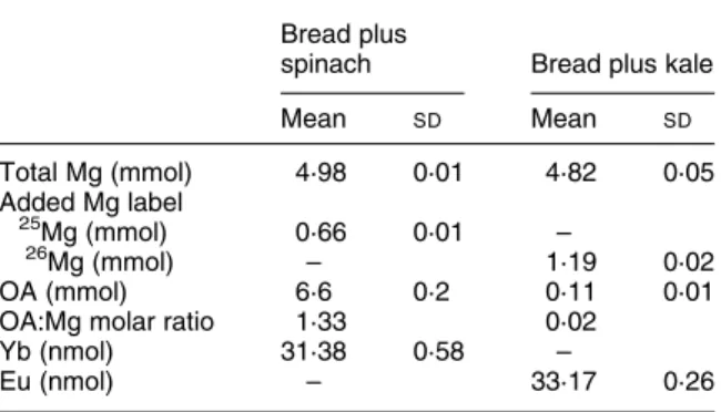
The Mg and oxalate contents of the test meals are presented in Table 1. The native Mg content of the bread, spinach, and kale was 0·96 (SD 0·03), 1·12 (SD 0·09), and 0·89 (SD 0·01) mmol/100 g, respectively (n 3). Of total oxalates, 87·5 (SD2·8) % were found to be soluble in spinach (n 3) and 51·8 (SD 11·7) % in kale (n 3). The phytic acid content was below the detection limit in wheat bread (, 0·5 mmol/100 g).
Mg absorption from the test meal served with spinach (26·7 (SD 10·4) %) was significantly lower (P¼ 0·01) than from the test meal served with kale (36·5 (SD 11·8) %) as shown in Fig.1. The within-subject difference in Mg absorption between the two test meals was 9·8 (SD 7·2) %. Absorption ratios (test meal served with spinach/test meals served with kale) were 0·73 (SD 0·19). One subject
was excluded from the evaluation due to a low recovery of Yb (, 85 %). For all other subjects, mean Yb recovery was 98·8 (range 90·6 – 115·4) % and mean Eu recovery was 95·0 (range 90·0 – 99·1) %.
Complete faecal collections were made during 8·9 (SD 0·6) d. The mean loss of faecal material during sample preparation, determined by weighing faecal pools before and after milling, was 1·1 (SD0·6) %. The measured isotopic enrichment of the stool pools was 5·6 (SD 1·8) % (24Mg:25Mg range 2·5 – 9·3 %) and 8·1 (SD 2·4) % (24Mg:26Mg range 5·3 – 13·1 %) based on differences of the measured isotope ratios of faecal pools and natural isotope ratios of a standard (Titrisol; Merck, Darmstadt, Germany), divided by the measured isotope ratio of the standard.
Discussion
The present study is, to our knowledge, the first report clearly demonstrating an inhibitory effect of an oxalate-rich vegetable on Mg absorption in human subjects, based on the stable-isotope technique. The mean fractional apparent Mg absorption from the labelled test meal served with spinach was about 35 % lower than from the test meal served with kale in the present study. However, these results should not be interpreted as suggesting that spinach is a poor source of dietary Mg. The observed decrease in fractional apparent Mg absorption can be assumed to be, at least partly, counterbalanced by the approximately 30 % higher Mg content of spinach as compared with kale (Holland et al. 1994; Souci et al. 1994); native Mg content of spinach was 26 % higher than that of kale in the test meals evaluated in the present study.
In the present study, we evaluated Mg absorption from extrinsically labelled test meals based on phytate-free wheat bread, served with spinach or kale. Limited infor-mation is available on the validity of the extrinsic labelling technique for studies of Mg absorption in human subjects although an earlier study reported no significant differences in Mg absorption from intrinsically and extrinsically labelled leafy vegetables, including spinach and collards, in man (Schwartz et al. 1984). Similar findings have also been reported in rats (Schwartz et al. 1980); however, it should be stressed that the usefulness of animal models in these evaluations is not clear. Contrary to the data (which are rather limited) on the validity of extrinsic label-ling of leafy vegetables with Mg isotopes, the extrinsic labelling technique has been demonstrated not to be valid for Ca absorption from spinach (Weaver & Heaney, 1991). Interestingly, the study by Weaver & Heaney (1991) reported Ca absorption from calcium oxalate to be twice as well absorbed as from spinach oxalate in healthy adult women. These data thus indicate different absorption mechanisms for, or different physical and chemical proper-ties of, pure calcium oxalate and spinach Ca and highlight the limited usefulness of evaluating the influence of pure calcium oxalate on Ca absorption from complex food matrices such as spinach.
Oxalates in plants are present as water soluble, bound to Na or K or present as free oxalates, as well as water-inso-luble compounds, i.e. calcium oxalate and magnesium Fig. 1. Apparent magnesium absorption from a meal based on
100 g white wheat bread served with 300 g spinach (6·6 mmol oxa-late; test meal A) or 300 g kale (0·11 mmol oxaoxa-late; test meal B). (K), Individual data (n 9); (———), mean absorption values.
Table 1. Test meal contents of total magnesium, stable-iso-tope labels (25Mg or 26Mg), oxalic acid (OA), oxalic acid: magnesium molar ratios and non-absorbable faecal markers, ytterbium and europium*
(Mean values and standard deviations) Bread plus
spinach Bread plus kale
Mean SD Mean SD Total Mg (mmol) 4·98 0·01 4·82 0·05 Added Mg label 25Mg (mmol) 0·66 0·01 – 26 Mg (mmol) – 1·19 0·02 OA (mmol) 6·6 0·2 0·11 0·01
OA:Mg molar ratio 1·33 0·02
Yb (nmol) 31·38 0·58 –
Eu (nmol) – 33·17 0·26
* Test meals consisted of 100 g phytate-free white wheat bread served with 300 g spinach or kale.
oxalate (Hodgkinson, 1977b; Souci et al. 1994). It is not clear to what extent insoluble oxalates dissolve in the gas-tric juice and it could be speculated that soluble oxalates would have a more pronounced negative impact on mineral and trace element absorption due to their ability to form complexes with minerals and trace elements in the gastro-intestinal tract. In addition, calcium oxalate has been suggested to be absorbed intact (Hanes et al. 1999) and it could be hypothesised that the more soluble magnesium oxalate could also be absorbed intact. In the present study, a large proportion (88 %) of total oxalates was present as water-soluble oxalates in spinach. However, at the present time, there is no information about the relative importance of soluble and insoluble oxalates on mineral and trace element absorption in man. Published data on the pro-portion of soluble oxalates relative to total oxalates also differ considerably; for example data for spinach vary from 15 – 20 % (Toma & Tabekhia, 1979; Souci et al. 1994) up to 93 % (Hodgkinson, 1977b).
Although based on indirect evidence, the observed difference in fractional Mg absorption in the present study is attributed to the differences in oxalate content; 6·6 mmol in the meal served with spinach and 0·1 mmol in the meal served with kale. Based on present knowledge, it is improbable that other plant components present in green leafy vegetables such as fermentable fibre or pheno-lic compounds would have influenced the results. For example, kale contains about 2 % fibre as pentosans and hexosans, about double the amount found in spinach (Souci et al. 1994). These partly soluble hemicelluloses are non-digestible and could be expected to be fermented by bacteria in the large intestine, similarly to fructo-oligo-saccharides, which were reported to increase fractional Mg absorption in human subjects significantly when consumed in amounts of 10 g/d (Tahiri et al. 2001). Recently, the enhancing effect of polyols (100 g/d) on apparent Mg absorption in man was also demonstrated (Coudray et al. 2003a). However, information about the effect of dietary fibre on Mg absorption is not conclusive, as several human studies have not demonstrated statistically signifi-cant differences in Mg absorption after an increased intake of fermentable fibre (for a review, see Coudray et al. 2003b). In addition, a study based on the chemical balance technique reported a significant negative effect on Mg absorption after the intake of a mixture of galactans, pentosans, and hexosans (14 g/d) by adolescent boys (Drews et al. 1979). Furthermore, phenolic compounds have been reported to have a strong negative effect on Fe absorption (Gillooly et al. 1983; Brune et al. 1989). How-ever, although phenolic compounds have not been evalu-ated for their effect on Mg absorption, the amount of total phenolic compounds has been reported to be similar in spinach and kale (Lucarini et al. 2000).
In conclusion, the results from the present study demon-strate that the mean fractional apparent Mg absorption from a test meal served with spinach was about 35 % lower than from a test meal served with kale. It is suggested that this reduction in Mg absorption is due to the higher oxalate content of spinach. However, the signifi-cantly lower fractional apparent Mg absorption from spi-nach can be assumed to be, at least partly,
counterbalanced by the higher native Mg content of spi-nach as compared with kale.
References
Anderson W, Hollins JG & Bond PS (1971) Composition of tea infusions examined in relation to association between mortality and water hardness. J Hyg Cambr 69, 1 – 15.
Berg W, Bothor C, Pirlich W & Janitzky V (1986) Influence of magnesium on the absorption and excretion of calcium and oxalate ions. Eur Urol 12, 274 – 282.
Brune M, Rossander L & Hallberg L (1989) Iron absorption and phenolic compounds: importance of different phenolic struc-tures. Eur J Clin Nutr 43, 547 – 557.
Coudray C, Bellanger J, Vermorel M, Sinaud S, Wils D, Feillet-Coudray C, Brandolini M, Bouteloup-Demange C & Rayssiguier Y (2003a) Two polyol, low digestible carbo-hydrates improve the apparent absorption of magnesium but not of calcium in healthy young men. J Nutr 133, 90 – 93. Coudray C., Demigne´ C & Rayssiguier Y (2003b) Effects of
diet-ary fibers on magnesium absorption in animals and humans. J Nutr 133, 1 – 4.
Drews LM, Kies C & Fox HM (1979) Effect of dietary fiber on copper, zinc, and magnesium utilization by adolescent boys. Am J Clin Nutr 32, 1893 – 1897.
Gillooly M, Bothwell TH, Torrance JD, MacPhail AP,
Derman DP, Bezwoda WR, Mills W, Charlton RW & Mayet F (1983) The effects of organic-acids, phytates and poly-phenols on the absorption of iron from vegetables. Br J Nutr 49, 331 – 342.
Hanes DA, Weaver CM, Heaney RP & Wastney M (1999) Absorption of calcium oxalate does not require dissociation in rats. J Nutr 129, 170 – 173.
Hanson CF, Frankos VH & Thompson WO (1989) Bioavailability of oxalic acid from spinach, sugar beet fibre and a solution of sodium oxalate consumed by female volunteers. Food Chem Toxicol 27, 181 – 184.
Heaney RP & Weaver CM (1990) Calcium absorption from kale. Am J Clin Nutr 51, 656 – 657.
Heaney RP, Weaver CM & Recker RR (1988)
Calcium absorbability from spinach. Am J Clin Nutr 47, 707 – 709.
Hodgkinson A (1977a) Oxalate metabolism in animals and man. In Oxalic Acid in Biology and Medicine, pp. 160 – 192 [A Hodgkinson, editor]. London: Academic Press.
Hodgkinson A (1977b) Oxalate content of foods and nutrition. In Oxalic Acid in Biology and Medicine, pp. 193 – 212 [A Hodg-kinson, editor]. London: Academic Press.
Holland B, Welch AA, Unwin ID, Buss DH, Paul AA & Southgate DAT (1994) The Composition of Foods, 5th ed., Cambridge, UK: Xerox Ventura.
Kelsay JL & Prather ES (1983) Mineral balances of human subjects consuming spinach in a low-fiber diet and in a diet containing fruits and vegetables. Am J Clin Nutr 38, 12 – 19.
Lowenstein FW & Stanton MF (1986) Serum magnesium levels in the United States, 1971 – 1974. J Am Coll Nutr 5, 399 – 414.
Lucarini M, Di Lullo G, Cappelloni M & Lombardi-Boccia G (2000) In vitro estimation of iron and zinc dialysability from vegetables and composite dishes commonly consumed in Italy: effect of red wine. Food Chem 70, 39 – 44.
Sabatier M, Arnaud MJ, Kastenmayer P, Rytz A & Barclay DV
(2002) Meal effect on magnesium bioavailability
from mineral water in healthy women. Am J Clin Nutr 75, 65 – 71.
Sandberg AS & Ahderinne R (1986) HPLC method for determination of inositol triphosphates, tetraphosphates, penta-phosphates and hexapenta-phosphates in foods and intestinal contents. J Food Sci 51, 547 – 550.
Schwartz R, Grunes DL, Wentworth RA & Wien EM (1980) Magnesium absorption from leafy vegetables, intrinsically labeled with stable26Mg. J Nutr 110, 1365 – 1371.
Schwartz R, Spencer H & Welsh JJ (1984) Magnesium absorption in human subjects from leafy vegetables, intrinsically labeled with stable26Mg. Am J Clin Nutr 39, 571 – 576.
Souci SW, Fachmann W & Kraut H (1994) Food Composition and Nutrition Tables, 5th ed., Stuttgart, Germany: CRC Press. Tabekhia MM (1980) Total and free oxalates calcium, magnesium and iron contents of some fresh vegetables. Dtsch Lebensmitt Rundsch 76, 280 – 282.
Tahiri M, Tressol JC, Arnaud J, et al. (2001) Five-week intake of short-chain fructo-oligosaccharides increases intestinal
absorption and status of magnesium in postmenopausal women. J Bone Miner Res 16, 2152 – 2160.
Toma RB & Tabekhia MM (1979) Trials to reduce soluble oxalates in home prepared spinach. Dtsch Lebensmitt Rundsch 75, 212 – 215.
Walczyk T, Davidsson L, Zavaleta N & Hurrell RF (1997) Stable isotope labels as a tool to determine the iron absorption by Peruvian school children from a breakfast meal. Fresenius J Anal Chem 359, 445 – 449.
Weast RC (1989) Handbook of Chemistry and Physics.
Boca Raton, FL: CRC.
Weaver MC & Heaney RP (1991) Isotopic exchange of ingested calcium between labelled sources Evidence that ingested calcium does not form a common absorptive pool. Calcif Tissue Int 49, 244 – 247.
Zarembski PM & Hodgkinson A (1962) Oxalic acid content of English diets. Br J Nutr 16, 627 – 634.