A review on bovine besnoitiosis: a disease with economic
impact in herd health management, caused by Besnoitia
besnoiti (Franco and Borges, 1916)
HELDER CORTES1, ALEXANDRE LEITÃO2*, BRUNO GOTTSTEIN3 and ANDREW
HEMPHILL3*
1
Victor Caeiro Laboratory of Parasitology, ICAAM– Instituto de Ciências Agrárias e Ambientais Mediterrânicas – Universidade de Évora– Núcleo da Mitra, Ap. 94, 7002-554, Évora, Portugal
2
Instituto de Investigação Científica Tropical, CVZ, CIISA Faculdade de Medicina Veterinária, Universidade de Lisboa, Av. Universidade Técnica, 1300-447 Lisboa, Portugal
3
Institute of Parasitology, Vetsuisse Faculty, University of Bern, Länggass-Strasse 122, CH-3012 Bern, Switzerland
(Received 15 January 2014; revised 9 February 2014; accepted 9 February 2014; first published online 2 April 2014)
S U M M A R Y
Bovine besnoitiosis is caused by the largely unexplored apicomplexan parasite Besnoitia besnoiti. In cows, infection during pregnancy often results in abortion, and chronically infected bulls become infertile. Similar to other apicomplexans B. besnoiti has acquired a largely intracellular lifestyle, but its complete life cycle is still unknown, modes of transmission have not been entirely resolved and the definitive host has not been identified. Outbreaks of bovine besnoitiosis in cattle were described in the 1990s in Portugal and Spain, and later several cases were also detected in France. More cases have been reported recently in hitherto unaffected countries, including Italy, Germany, Switzerland, Hungary and Croatia. To date, there is still no effective pharmaceutical compound available for the treatment of besnoitiosis in cattle, and progress in the identification of novel targets for intervention through pharmacological or immunological means is hampered by the lack of molecular data on the genomic and transcriptomic level. In addition, the lack of an appropriate small animal laboratory model, and wide gaps in our knowledge on the host-parasite interplay during the life cycle of this parasite, renders vaccine and drug development a cost- and labour-intensive undertaking.
Key words: Besnoitia besnoiti, bovine besnoitiosis, Apicomplexa, vaccines, diagnosis, chemotherapy.
I N T R O D U C T I O N A N D H I S T O R I C A L B A C K G R O U N D
The first report related to bovine besnoitiosis was published in 1884, when Cadéac described a skin disease in cattle he named elephantiasis (Cadéac,
1884). In 1912, the French parasitologists Besnoit and Robin found that the disease described by Cadéac was caused by a parasite, and described it as sarcosporidiosis. However, they noted morphological differences from Sarcocystis blanchardi (the only known Sarcocystis species in cattle at that time, a synonym of Sarcocystis fusifirmis (Besnoit and Robin,
1912). In the same year two papers commented on this discovery (Henry,1912; Marotel,1912). In 1916 Franco and Borges published an epidemiological and histological study of this cattle disease in Portugal covering a time span of 30 years. They proposed the genus Besnoitia and named the aetiological agent Besnoitia besnoiti (Franco and Borges,1916), leading
to the introduction of the term besnoitiosis for the corresponding disease caused by these parasites.
Bovine besnoitiosis has been reported largely in sub-Saharan Africa (Bigalke and Prozesky,2004) and Asia (Olias et al. 2011). In Europe, after the earlier reports from France and Portugal, bovine besnoitio-sis has been spreading on a larger scale (reviewed in Alvarez-Garcia et al. 2013), such as in the south of Portugal (Cortes et al. 2005, 2006a), in Spain (Irigoien et al.2000; Fernandez-Garcia et al.2009a,
2010), France (Alzieu et al. 2007; Jacquiet et al.
2010; Liénard et al.2011) and recent outbreaks have been reported in Germany (Mehlhorn et al. 2009; Rostaher et al. 2010), Italy (Gollnick et al. 2010; Gentile et al. 2012) and Central Eastern Europe (Hornok et al.2014).
Frequently, the occurrence of bovine besnoitiosis coincides with the introduction of animals in farms. Basically these are males to be used for reproduction purposes, either avoiding consanguineous situations in herds or promoting heterosis. By buying sub-clinically infected animals that are introduced to farms without an appropriate diagnosis of infection for relevant parasitic diseases, owners are introducing a disease that can spread within the herd. When this occurs with Besnoitia-infected cattle, 10% of animals are expected to acquire the disease and to have lost * Corresponding authors: Institute of Parasitology,
Vetsuisse Faculty, University of Bern, Länggass-Strasse
122, CH-3012 Bern, Switzerland. E-mail: andrew.
hemphill@vetsuisse.unibe.ch; Alexandre Leitão, Instituto de Investigação Científica Tropical, CVZ, CIISA Faculdade de Medicina Veterinária, Universidade de Lisboa, 1300-447 Lisboa, Portugal. E-mail: alexandre@ fmv.ulisboa.pt
1406 SPECIAL ISSUE ARTICLE
their commercial value within the next 3 years (Pols,1960; Bigalke, 1968). After 3 years the intra-herd prevalence is high (frequently higher than 80%) and from time to time an animal may develop clinical symptoms. When an infected herd has a high prevalence of sub-clinically infected animals, com-monly the diseased animals are the naïve ones being introduced for reproduction purposes (Cortes et al.
2005, 2006b). Overall, this poses a significant economic burden in animal production.
Until some years ago, most veterinarians in Europe, when confronted with cattle suffering from excessive skin disorders, would not include bovine besnoitiosis in their differential diagnosis. This has changed. Information on the occurrence of bovine besnoitiosis in previously unaffected areas has been disseminated and schemes developed that aim at the prevention of the introduction of the parasite into infection-free herds. The European Food and Safety Authority (EFSA) has now recognized bovine besnoitiosis as a re-emerging disease and endemic in Europe (EFSA Journal,2010).
B I O L O G I C A L F E A T U R E S O F T H E G E N U S B E S N O I T I A A N D B . B E S N O I T I
The genus Besnoitia represents mandatory intra-cellular protozoan parasites belonging to the phylum Apicomplexa. The species in this genus are closely related to Neospora caninum, which causes abortion and stillbirth in cattle and neuromuscular disease in dogs, and the anthropozoonotic parasite Toxoplasma gondii. To the present date, no member of the genus Besnoitia has been found to be infective for humans. At least ten Besnoitia species have been recognized so far worldwide, most of them have been reported from marsupials and micro mammals, where the cysts are more prevalent internally, mainly in the mesenter-ium, and for these species the cat has been identified as the definitive host. In contrast, tissue cysts with a high tropism for the skin are presented during
infections with Besnoitia benneti in equines
(van Heerden et al. 1993; Elsheikha et al. 2005), Besnoitia tarandi in deer (Ayroud et al.1995; Dubey et al. 2004; Ducrocq et al. 2012) and B. besnoiti in cattle.
Those Besnoitia species found in rodents and lagomorphs from the American continent (Besnoitia neotomofelis, Besnoitia darlingi and Besnoitia orycto-fellisi) have been well characterized, their life cycles have been described and the cat was found to act as the definitive host (Olias et al. 2011). Besnoitia tarandi and Besnoitia bennetti were isolated in Europe and USA respectively, and B. besnoiti was isolated from infected cattle in South Africa, Israel and different European countries, and for these three species, the respective definitive host has not been identified to date. Thus, it is not known where and how, or if, in their life cycle sexual reproduction takes
place. There have been several attempts to generate B. besnoiti oocysts in cats. There is only one report which claims that oocyst formation has occurred in cats fed on cattle tissues supposedly infected with B. besnoiti (Peteshev et al. 1974). However, others have not confirmed these results (Dubey, 1976; Soulsby, 1982; Rommel, 1989), and attempts to infect domestic cats by feeding tissue cysts have failed (Diesing et al. 1988; Ng’ang’a and Kasigazi,1994; Ayroud et al.1995; Basso et al.2011).
All phylogenetic analyses on the genus Besnoitia have been based on the availability of ITS1 gene sequences. Although sequence information is not entirely complete for all species, it was possible to identify a Besnoitia genus-specific cluster within the ITS1 region which, by multiple alignments, was demonstrated to be independent from other apicom-plexans. Phylogeny suggests that the Besnoitia genus is comprised of two distinct groups. One group includes all Besnoitia isolates from cattle, equines and deer, while the other contains Besnoitia isolates from rodents and lagomorphs from the American conti-nent (Kiehl et al. 2010). The latter exhibits a higher degree of ITS1 sequence variation. This may be related to the fact that the diploid phase contributes to a larger extent during differentiation and the life cycle, with a higher chance of recombi-nation occurring. The ITS regions have been used both to distinguish species (Coleman and Mai,1997) and to differentiate between populations of the same species (Rinder et al. 1997). Isolates from infected cattle in Portugal, Spain, Germany and France share the same clinical manifestations and histopathologi-cal characteristics of the reported B. besnoiti isolated in South Africa and Israel (Basson et al. 1970; Diesing et al.1988), and the ITS1 transcribed region shows a high level of conservation in these species (Kiehl et al. 2010). The ITS1 transcribed region-based grouping of Besnoitia species does not seem to be related to the geographic origin of the respective isolates but is more likely to be connected with the nature of the respective intermediate hosts.
For B. besnoiti in cattle, the oral route of infection
through oocysts shed by a definitive host, as
established for T. gondii and cats, or N. caninum and dogs, has not been identified to date. However, it was demonstrated that one form of transmission occurs by blood feeding insects such as horse flies and the stable fly, Stomoxys calcitrans (Bigalke,
1968), therefore simply bypassing the sexual cycle and promoting clonality of B. besnoiti. This was also suggested by comparison of the sequences of ITS1-transcribed regions of B. besnoiti from different geographic regions, showing a lower degree of sequence variation for mechanically transmitted Besnoitia species (Kiehl et al.2010). Nevertheless, it is important to investigate more Besnoitia isolates from different species and geographic areas, and respective molecular characterization will allow more
insights related to transmission and host specificity. It is, however, conceivable that besnoitiosis in cattle simply represents a disease caused by accidental infection, and that the actual life cycle of B. besnoiti has not yet been discovered because it takes place in wildlife (Franco and Borges,1916; Kiehl et al.2010; Basso et al.2011).
M O R P H O L O G I C A L F E A T U R E S O F B . B E S N O I T I
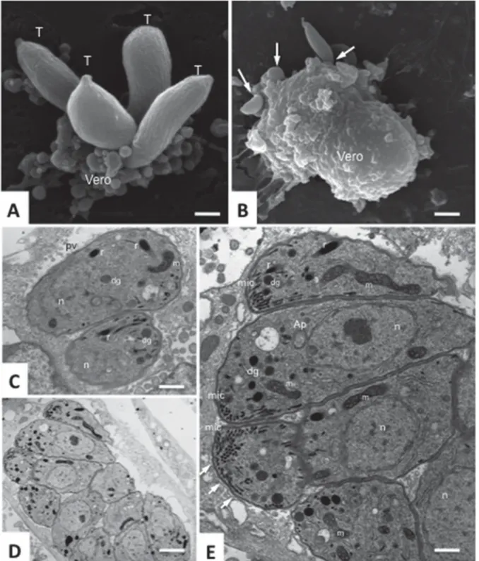
Besnoitia besnoiti tachyzoites represent the prolifer-ative stage responsible for acute disease. They are 6–7·5×2·5–3·9 μm (Reis et al. 2006) and can be cultured in vitro employing various mammalian cell types, using similar techniques as for N. caninum and T. gondii tachyzoites (Shkap et al. 1987b). By light microscopy, B. besnoiti tachyzoites cannot be di ffer-entiated from Neospora or Toxoplasma tachyzoites. By TEM, the hallmarks of apicomplexans are clearly visible. These include the invasion-relevant secretory organelles such as micronemes, rhoptries and dense granules, and the conoid and the microtubular cytoskeleton underlying the plasma membrane (see
Fig. 1). As for T. gondii and N. caninum, these tachyzoites can reside within a wide range of host cells, and they occupy a cytoplasmic compartment called the parasitophorous vacuole, which is sur-rounded by a parasitophorous vacuole membrane. Proliferation occurs through endodyogeny, with a replication rate of tachyzoites, depending on cell lines, from 0·14 and 20 tachyzoites per hour (Schares et al.2009). As with other apicomplexans, B. besnoiti tachyzoites enter their host cells with the apical part first, most likely employing similar mechanisms as described for T. gondii involving actin/myosin motor proteins. However, clear differences exist with respect to the structure and dynamics of tubulin polymers in Toxoplasma and Besnoitia, and with respect to their interaction with the host microtub-ular cytoskeleton once host cell invasion is achieved (Reis et al. 2006). In addition, the two species also differentially modulate the host cell centrosome and the Golgi apparatus (see paper by Cardoso et al. in this Special Issue).
As with the other apicomplexans, tissue cysts of B. besnoiti harbouring bradyzoites persist within chronically infected animals for extended periods of time. Bradyzoites are 6·0–7·5 ×1·9–2·3 μm (Dubey et al.2003), and they form spectacular tissue cysts often containing hundreds of parasites. These cysts are characterized by two distinct cyst walls (Fig. 2). The inner cyst wall is, similar to T. gondii and N. caninum, located intracellularly within a large multinuclear host cell, surrounded by the membrane of the parasitophorous vacuole, and is most likely synthesized by the parasite. The second cyst wall surrounds the entire host cell and is 10–12 μm in thickness (Dubey et al. 2003; Cortes et al. 2006c). The outer cyst wall is comprised of collagenous
material (Ayroud et al. 1995) and seems to be a product of a physiological reaction on the part of the host (Dubey et al. 2013). In any case, it is this double-layered cyst wall which clearly distinguishes B. besnoiti from its related apicomplexans.
A E T I O L O G Y , P A T H O G E N E S I S A N D C L I N I C A L D I A G N O S I S O F B O V I N E B E S N O I T I O S I S
Bovine besnoitiosis may have two different presenta-tions: an acute stage occurring 11–13 days after infection and lasting 6–10 days, followed by a chronic stage, which develops while the animals recover from the acute stage, and is lifelong (Pols, 1960; Bigalke,
1968).
The acute stage is characterized by rapid prolifer-ation of tachyzoites and subsequent immunopathol-ogy and tissue destruction, all within 1–2 weeks post-infection. Tachyzoites proliferate in macro-phages, fibroblasts and endothelial cells within blood vessels. The result is vasculitis and thrombosis, especially of capillaries and small veins in the dermis, subcutis, fascia, testes and upper respiratory mucosae (Basson et al. 1970). Acute stage clinical signs are increased body temperature (above 40 °C), intensive respiratory disorder, increased heart rates, serous nasal and ocular discharges, anorexia, generalized weakness, loss of milk production, reluctance to move, rapid loss of weight and declining body condition due to diminished food uptake, swelling of superficial lymph nodes, generalized oedema of the skin, acute orchitis with swollen, painful testes and, in some cases, anasarca (Schulz,1960; McCully et al.
1966; Cortes et al.2005). Inspiratory dyspnoea may result from inflammation of the upper respiratory mucosae (McCully et al. 1966). The acute stage does not always lead to death, but these processes can result in dramatically decreased body condition scores (Cortes et al.2005). In pregnant animals, this pathophysiological disorder can lead to abortion (Pols,1960; Juste et al.1990).
One to 2 weeks after the onset of acute stage symptoms, the oedema reduces and the infection reaches the chronic stage, which is characterized by the formation of tissue cysts of up to 0·5 mm in diameter by slowly proliferating bradyzoites, which can persist for several years in different tissues of the host (Fig. 2A). These characteristic cysts form especially in cutaneous and subcutaneous tissues, and in intermuscular fascia (McCully et al.1966; Basson et al.1970). A low-grade, intermittent febrile reaction may be observed, causing anorexia and weight loss. Dermal lesions, presenting different extents of hair loss, are always present during chronic disease. These consist of rather dramatic thickening, hardening and folding or wrinkling of the skin, especially around the neck, shoulders and rump, always accompanied by hyperkeratosis, hyperpigmentation and alopecia (Pols,1960). The thickening of the skin is caused by
scleroderma (Basson et al. 1970). Scleroderma and alopecia are permanent disfigurements in surviving animals (Bigalke, 1960). There may also be pro-nounced thickening of the limbs, and locomotion may be difficult and painful (Pols, 1960). A muco-purulent nasal discharge may be accompanied by
inspiratory dyspnoea (McCully et al. 1966). Small cysts that are visible by eye are usually apparent upon close visual inspection of the scleral conjunctiva and vulvae (Rostaher et al. 2010; Frey et al. 2013; see
Fig. 3), a feature that is of considerable value in clinical diagnosis (Sannusi,1991).
Fig. 1. Besnoitia besnoiti tachyzoites. (A) and (B) show scanning electron micrographs of tachyzoites (T) establishing contact with the surface of Vero cells. Arrows point to parasites in the process of host cell invasion. Following entry, tachyzoites proliferate rapidly within a parasitophorous vacuole (C–E). Arrows in (E) point towards the parasitophorous vacuole membrane. E is a higher magnification view of (D). Micronemes (mic), dense granules (dg), rhoptries (dg) and the parasite mitochondrion are clearly visible, n indicates the nucleus, and the apicoplast (Ap) is visible in one of the tachyzoites in (E). Note the parasitophorous vacuole membrane indicated by arrows. Bars in A = 1·8μm; in B = 3·6 μm; in C = 1μm; in D = 1·9 μm; in E = 0·65 μm.
All cattle breeds, both sexes and animals of all ages can be affected, except that acute and chronic disease seldom occurs in calves of less than 6 months of age (Bigalke,1968; Alzieu et al.2007). Animals with poor body condition are usually culled without any commercial value or die in thefield. In cases where the chronic stage does not dramatically decrease the health status of the animal, the parasite will persist and will present a large number of cysts in any part of the skin during the entire lifespan, even if the animal recovers body condition. Cows at this stage are still
fertile and frequently become pregnant and give birth. There are no reports of vertical transmission of B. besnoiti. The disease, however, has a negative impact on milk production, and this often negatively affects calves in their growth capacity (Cortes et al.
2006c). However, due to the high number of skin cysts in the mucosae of teats and udder, and the loss of elasticity, the teats will present large wounds and will bleed (Fig. 3). Newborn calves do not have antibodies against B. besnoiti, and receive them through suckling from the dam, soon acquiring titres Fig. 2. Besnoitia besnoiti bradyzoites. (A) Shows a histological section cut through a biopsy of the dermis and epidermis of a cow with chronic besnoitiosis. Tissue cysts with a double-layered cyst wall are present at a high density. (B) Shows a section through the interior part of a tissue cyst, with numerous bradyzoites embedded in a granular matrix (m). Note the high numbers of micronemes (m) and the parasite nucleus (n), which is located at the posterior end of the parasite. (C) and (D) are sections showing peripheral areas of tissue cysts. Bradyzoites at the cyst periphery are marked with a b, and the asterisk indicates a dead and partially lysed bradyzoite. The inner cyst wall (icw) is located within a host cell and presents a rather amorphous appearance. Arrows in (C) and (D) point towards the parasitophorous vacuole membrane. The outer cyst wall (ocw) surrounds the entire host cell and is a structure rich in densely packedfilaments. In (D) aflattened host cell nucleus (hcn) is visible in the space between inner and outer cyst wall (hcc = host cell cytoplasm). Bars in A = 600μm; in B = 0·9 μm; in C and D = 1·5 μm.
comparable to the dam up to the age of 6–8 months (Shkap et al.1994).
Infected bulls present the same symptoms in the acute and chronic stages of the disease as cows. Severely affected bulls often develop irreversible intratesticular lesions of vasculitis, focal necrosis, sclerosis and atrophy, which usually result in permanent infertility (Kumi-Diaka et al. 1981). The case fatality rate during the chronic stage is usually in the order of 10% (Pols,1960).
Although some of the typical clinical aspects of bovine besnoitiosis have been well described, the di-agnosis based on clinical signs is difficult, and the disease may be misdiagnosed as a fungal infection, rinderpest, scabies, mineral deficiency, photosensi-tivity or even blue tongue virus infection, and differential laboratory diagnostics need to be further improved.
L A B O R A T O R Y D I A G N O S I S
Presently, different diagnostic methods are available for the detection of B. besnoiti infection in cattle. These include direct detection of the parasite and/or its DNA in tissue samples (histopathology, PCR) and indirect detection based on serology. Application of these methods or combinations thereof depends on the clinical status of the animal and the status of the herd it comes from.
Direct diagnosis of B. besnoiti infection
Histopathological diagnosis of besnoitiosis is achieved in skin biopsies of cattle suffering from chronic disease. Typically, a biopsy punch of 8 mm diameter is suitable, and should be obtained at a location where the skin exhibits the corresponding pathological alterations described above. In a densely infected skin biopsy, tissue cysts and bradyzoites can also be visualized by TEM (Cortes et al.2006c; see
Fig. 2). As a differential diagnostic approach a skin scrape should be performed, and stained with Giemsa or May-Grünwald Giemsa for the detection of ectoparasites such as mites and in order to visualize tachyzoites and/or bradyzoites. For the diagnosis of sub-clinically infected animals, histopathology is not suitable, and more sensitive methods such as PCR are required (Cortes et al. 2007a; Schares et al. 2011b). Nevertheless, sub-clinically infected animals with a low number of cysts in the skin may still not present B. besnoiti DNA in the sample and might be PCR-negative. Despite this limitation, PCR was shown to be appropriate for the diagnosis of bovine besnoi-tiosis in the acute stage of the disease, during which no anti-B. besnoiti antibodies are found yet (Jacquiet et al. 2010). Quantitative real-time PCR has been applied not only in diagnosis, but also to detect the inhibition of tachyzoite proliferation in vitro using chemotherapeutically interesting compounds (Cortes et al.2007b,2011).
Indirect detection of Besnoitia infection
Assessment of antibodies against B. besnoiti in sera of infected animals is an appropriate approach for the diagnosis of clinical and subclinical disease. As a prerequisite, B. besnoiti antigen had to be generated, and the in vitro culture of the tachyzoite stage has been, and still is, an important step. Several isolates Fig. 3. Selected clinical features of bovine besnoitiosis.
(A) Shows an udder of a chronically infected cow presenting wounds in the teats, resulting from repeated lesions caused by nursing the calf. Individual and/or clusters of cysts can be detected by eye in the conjunctive (B) and in the vulva (C).
were produced by the primary passage of B. besnoiti through rabbits (Bigalke and Naude,1962; Bigalke et al.1974) or gerbils (Neuman, 1962; Shkap et al.
1987b) followed by in vitro culture in various cell lines. The isolation of the parasite directly from infected cattle to in vitro cultures was achieved by Cortes et al. (2006c) and this is the common approach also used by others (Fernandez-Garcia et al.2009a; Schares et al.2009; Gentile et al.2012). The existence of in vitro cultures has been the starting point for the development and application of different serological techniques that are being used in the diagnosis of infected animals and in epidemiological studies (see below).
Sub-clinically infected animals may represent a major factor in the transmission of B. besnoiti infection to new herds, thus ideally these should be detected as early as possible. Indirect fluorescence antibody test (IFAT) is considered the gold standard for serology (Shkap et al. 2002). When performed with the appropriate expertise, there is no cross-re-action of anti-B. besnoiti antibodies with N. caninum and T. gondii tachyzoites and vice versa. The cut-off dilution for Besnoitia IFAT ranges from 1/200 to 1/256 (Shkap et al.2002; Cortes et al.2006a; Schares et al.2010). However, assessments are often highly dependent on the expertise and experience of the individual analysing the results.
Western blotting for the diagnosis of B. besnoiti infection in cattle has been applied by different lab-oratories (Cortes et al.2006a; Fernandez-Garcia et al.
2009b; Schares et al.2010), but is clearly more ex-pensive and time consuming compared with IFAT. Although it has been shown that cross-reactions with related protozoans, especially N. caninum occur, Western blotting has been recommended as a con-firmation test in combination with other methods (Cortes et al. 2006a; Schares et al. 2010, 2011a; Millan et al.2012; Garcia-Lunar et al.2013a).
The enzyme-linked immunosorbent assay
(ELISA) is more appropriate for analysing larger numbers of samples. Several in-house ELISAs have been developed and used for the diagnosis of B. besnoiti infection (Neuman, 1972; Janitschke et al. 1984; Cortes et al. 2006a; Fernandez-Garcia et al.2010; Jacquiet et al.2010; Schares et al.2010,
2011a; Liénard et al. 2011; Garcia-Lunar et al.
2013a; Rinaldi et al. 2013), and have also been applied in epidemiological studies (Shkap et al.1984,
1985a,1989,1990; Gentile et al.2012; Garcia-Lunar et al. 2013a). A commercially available ELISA
(PrioCHECK® Besnoitia Ab; Prionics AG,
Schlieren, Switzerland) has been developed and used in the identification of infected herds and/or individual animals. Nevertheless, for the serodiag-nosis of individual cases at least one other con firma-tion test, IFAT or western blot, should be used (Cortes et al.2006a; Jacquiet et al.2010; Schares et al.
2011a; Garcia-Lunar et al. 2013a). A multicentre
study (Garcia-Lunar et al. 2013a) showed that the sensitivity of serological detection in sub-clinically infected animals was low, and some of the currently applied tests exhibited a reduced sensitivity after a season of low or no insect activity with low or even absent mechanical transmission (Liénard et al.2011; Schares et al. 2011b). Several tests also exhibit suboptimal specificity due to cross-reactions with related apicomplexans, resulting in false positive results (Nasir et al.2012).
A major breakthrough for the differential diagnosis of acute and chronic bovine besnoitiosis has recently been achieved by Schares et al. (2013). They developed serological tests employing affinity pur-ified antigens of B. besnoiti tachyzoites in western blots, conventional ELISA and avidity ELISA. They identified two B. besnoiti surface antigens of 39 and 42 kDa which were efficiently recognized by sera from infected animals. These two antigens were affinity purified and were used in ELISA, showing 100% sensitivity for sera of chronically infected animals, and 99·8% specificity when tested with sera from N. caninum associated abortions. Acute cases of besnoitiosis were confirmed by low-avidity IgG (Schares et al. 2013). In any case, however, the detection of parasite DNA in skin by real-time PCR is clearly superior to serological analysis for the detection of infected cattle during acute (early) besnoitiosis.
The direct agglutination test, based on the complex formation of formalin-treated parasites in the
pres-ence of diluted serum containing specific IgG
antibodies, has been used for the diagnosis of closely related apicomplexans such as T. gondii (Fulton and Turk,1959) and N. caninum (Packham et al. 1998; Romand et al.1998). Waap et al. (2011) developed a modified agglutination test for B. besnoiti. This method avoids the use of secondary host-specific antisera, and can thus be used to test different species which may be implicated in the life cycle and could therefore specifically contribute to the elucidation of the definitive host for B. besnoiti.
T R A N S M I S S I O N O F B . B E S N O I T I T O C A T T L E
Cattle are the only known intermediate host of B. besnoiti. Elucidation of the entire life cycle would be important to define the role of the putative definitive host in transmission, and would aid in the development of appropriate prophylaxis and control measures. It is not even known, however, whether a definitive host exists, and if so, how important it is in transmitting the parasite. In fact, it has been suggested that some T. gondii populations represent clonal lineages, indicating that transmission is not dependent on the diploid infective stage in the definitive host (Johnson,1997).
Similar to other cyst-forming coccidians, natural exposure of cattle to sporulated oocysts suggests the
presence of a definitive host in the immediate environment (most likely a carnivore) or an infective feed source (infected or contaminated with tachy-zoites or bradytachy-zoites of B. besnoiti) to which the definitive host has access (Tenter and Johnson,1997). On many farms where bovine besnoitiosis is endemic, dead cattle in farms are conscientiously disposed of by rendering, incineration, deep burial or other means that do not allow for recycling of infectious materials into potential definitive hosts. On the other hand, it is possible that a sylvatic cycle exists and as a consequence oocysts could be produced by a de fini-tive host in wildlife. That this possibility is viable was suggested in South Africa (Basson et al.1965,1970; McCully et al.1966; Bigalke et al.1967,1974), where B. besnoiti has been shown to affect wildebeest, kudu and impala. However, Bigalke et al. (1967) registered differences among isolates from wildebeest, impala and cattle, and concluded that they should be regarded as distinct strains or biologically different isolates of B. besnoiti. There is also serological evidence for B. tarandi and/or B. besnoiti infection in wild ruminants in Canada (caribou, reindeer, mule deer and musk ox with similar characteristic clinical signs and lesions) and in Spain (red deer and roe deer) (Gutiérrez-Expósito et al. 2012, 2013), but the meaning of these observations for the explanation of the transmission of B. besnoiti to cattle remains to be elucidated.
Within endemic areas there is evidence that biting flies can mechanically transmit B. besnoiti (Pols,
1960; Alzieu et al.2007; Liénard et al.2011,2013). Outbreaks of bovine besnoitiosis mostly occur during seasons of the year when bitingflies are active. Several biting insects such as Stomoxys, Glossina and Chrysops have been implicated in the mechanical transmission of infection (Pols,1960; Bigalke,1968). They transport bradyzoites in their mouthparts and abdominal contents from one host to the other and deposit them onto and/or into the skin. Since tachyzoites have been demonstrated in lacrimal secretions (Bigalke,1968; Cortes et al.2003), there is a possibility that non-biting flies, such as Musca autumnalis and Musca domestica, and other insects, might also be capable of mechanical transmission by having access to B. besnoiti in the lacrimal fluid. Nevertheless mechanical transmission would in this case result in parasite transfer from disrupted cysts in wounds of infected animals to wounds of susceptible ones, through direct contact of legs and licking mouthparts, but this is expected to be extremely rare in field conditions (Liénard et al. 2013). Introduction of sub-clinically or clinically infected animals into herds that had not been exposed to Besnoitia before may often result in an outbreak of disease. Thus, acquisition of infected animals should be avoided. Conversely, the fact that clinical signs occurred rapidly in animals that have been intro-duced into herds known to be infected suggested
that mechanical transmission occurred within a short period, most likely due to a high population of potential mechanical vectors (Pols, 1960; Bigalke,
1968; Liénard et al.2011). In addition, B. besnoiti can also be transmitted iatrogenically during prophylac-tic or treatment procedures within the herd (Pols,
1954, 1960; Bigalke, 1968). Finally, another mode, although speculative, might be a direct transmission from animal to animal, e.g. via mucosal contact by licking. If a chronically infected host presents many mucosal surface cysts such as frequently seen on the eye (and then maybe also other mucosal surfaces accessible for licking), contact with mechanically released bradyzoites from one animal to the oral mucosa of another animal may be a possibility.
T R E A T M E N T O P T I O N S F O R B O V I N E B E S N O I T I O S I S
The first reported attempts for the treatment of bovine besnoitiosis were based on a single intra-venous injection of 30 mL of a 1% solution of formalin during the acute stage of the disease (Herin, 1952). For the treatment of chronically in-fected animals, the same author recommended administering 30–40 mL Lugol’s iodine solutions 5 times intravenously at intervals of 4–7 days. However, these treatment approaches were not further developed, due to obvious adverse events associated with such treatments. Other treatment approaches were investigated in rabbits, including the administration of pentamidine, aureomicin, for-malin, sodium iodide, sulfamerazine, mycostatin and terramycin, but were not successful (Pols, 1960). Shkap et al. (1985b) tested several compounds in rabbits experimentally infected with B. besnoiti tachyzoites obtained from cell culture. Oxytetra-cycline, given at 30 mg kg− 1i.m. simultaneously with infection, prevented the development of orchitis. All infected animals showed a transient febrile reaction, and the authors concluded that oxytetracycline had some therapeutic potential against B. besnoiti and that rabbits are a suitable model for therapeutic trials of acute disease. Subsequent studies were performed in vitro and in experimentally infected gerbils. Drugs investigated in gerbils included oxytetracycline, sulfonamides, trimethoprim, halofunginone, diami-nazene aceturate and pentamidine. Of these com-pounds, only oxytetracycline prevented death in gerbils, but only if administered at the time of infection (Shkap et al.1987a).
More recently, the nitro-thiazolide nitazoxanide and a range of bromo-derivatives were shown to inhibit B. besnoiti tachyzoite proliferation in Vero cells, and induced severe ultrastructural alterations. Bromo-derivatives were found to be equally effective as nitro-compounds, indicating that they could be a safer alternative to compounds containing a poten-tially harmful nitro group (Cortes et al. 2007b). Another in vitro study demonstrated that a series of
new-generation pentamidine derivatives, arylimida-mides, exhibit profound activity against B. besnoiti tachyzoites, but corresponding studies in animals have not yet been undertaken (Cortes et al.2011).
V A C C I N E S
Live vaccines have been used in South Africa and in Israel. In South Africa, the vaccine is based on tachyzoites of an isolate from blue wildebeest grown in cell cultures (Bigalke et al. 1974) and is recom-mended for use in weaners and older animals. It protects cattle against clinical besnoitiosis, although it does not entirely prevent sub-clinical infection (Bigalke and Prozesky,2004). The vaccine in use in Israel contains live attenuated parasites derived from cell cultures (Pipano,1997) and there are no data in the scientific literature that allows for any judgement on its efficacy and safety. However, the use of any of these vaccines is geographically limited. Live-attenuated vaccines pose risks of introducing the parasite into uninfected herds and of inducing carriers among the vaccinated animals. This is of particular concern when, as in the case of B. besnoiti, the knowledge on the biology, transmission and life cycle of the parasite is scarce. This lack of knowledge has hampered the development of new vaccines. Little is known about the chain of infection and the infective stage and portal of entry for bovines under natural conditions. These aspects are of major relevance for the development and evaluation of immunization protocols and, in a broader sense, for the development of studies towards a better under-standing of the bovine immune response against B. besnoiti infection.
Also crucial for the development of new vaccines against the disease is the characterization of the antigenic mosaic of the parasite. An important contribution can be taken from the studies mentioned above on the development of diagnostic tests based on western blot. The pattern of antigens with high diagnostic value (e.g. Schares et al. 2013) certainly includes those antigens that are serologically immu-nodominant, and potentially suitable as vaccine targets. However, their molecular identification and characterization remains largely incomplete. Njagi et al. (2004) characterized a panel of four monoclonal antibodies, obtained from mice immunized with live tachyzoites, two of which have been shown to significantly inhibit the invasion of cell culture monolayers by B. besnoiti tachyzoites. However, no subsequent studies were published and the antigens recognized by these monoclonal antibodies remain to be identified.
So far, the only B. besnoiti protein characterized and expressed in a heterologous system has been disulphide isomerase (BbPDI) (Marcelino et al.
2011). Toxoplasma gondii and N. caninum had been previously shown to express highly homologous
molecules (Meek et al.2002a,b; Naguleswaran et al.
2005) that were shown to be targeted, respectively, by human (Meek et al. 2002a,b) and bovine (Liao
et al. 2006) IgA. The N. caninum PDI was
demonstrated to be important for the host cell invasion (Naguleswaran et al.2005) and turned out to be an interesting candidate for vaccine develop-ment (Debache et al.2010). Interestingly, conversely to what has been observed for N. caninum (Shin et al.
2004, 2005), a recent study tackling the charac-terization of the immunome of B. besnoiti tachyzoites using pooled sera from seven naturally infected cattle (Garcia-Lunar et al. 2013b) has not confirmed BbPDI as a serologically dominant antigen. In this study, out of the 20 immunogenic proteins identified based on homology with sequences available in databases from other members of the Toxoplasma-tinae sub-family, were four proteins related to energy metabolism (fructose-1,6-bisphosphate aldolase, LDH, pyruvate kinase and ENO), three heat shock proteins (Hsp60, Hsp70 and Hsp90), and actin and profilin.
Fernandez-Garcia et al. (2013) reported on the identification of antigens that are differentially expressed in B. besnoiti tachyzoites and bradyzoites. They performed comparative 2D-gel electrophoresis on extracts of both stages, followed by mass spectro-metry (MS) analysis. A total of 130 and 132 spots were differentially expressed in bradyzoites and tachyzoites, respectively, and 25 differentially
ex-pressed spots were selected and analysed by
MALDI-TOF/MS. As a result, five up-regulated
bradyzoite proteins (GAPDH, ENO1, LDH, SOD and RNA polymerase) andfive up-regulated tachy-zoite proteins (ENO2; LDH; ATP synthase; HSP70 and PDI) were identified. These proteins can now be further studied with regard to their role in host-cell parasite interactions, tachyzoite-bradyzoite tran-sition, and in terms of a putative role as drug targets or vaccine candidates.
Clearly, the contribution of these studies for the development of safer subunit vaccines is hampered by two main obstacles. First, the scarce knowledge currently available on the molecular aspects of this parasite, and second the absence of a good exper-imental animal model. Thus, B. besnoiti genome and transcriptome sequencing as a basis for the systematic molecular analysis of its biology must be a priority, as well as the development of an experimental bovine model that allows assessment of the efficacy of potential vaccine candidates directly in the most relevant target host.
C O N C L U S I O N S
Bovine besnoitiosis is an emerging disease in Europe and an economic concern to the cattle breeding industry. Efficient measures must be undertaken to avoid further spread of the disease. Many features
of the basic biology of B. besnoiti are waiting to be discovered, and these discoveries are a prerequisite for the development of efficient control options. Primary concerns are the development of standar-dized laboratory diagnostic techniques for the rapid identification of sub-clinically infected animals. With this in mind, the responsible authorities and farmers should consider taking action to avoid introduction of bovine besnoitiosis into countries and regions of the EU where it is not yet present. In addition, it will be important to generate genomics and transcriptomics data of the different life cycle stages of B. besnoiti. This will allow the identification of novel targets for vaccination and therapeutic intervention, and will generate invaluable tools to elucidate the molecular and physiological aspects of the host-parasite re-lationship.
A C K N O W L E D G E M E N T S
The authors acknowledge thefinancial support of the Swiss National Science Foundation (grant No. grant 310030-146162) and of the Portuguese Fundação para a Ciência e a Tecnologia (grant No. PTDC/CVT/65674/2006, PTDC/ CVT/71630/2006), and are thankful to COST actions 854 and 857 for contributing to a research network.
R E F E R E N C E S
Alvarez-Garcia, G., Frey, C., Ortega-Mora, L. M. and Schares, G. (2013). A century of bovine besnoitiosis: an unknown disease re-emerging in Europe. Trends in Parasitology29, 407–415. doi: 10.1016/j.pt.2013.06.002. Alzieu, J. P., Cortes, H., Gottstein, B., Jacquiet, P., Dorchies, P., Schelcher, F. and L’Hostis, M. (2007). La besnoitiose bovine: actualités épidémiologiques et diagnostiques. Bulletin des G.T.V. – Hors-série parasitisme des bovins, 41–49.
Ayroud, M., Leighton, F. A. and Tessaro, S. V. (1995). The morphology and pathology of Besnoitia sp. in reindeer (Rangifer tarandus tarandus). Journal of Wildlife Diseases31, 319–326. doi: 10.7589/0090-3558-31.3.319. Basso, W., Schares, G., Gollnick, N. S., Rütten, M. and Deplazes, P. (2011). Exploring the life cycle of Besnoitia besnoiti– experimental infection of putative definitive and intermediate host species. Veterinary Parasitology 178, 223–234. doi: 10.1016/j.vetpar.2011.01.027.
Basson, P. A., van Niekerk, J. W., McCully, R. M. and Bigalke, R. D. (1965). Besnoitiosis in South African antelopes: a preliminary note on the occurrence of Besnoitia cysts in the cardiovascular system. Journal of the South African Veterinary Medical Association36, 578.
Basson, P. A., McCully, R. M. and Bigalke, R. D. (1970). Observations on the pathogenesis of bovine and antelope strains of Besnoitia besnoiti (Marotel, 1912) infection in cattle and rabbits. Onderstepoort Journal of Veterinary Research37, 105–126.
Besnoit, C. and Robin, V. (1912). Sarcosporidioses cutanée chez une vache. Revue Vétérinaire37, 649–663.
Bigalke, R. D. (1960). Preliminary observation on the mechanical transmission of cyst organisms of Besnoitia besnoiti (Marotel, 1912) from a chronically infected bull to rabbits by Glossina brevipalpis Newstead, 1910. Journal of the South African Veterinary Association31, 37–44.
Bigalke, R. D. (1968). New concepts on the epidemiological features of bovine besnoitiosis as determined by laboratory andfield investigations. Onderstepoort Journal of Veterinary Research35, 3–138.
Bigalke, R. D. and Naude, T. W. (1962). The diagnostic value of cysts in the scleral conjunctiva in bovine besnoitiosis. Journal of the South African Veterinary Medical AssociationXXXIII, 21–27.
Bigalke, R. D. and Prozesky, L. (2004). Besnoitiosis. In Infectious Diseases of Livestock, Vol. 1 (ed. Coetzer, J. A. W. and Tustin, R. C.), pp. 331–359. Oxford University Press, Cape Town, South Africa.
Bigalke, R. D., van Niekerk, J. W., Basson, P. A. and McCully, R. M. (1967). Studies on the relationship between Besnoitia of blue wildebeest and
impala, and Besnoitia besnoiti of cattle. Onderstepoort Journal of Veterinary Research34, 7–28.
Bigalke, R. D., Schoeman, J. H. and McCully, R. M. (1974). Immunization against bovine besnoitiosis with a live vaccine prepared from a blue wildebeest strain of Besnoitia besnoiti grown in cell cultures. 1. Studies on rabbits. Onderstepoort Journal of Veterinary Research41, 1–5.
Cadéac, C. (1884). Identit‚ de l‚Elephantiasis et de l’anasarque du boeuf. Description de cette maladie. Revue Vétérinaire521, 521–540.
Coleman, A. W. and Mai, J. C. (1997). Ribosomal DNA ITS-1 and ITS-2 sequence comparisons as a tool for predicting genetic relatedness. Journal of Molecular Evolution45, 168–177. doi: 10.1007/PL00006217.
Cortes, H., Ferreira, M. L., Silva, J. F., Vidal, R., Serra, P. and Caeiro, V. (2003). Contribuição para o estudo da besnoitiose bovina em Portugal. Revista Portuguesa de Ciências Veterinárias98, 43–46.
Cortes, H., Leitão, A., Vidal, R., Vila-Vicosa, M. J., Ferreira, M. L., Caeiro, V. and Hjerpe, C. A. (2005). Besnoitiosis in bulls in Portugal. Veterinary Record157, 262–264. doi: 10.1136/vr.157.9.262.
Cortes, H. C. E., Nunes, S., Reis, Y., Staubli, D., Vidal, R., Sager, H., Leitão, A. and Gottstein, B. (2006a). Immunodiagnosis of Besnoitia besnoiti infection by ELISA and Western blot. Veterinary Parasitology141, 216–225. doi: 10.1016/j.vetpar.2006.05.023.
Cortes, H., Chagas e Silva, J., Baptista, M. C., Pereira, R. M., Leitão, A., Horta, A. E. M., Vasques, M. I., Barber, J. and Marques, C. C. (2006b). Besnoitia besnoiti impact on fertility of cattle exploited in mediterranean pastures (Alentejo). In EAAP Publication 119 (ed. Ribeiro, J. M. R.), pp. 323–329. Wageningen Academic Press, Wageningen, the Netherlands.
Cortes, H. C., Reis, Y., Waap, H., Vidal, R., Soares, H., Marques, I., da Pereira, F., Fazendeiro, I., Ferreira, M. L., Caeiro, V., Shkap, V., Hemphill, A. and Leitão, A. (2006c). Isolation of Besnoitia besnoiti from infected cattle in Portugal. Veterinary Parasitology 141, 226–233. doi: 10.1016/j.vetpar.2006.05.022.
Cortes, H. C. E., Reis, Y., Gottstein, B., Hemphill, A., Leitão, A. and Müller, N. (2007a). Application of conventional and real-timefluorescent ITS1 rDNA PCR for detection of Besnoitia besnoiti infections in bovine skin biopsies. Veterinary Parasitology 146, 352–356. doi: 10.1016/j.vet-par.2007.03.003.
Cortes, H. C. E., Mueller, N., Esposito, M., Leitão, A., Naguleswaran, A. and Hemphill, A. (2007b). In vitro efficacy of nitro-and bromo-thiazolyl-salicylamide compounds (thiazolides) against Besnoitia besnoiti infection in Vero cells. Parasitology134, 975–985. doi: 10.1017/S0031182007002417.
Cortes, H. C., Muller, N., Boykin, D., Stephens, C. E. and Hemphill, A. (2011). In vitro effects of arylimidamides against Besnoitia besnoiti infection in Vero cells. Parasitology138, 583–592. doi: 10.1017/ S0031182011000114.
Debache, K., Guionaud, C., Alaeddine, F. and Hemphill, A. (2010). Intraperitoneal and intra-nasal vaccination of mice with three distinct recombinant Neospora caninum antigens results in differential effects with regard to protection against experimental challenge with Neospora caninum tachyzoites. Parasitology137, 229–240. doi: 10.1017/S0031182009991259. Diesing, L., Heydorn, A. O., Matuschka, F. R., Bauer, C., Pipano, E., de Waal, D. T. and Potgieter, F. T. (1988). Besnoitia besnoiti: studies on the definitive host and experimental infections in cattle. Parasitology Research75, 114–117. doi: 10.1007/BF00932710.
Dubey, J. P. (1976). A review of Sarcocystis of domestic animals and of other coccidia of cats and dogs. Journal of the American Veterinary Medical Association169, 1061–1078.
Dubey, J. P., Shkap, V., Pipano, E., Fish, L. and Fritz, D. L. (2003). Ultrastructure of Besnoitia besnoiti tissue cysts and bradyzoites. Journal of Eukaryotic Microbiology 50, 240–244. doi: 10.1111/j.1550-7408.2003. tb00127.x.
Dubey, J. P., Sreekumar, C., Rosenthal, B. M., Vianna, M. C. B., Nylund, M., Nikander, S. and Oksanen, A. (2004). Redescription of Besnoitia tarandi (Protozoa: Apicomplexa) from the reindeer (Rangifer tarandus). International Journal for Parasitology 34, 1273–1287. doi: 10.1016/j.ijpara.2004.07.002.
Dubey, J. P., van Wilpe, E., Blignaut, D. J. C., Schares, G. and Williams, J. H. (2013). Development of early tissue cysts and associated pathology of Besnoitia besnoiti in a naturally infected bull (Bos taurus) from South Africa. Journal of Parasitology99, 459–466. doi: 10.1645/12-128.1. Ducrocq, J., Beauchamp, G., Kutz, S., Simard, M., Elkin, B., Croft, B., Taillon, J., Cote, S. D., Brodeur, V., Campbell, M., Cooley, D., Cuyler, C. and Lair, S. (2012). Comparison of gross visual and microscopic assessment of four anatomic sites to monitor Besnoitia tarandi in barren-ground caribou (Rangifer tarandus). Journal of Wildlife Diseases 48, 732–738. doi: 10.7589/0090-3558-48.3.732.
EFSA Journal (2010). Bovine besnoitiosis: an emerging disease in Europe. EFSA Journal8, 1499 [15 pp.]. doi: 10.2903/j.efsa.2010.1499.
Elsheikha, H. M., Mackenzie, C. D., Rosenthal, B. M., Marteniuk, J. V., Steficek, B., Windsor, S., Saeed, A. M. and Mansfield, L. S. (2005). An outbreak of besnoitiosis in miniature donkeys. Journal of Parasitology91, 877–881. doi: 10.1645/GE-3277.1.
Fernandez-Garcia, A., Risco-Castillo, V., Pedraza-Diaz, S., Aguado-Martinez, A., Alvarez-Garcia, G., Gomez-Bautista, M., Collantes-Fernandez, E. and Ortega-Mora, L. M. (2009a). First isolation of Besnoitia besnoiti from a chronically infected cow in Spain. Journal of Parasitology95, 474–476.
Fernandez-Garcia, A., Alvarez-Garcia, G., Risco-Castillo, V., Aguado-Martinez, A., Marugan-Hernandez, V. and Ortega-Mora, L. M. (2009b). Pattern of recognition of Besnoitia besnoiti tachyzoite and bradyzoite antigens by naturally infected cattle. Veterinary Parasitology 164, 104–110. doi: 10.1016/j.vetpar.2009.06.020.
Fernandez-Garcia, A., Alvarez-Garcia, G., Risco-Castillo, V., Aguado-Martinez, A., Marcen, J. M., Rojo-Montejo, S., Castillo, J. A. and Ortega-Mora, L. M. (2010). Development and use of an indirect ELISA in an outbreak of bovine besnoitiosis in Spain. Veterinary Record166, 818–822. doi: 10.1136/vr.b4874.
Fernandez-García, A., Alvarez-García, G., Marugán-Hernández, V., García-Lunar, P., Aguado-Martínez, A., Risco-Castillo, V. and Ortega-Mora, L. M. (2013). Identification of Besnoitia besnoiti proteins that showed differences in abundance between tachyzoite and bradyzoite stages by difference gel electrophoresis. Parasitology 140, 999–1008. doi: 10.1017/S003118201300036X.
Franco, E. E. and Borges, I. (1916). Sur la sarcosporidiose Bovine. Arquivos do Instituto Bacteriologico Câmara Pestana4, 269–289.
Frey, C. F., Gutiérrez-Expósito, D., Ortega-Mora, L. M., Benavides, J., Marcén, J. M., Castillo, J. A., Casasús, I., Sanz, A., García-Lunar, P., Esteban-Gil, A. and Álvarez-García, G. (2013). Chronic bovine besnoitiosis: intra-organ parasite distribution, parasite loads and parasite-associated lesions in subclinical cases. Veterinary Parasitology 197, 95–103. doi: 10.1016/j.vetpar.2013.04.023.
Fulton, J. D. and Turk, J. L. (1959). Direct agglutination test for Toxoplasma gondii. Lancet2, 1068–1069.
Garcia-Lunar, P., Ortega-Mora, L. M., Schares, G., Gollnick, N. S., Jacquiet, P., Grisez, C., Prevot, F., Frey, C. F., Gottstein, B. and Alvarez-Garcia, G. (2013a). An inter-laboratory comparative study of serological tools employed in the diagnosis of Besnoitia besnoiti infection in bovines. Transboundary and Emerging Diseases60, 59–68. doi: 10.1111/ j.1865-1682.2012.01318.x.
Garcia-Lunar, P., Regidor-Cerrillo, J., Gutiérrez-Expósito, D., Ortega-Mora, L. and Alvarez-García, G. (2013b). First 2-DE approach towards characterising the proteome and immunome of Besnoitia besnoiti in the tachyzoite stage. Veterinary Parasitology195, 24–34. doi: 10.1016/j. vetpar.2012.12.040.
Gentile, A., Militerno, G., Schares, G., Nanni, A., Testoni, S., Bassi, P. and Gollnick, N. S. (2012). Evidence for bovine besnoitiosis being endemic in Italy– first in vitro isolation of Besnoitia besnoiti from cattle born in Italy. Veterinary Parasitology 184, 108–115. doi: 10.1016/j. vetpar.2011.09.014.
Gollnick, N. S., Gentile, A. and Schares, G. (2010). Diagnosis of bovine besnoitiosis in a bull born in Italy. Veterinary Record166, 599. doi: 10.1136/ vr.b4874.
Gutiérrez-Expósito, D., Ortega-Mora, L. M., Gajadhar, A. A., García-Lunar, P., Dubey, J. P. and Alvarez-García, G. (2012). Serological evidence of Besnoitia spp. infection in Canadian wild ruminants and strong cross-reaction between Besnoitia besnoiti and Besnoitia tarandi. Veterinary Parasitology 190, 19–28. doi: 10.1016/j. vetpar.2012.06.017.
Gutiérrez-Expósito, D., Ortega-Mora, L. M., Marco, I., Boadella, M., Gortázar, C., San Miguel-Ayanz, J. M., García-Lunar, P., Lavín, S. and Alvarez-García, G. (2013). First serosurvey of Besnoitia spp. infection in wild European ruminants in Spain. Veterinary Parasitology 197, 557–564. doi: 10.1016/j.vetpar.2013.05.017.
Henry (1912). Sarcosporidiose cutanée chez une Vache. Recueil de médecine vétérinaireXC, 327.
Herin, V. V. (1952). Bovine globidiosis in Ruanda-Urundi. Annales de la Société Belge de Médecine Tropicale32, 155–159.
Hornok, S., Fedák, A., Baska, F., Hofmann-Lehmann, R. and Basso, W. (2014). Bovine besnoitiosis emerging in Central-Eastern Europe, Hungary. Parasites and Vectors7, 20. doi: 10.1186/1756-3305-7-20. Irigoien, M., Del Cacho, E., Gallego, M., López-Bernad, F., Quílez, J. and Sánchez-Acedo, C. (2000). Immunohistochemical study of the cyst of Besnoitia besnoiti. Veterinary Parasitology91, 1–6. doi: 10.1016/S0304-4017 (00)00260-0.
Jacquiet, P., Liénard, E. and Franc, M. (2010). Bovine besnoitiosis: epidemiological and clinical aspects. Veterinary Parasitology174, 30–36. doi: 10.1016/j.vetpar.2010.08.013.
Janitschke, K., De Vos, A. J. and Bigalke, R. D. (1984). Serodiagnosis of bovine besnoitiosis by ELISA and immunofluorescence tests. Onderstepoort Journal of Veterinary Research51, 239–243.
Johnson, A. M. (1997). Speculation on possible life cycles for the clonal lineages in the genus Toxoplasma. Parasitology Today13, 393–397. Juste, R. A., Cuervo, L. A., Marco, J. C. and Oregui, L. M. (1990). La besnoitiosis bovina: desconocida en España? Medicina Veterinária 7, 613–618.
Kiehl, E., Heydorn, A. O., Schein, E., Al-Rasheid, K. A. S., Selmair, J., Abdel-Ghaffar, F. and Mehlhorn, H. (2010). Molecular biological comparison of different Besnoitia species and stages from different countries. Parasitology Research106, 889–894. doi: 10.1007/s00436-010-1770-9. Kumi-Diaka, J., Wilson, S., Sannusi, A., Njoku, C. E. and Osoru, D. I. K. (1981). Bovine besnoitiosis and its effect on the male reproductive system. Theriogenology16, 523–530. doi: 10.1016/0093-691X (81)90037-6.
Liao, M., Ma, L., Bannai, H., Lee, E.-G., Xie, Z., Tang, X., Zhang, H., Xuan, X. and Fujisaki, K. (2006). Identification of a protein disulfide isomerase of Neospora caninum in excretory-secretory products and its IgA binding and enzymatic activities. Veterinary Parasitology139, 47–56. doi: 10.1016/j.vetpar.2006.02.029.
Liénard, E., Salem, A., Grisez, C., Prévot, F., Bergeaud, J. P., Franc, M., Gottstein, B., Alzieu, J. P., Lagalisse, Y. and Jacquiet, P. (2011). A longitudinal study of Besnoitia besnoiti infections and seasonal abundance of Stomoxys calcitrans in a dairy cattle farm of southwest France. Veterinary Parasitology177, 20–27. doi: 10.1016/j.vetpar.2010.11.030. Liénard, E., Salem, A., Jacquiet, P., Grisez, C., Prévot, F., Blanchard, B., Bouhsira, E. and Franc, M. (2013). Development of a protocol testing the ability of Stomoxys calcitrans (Linnaeus, 1758) (Diptera: Muscidae) to transmit Besnoitia besnoiti (Henry, 1913) (Apicomplexa: Sarcocystidae). Parasitology Research112, 479–486. doi: 10.1007/s00436-012-3157-6.
Marcelino, E., Martins, T. M., Morais, J. B., Nolasco, S., Cortes, H., Hemphill, A., Leitão, A. and Novo, C. (2011). Besnoitia besnoiti protein disulfide isomerase (BbPDI): molecular characterization, expression and in silico modelling. Experimental Parasitology129, 164–174. doi: 10.1016/j. exppara.2011.06.012.
Marotel, M. (1912). Discussion of paper by Besnoit and Robin. Bullet Mémoires de la Société de Sciences Veterinaires de Lyon et de la Société de Medicine Veterinaire de Lyon et du Sud-Est15, 196–217.
McCully, R. M., Basson, P. A., van Niekerk, J. W. and Bigalke, R. D. (1966). Observations on Besnoitia cysts in the cardiovascular system of some wild antelopes and domestic cattle. Onderstepoort Journal of Veterinary Research33, 245–276.
Meek, B., Back, J. W., Klaren, V. N., Speijer, D. and Peek, R. (2002a). Protein disulfide isomerase of Toxoplasma gondii is targeted by mucosal IgA antibodies in humans 470. FEBS Letters522, 104–108. doi: 10.1016/S0014-5793(02)02911-3.
Meek, B., Back, J. W., Klaren, V. N. A., Speijer, D. and Peek, R. (2002b). Conserved regions of protein disulfide isomerase are targeted by natural IgA antibodies in humans. International Immunology14, 1291–1301. doi: 10.1093/intimm/dxf091.
Mehlhorn, H., Klimpel, S., Schein, E., Heydorn, A. O., Al-Quraishy, S. and Selmair, J. (2009). Another African disease in Central Europe: Besnoitiosis of cattle. I. Light and electron microscopical study. Parasitology Research 104, 861–868. doi: 10.1007/s00436-008-1267-y.
Millan, J., Sobrino, R., Rodriguez, A., Oleaga, A., Gortazar, C. and Schares, G. (2012). Large-scale serosurvey of Besnoitia besnoiti in free-living carnivores in Spain. Veterinary Parasitology 190, 241–245. doi:10.1016/j.vetpar.2012.06.014.
Naguleswaran, A., Alaeddine, F., Guionaud, C., Vonlaufen, N., Sonda, S., Jenoe, P., Mevissen, M. and Hemphill, A. (2005). Neospora caninum protein disulfide isomerase is involved in tachyzoite-host cell interaction. International Journal for Parasitology 35, 1459–1472. doi: 10.1016/j.ijpara.2005.06.006.
Nasir, A., Lanyon, S. R., Schares, G., Anderson, M. L. and Reichel, M. P. (2012). Sero-prevalence of Neospora caninum and Besnoitia besnoiti in South Australian beef and dairy cattle. Veterinary Parasitology186, 480–485. doi: 10.1016/j.vetpar.2011.11.032.
Neuman, M. (1962). The experimental infection of the gerbil (Meriones tristrami shanii) with Besnoitia besnoiti. Refuah Veterinarith19, 184–188. Neuman, M. (1972). Serological survey of Besnoitia besnoiti (Marotel 1912) infection in Israel by immunofluorescence. Zentralblatt Veterinarmedizin Reihe B19, 391–396.
Ng’ang’a, C. J. and Kasigazi, S. (1994). Caprine besnoitiosis: studies on the experimental intermediate hosts and the role of the domestic cat in transmission. Veterinary Parasitology52, 207–210. doi: 10.1016/0304-4017 (94)90112-0.
Njagi, O. N., Entzeroth, R., Nyaga, P. N. and Musoke, A. J. (2004). Monoclonal antibodies identify two neutralization-sensitive epitopes in Besnoitia besnoiti endocytes. Parasitology Research94, 247–253. doi: 10.1007/s00436-004-1210-9.
Olias, P., Schade, B. and Mehlhorn, H. (2011). Molecular pathology, taxonomy and epidemiology of Besnoitia species (Protozoa: Sarcocystidae). Infection, Genetics and Evolution 11, 1564–1576. doi: 10.1016/j. meegid.2011.08.006.
Packham, A. E., Sverlow, K. W., Conrad, P. A., Loomis, E. F., Rowe, J. D., Anderson, M. L., Marsh, A. E., Cray, C. and Barr, B. C. (1998). A modified agglutination test for Neospora caninum: development, optimization, and comparison to the indirectfluorescent-antibody test and enzyme-linked immunosorbent assay. Clinical and Diagnostic Laboratory Immunology5, 467–473.
Peteshev, V. M., Galuzo, I. G. and Polomoshov, A. P. (1974). Cats– definitive hosts Besnoitia (Besnoitia besnoiti) (in Russian). Izvestiae Akademii Nauk Kazakheskan SSRB, 33–38.
Pipano, E. (1997). Vaccines against hemoparasitic diseases in Israel with special reference to quality assurance. Tropical Animal Health and Production29, 86S–90S.
Pols, J. W. (1954). The artificial transmission of Globidium besnoiti Marotel, 1912, to cattle and rabbits. Journal of the South African Veterinary Medical Association25, 37–44.
Pols, J. W. (1960). Studies on bovine besnoitiosis with special reference to the aetiology. Onderstepoort Journal of Veterinary Research28, 265–356. Reis, Y., Cortes, H., Viseu, M. L., Fazendeiro, I., Leitão, A. and Soares, H. (2006). Microtubule cytoskeleton behavior in the initial steps of host cell invasion by Besnoitia besnoiti. FEBS Letters580, 4673–4682. doi: 10.1016/j.febslet.2006.07.050.
Rinaldi, L., Maurelli, M. P., Musella, V., Bosco, A., Cortes, H. and Cringoli, G. (2013). First cross-sectional serological survey on Besnoitia besnoiti in cattle in Italy. Parasitology Research 112, 1805–1807. doi: 10.1007/s00436-012-3241-y.
Rinder, H., Rausch, R. L., Takahashi, K., Kopp, H., Thomschke, A. and Loscher, T. (1997). Limited range of genetic variation in Echinococcus multilocularis. Journal of Parasitology83, 1045–1050.
Romand, S., Thulliez, P. and Dubey, J. P. (1998). Direct agglutination test for serologic diagnosis of Neospora caninum infection. Parasitology Research84, 50–53. doi: 10.1007/s004360050355.
Rommel, M. (1989). Recent advances in the knowledge of the biology of the cyst-forming coccidian. Angewandte Parasitologie30, 173–183. Rostaher, A., Mueller, R. S., Majzoub, M., Schares, G. and Gollnick, N. S. (2010). Bovine besnoitiosis in Germany. Veterinary Dermatology21, 329–334. doi: 10.1111/j.1365-3164.2009.00813.x. Sannusi, A. (1991). A simple field diagnostic smear test for bovine besnoitiosis. Veterinary Parasitology39, 185–188. doi: 10.1016/0304-4017 (91)90073-5.
Schares, G., Basso, W., Majzoub, M., Cortes, H. C. E., Rostaher, A., Selmair, J., Hermanns, W., Conraths, F. J. and Gollnick, N. S. (2009). First in vitro isolation of Besnoitia besnoiti from chronically infected cattle in Germany. Veterinary Parasitology 163, 315–322. doi: 10.1016/j.vet-par.2009.04.033.
Schares, G., Basso, W., Majzoub, M., Rostaher, A., Scharr, J. C., Langenmayer, M. C., Selmair, J., Dubey, J. P., Cortes, H. C., Conraths, F. J. and Gollnick, N. S. (2010). Comparative evaluation of immunofluorescent antibody and new immunoblot tests for the specific detection of antibodies against Besnoitia besnoiti tachyzoites and bradyzoites in bovine sera. Veterinary Parasitology 171, 32–40. doi: 10.1016/j. vetpar.2010.03.017.
Schares, G., Basso, W., Majzoub, M., Rostaher, A., Scharr, J. C., Langenmayer, M. C., Selmair, J., Dubey, J. P., Cortes, H. C., Conraths, F. J., Haupt, T., Pürro, M., Raeber, A., Buholzer, P. and Gollnick, N. S. (2011a). Evaluation of a commercial ELISA for the specific detection of antibodies against Besnoitia besnoiti. Veterinary Parasitology 175, 52–59. doi: 10.1016/j.vetpar.2010.09.024.
Schares, G., Maksimov, A., Basso, W., Moré, G., Dubey, J. P., Rosenthal, B., Majzoub, M., Rostaher, A., Selmair, J., Langenmayer, M. C., Scharr, J. C., Conraths, F. J. and Gollnick, N. S. (2011b). Quantitative real time polymerase chain reaction assays for the sensitive detection of Besnoitia besnoiti infection in cattle. Veterinary Parasitology178, 208–216. doi: 10.1016/j.vetpar.2011.01.038. Schares, G., Langenmayer, M. C., Scharr, J. C., Minke, L., Maksimov, P., Maksimov, A., Schares, S., Bärwald, A., Basso, W., Dubey, J. P., Conraths, F. J. and Gollnick, N. S. (2013). Novel tools for the diagnosis and differentiation of acute and chronic bovine besnoitiosis. International Journal for Parasitology 43, 143–154. doi: 10.1016/j. ijpara.2012.10.011.
Schulz, K. C. A. (1960). A report on naturally acquired besnoitiosis in bovines with special reference to its pathology. Journal of the South African Veterinary Medical Association31, 21–35.
Shin, Y., Lee, E., Shin, G., Kim, Y., Lee, E., Kim, J., Jang, H., Gershwin, L. J., Kim, D., Kim, Y., Kim, G. S., Suh, M. D. and Jung, T. S. (2004). Identification of antigenic proteins from Neospora caninum recognized by bovine immunoglobulins M, E, A and G using immunoproteomics. Proteomics 4, 3600–3609. doi: 10.1002/ pmic.200400963.
Shin, Y.-S., Shin, G.-W., Kim, Y.-R., Lee, E.-Y., Yang, H.-H., Palaksha, K. J., Youn, H.-J., Kim, J.-H., Kim, D.-Y., Marsh, A. E., Lakritz, J. and Jung, T. S. (2005). Comparison of proteome and antigenic proteome between two Neospora caninum isolates. Veterinary Parasitology 134, 41–52. doi: 10.1016/j.vetpar.2005.06.021.
Shkap, V., Ungar-Waron, H., Pipano, E. and Greenblatt, C. (1984). Enzyme linked immunosorbent assay for detection of antibodies against Besnoitia besnoiti in cattle. Tropical Animal Health and Production 16, 233–238.
Shkap, V., Ungar-Waron, H., Pipano, E. and Greenblatt, C. (1985a). Specific antibodies to Besnoitia besnoiti precipitated from serum of cattle by live parasites and by soluble antigen. Veterinary Immunology and Immunopathology9, 53–57.
Shkap, V., De Waal, D. T. and Potgieter, F. T. (1985b). Chemotherapy of experimental Besnoitia besnoiti infection in rabbits. Onderstepoort Journal of Veterinary Research52, 289.
Shkap, V., Pipano, E. and Ungar-Waron, H. (1987a). Besnoitia besnoiti: chemotherapeutic trials in vivo and in vitro. Revue d’Élevage et de Médecine Vétérinaire des Pays Tropicaux40, 259–264.
Shkap, V., Pipano, E. and Greenblatt, C. (1987b). Cultivation of Besnoitia besnoiti and evaluation of susceptibility of laboratory animals to cultured parasites. Veterinary Parasitology23, 169–178.
Shkap, V., Ungar-Waron, H. and Pipano, E. (1989). Soluble antigens from culture-grown Besnoitia besnoiti endozoites. Veterinary Parasitology 34, 165–170.
Shkap, V., Ungar-Waron, H. and Pipano, E. (1990). Identification and partial purification of soluble antigens from culture-grown Besnoitia besnoiti endozoites. Revue d’Élevage et de Médecine Vétérinaire des Pays Tropicaux 43, 63–68.
Shkap, V., Pipano, E., Marcus, S. and Krigel, Y. (1994). Bovine besnoitiosis: transfer of colostral antibodies with observations possibly relating to natural transmission of the infection. Onderstepoort Journal of Veterinary Research61, 273–275.
Shkap, V., Reske, A., Pipano, E., Fish, L. and Baszler, T. (2002). Immunological relationship between Neospora caninum and Besnoitia besnoiti. Veterinary Parasitology106, 35–43.
Soulsby, E. J. L. (1982). Helminths, Arthropods and Protozoa of Domesticated Animals. Baillière Tindall, London, UK.
Tenter, A. M. and Johnson, A. M. (1997). Phylogeny of the tissue cyst-forming coccidia. Advances in Parasitology39, 69–139. doi: 10.1016/S0065-308X(08)60045-7.
Van Heerden, J., Els, H. J., Raubenheimer, E. J. and Williams, J. H. (1993). Besnoitiosis in a horse. Journal of the South African Veterinary Association64, 92–95.
Waap, H., Cardoso, R., Marcelino, E., Malta, J., Cortes, H. and Leitão, A. (2011). A modified agglutination test for the diagnosis of Besnoitia besnoiti infection. Veterinary Parasitology 178, 217–222. doi: 10.1016/j.vetpar.2011.01.035.