The Transareolar Incision for Breast Augmentation Revisited
Peter Kompatscher, M.D.,1Christine Schuler, M.D.,1and Gertrude M. Beer, M.D.2
1Feldkirch, Austria 2
Zu¨rich, Switzerland
Abstract. Of the various possible incisions for breast aug-mentation, the transareolar access has gained only limited popularity. The potential side effects of this incision are said to be altered nipple sensation, impaired lactation, an in-creased rate of infections with capsular fibrosis, well visible scar formation with hypopigmentation, and the need for an additional access in case a breast ptosis correction should prove necessary at a later date. The purpose of this retro-spective study was to judge advantages and limitations of transareolar breast augmentation, and to verify whether the reluctant attitude toward this surgical approach is justified. A sample of 18 patients with a transareolar, retropec-toral breast augmentation was selected for a retrospective evaluation. The suitability of the technique in general was examined together with early postoperative complications, sensory changes, and late complications on the basis of an evaluation system for cosmetic surgical results.
The study showed that only women with an areolar di-ameter of 3.5 cm or more without pronounced breast ptosis were suitable for the transareolar access. No early infections were noted. The rate of capsular fibrosis was 11%. Two years after breast augmentation, 16 women (89%) judged their breast sensation to be normal, but objective assessment showed that mean pressure and vibration sensation were moderately compromised in all parts of the breast. The scars were of good quality, with very little hypopigmentation.
With appropriate patient selection, respecting the advan-tages and limitations, the transareolar incision has its definite place among the different incisions for breast augmentation. Key words: Breast augmentation—Breast sensation— Scars—Transareolar approach
Breast augmentation can be performed by various surgical approaches. The most popular are the in-framammary, axillary, and periareolar approaches [2,8,12,16,23]. Among the remaining approaches [11], the transareolar incision has not gained widespread popularity although it was published already three decades ago by Pitanguy [13–15]. It is feared that this incision could lead to impaired lactation, an increased rate of infections with capsular fibrosis, well visible scar formation with hypopigmentation, and the need for an additional access in case a breast ptosis cor-rection should prove necessary at a later date.
One main reason for rejection of the transareolar incision is concern of the women that nipple sensa-tion will be compromised. Women feel that the nipple is the most sensitive part of the breast [24], and that this kind of incision could lead to greater impairment of nipple–areola complex (NAC) sensation than other surgical approaches.
Although there is general agreement that the breast receives its innervation medially and laterally in a converging way from branches of the upper (first to sixth or seventh) intercostal nerves [18], there is an ongoing controversy about which nerves supply the NAC as well as their course and distribution. Craig and Sykes [3], for instance, found that the anterior branch of the fourth lateral intercostal nerve is the unique nerve to the nipple passing deeply through the breast tissue. In contrast, Cooper [4], in one of the oldest descriptions on breast sensation, noted in 1840 that nerves travel on the surface of the gland, several of them reaching the areola and nipple from both the lateral and medial directions.
In most of the newer studies on breast specimens from cadavers [18], these observations are confirmed. The nipple and areola receive their nerve supply from the two branches of the fourth intercostal nerve in all cases, with additional branches from the third and fifth lateral and medial intercostal branches passing
Correspondence to Gertrude M. Beer M.D., Division for Plastic, Hand, and Reconstructive Surgery, Department of Surgery, University Hospital (Academic Medical Center), Zu¨rich, Switzerland; email: [email protected] DOI: 10.1007/s00266-004-3105-7
under the areola in a subdermal plane to reach the nipple. Given this superficial and converging course of the nerve branches from the entire periphery of the breast to the NAC, the transareolar incision, with a strictly vertical course toward the pectoralis major muscle, would theoretically be of great benefit in sparing many of the tiny nerve branches.
This retrospective review was undertaken to judge advantages, disadvantages, and limitations of trans-areolar breast augmentation, and to verify whether the reluctant attitude toward this surgical approach is justified. The assessment of complications included a subjective and quantitative evaluation of NAC sen-sory perception.
Patients and Methods
This study enrolled 18 patients who had undergone a primary bilateral transareolar, retropectoral breast augmentation with textured implants. The study ex-cluded patients with previous diseases or surgery of the breast who had impaired sensation and women who had breast ptosis [17].
Routine physical workup was performed. Com-orbidities, previous pregnancies and lactation, and the diameter of the areola were recorded. The volume of the breast was estimated by cup size. Preoperatively, a single-shot intravenous dose of antimicrobiologic prophylaxis (cephazoline 2 g) was administered [7]. Intraoperatively, the volume of the breast implants and postoperative early infections [7] were recorded.
Two years later, women were asked about their subjective feelings of breast sensation, new pregnan-cies, and lactation. Additionally, the sensory modal-ities of the breast were measured by testing the pressure threshold of slowly adapting nerve fibers [19] using the complete set of Semmes–Weinstein ‘‘Rol-yan’’ filaments (Smith & Nephew, Menomane Falls, WI, USA) in a descending way. The measurements were indicated in terms of log10F because these va-lues are more familiar to all surgeons than g/mm2. The vibratory threshold of rapidly adapting nerve fibers [24] was tested with a biotesiometer (Vibra-meterR; Somedic Sales AB, Farsta, Sweden) using slowly increasing amplitude. Temperature perception was measured qualitatively with cold–warm discrim-ination according to Terzis et al. [24]. Nipple erecti-bility also was assessed qualitatively to evaluate the intactness of the autonomic nervous system.
The tests were conducted in a room with a con-trolled temperature of 22°C. The patients were tested in a semisitting position. All the tests were performed by one observer. The nine coordinates of the breast according to Courtiss and Goldwyn [5] were tested: point 1 (the nipple) and points 2 through 5 (the mi-dareolar area at 12, 3, 6, and 9 o’clock). Coordinates 6 to 9 represented the four quadrants of the breast.
The resulting scars and pigmental changes were classified according to a system for evaluating cosmetic surgical results [21] with the subdivisions ‘‘noticeable,’’ ‘‘obvious,’’ and ‘‘deforming.’’ All the findings were recorded according to a standard clin-ical protocol.
The results were analyzed using SPSS 11.0 (SPSS, Chicago, IL, USA). Continuous variables were sum-marized as mean ± standard deviation. Comparison of our patient group with the two groups that had ‘‘normal’’ breasts without surgery was performed by the unpaired t test. A p value of 0.05 or less was considered significant.
Results
The age of the 18 patients ranged from 20 to 39 years (mean, 29 years). Preoperatively, all 18 were healthy (American Society of Anesthesiologists [ASA] score, 1) and had either a brassiere cup A or B. The diam-eter of the areola ranged from 3.5 to 4.9 cm (mean, 3.9 ± 0.8 cm), 3.5 cm being the smallest diameter possible for the transareolar augmentation. Previous pregnancies had been encountered in four patients: three women had experienced one previous nancy, and one women had experienced two preg-nancies. Three of the four women had breastfed their children from 1 to 4 1/2 months. Postoperatively, none of the women had delivered an additional child. The implants used ranged from 115 to 325 cm3.
No early infections were encountered. At the end of the first postoperative year, two patients experi-enced a mild to moderate capsular contracture (11%). No reoperations had been required. Scars and pig-mentation were rated satisfactorily for all but one woman. Ratings of the scars and the pigmental changes are presented in Table 1 and Figure 1.
Two years after augmentation, erectibility of the nipple was present in all the women, and 16 (79%) women felt (subjectively) that sensation had returned to normal after a short period (1 to 3 months) of hyposensitivity or hypersensitivity with or without pain in the NAC region. In contrast, sensation measurements for pressure and vibration showed sig-nificant impairment. The different sensation thresh-olds compared with normal values in the literature are presented in Table 2.
Table 1. The quality of scars and pigmental changes 2 years after transareolar breast augmentation (n = 18)a
Noticeable Obvious Deforming
Scars 3 1 —
Pigment changes 4 — —
a
The ranging is derived from a system for the evaluation of cosmetic surgical results [21].
Discussion
There is a striking discrepancy between the fact that most of the women felt normal sensation 2 years after breast augmentation and the increase in measured mean thresholds for pressure or vibration.
This shows that the final recovery and judgment of breast sensibility depends not only the regeneration of severed nerves or the cross-innervation from re-maining unharmed intercostal nerve branches [10], but also on other physiologic and psychological elements.
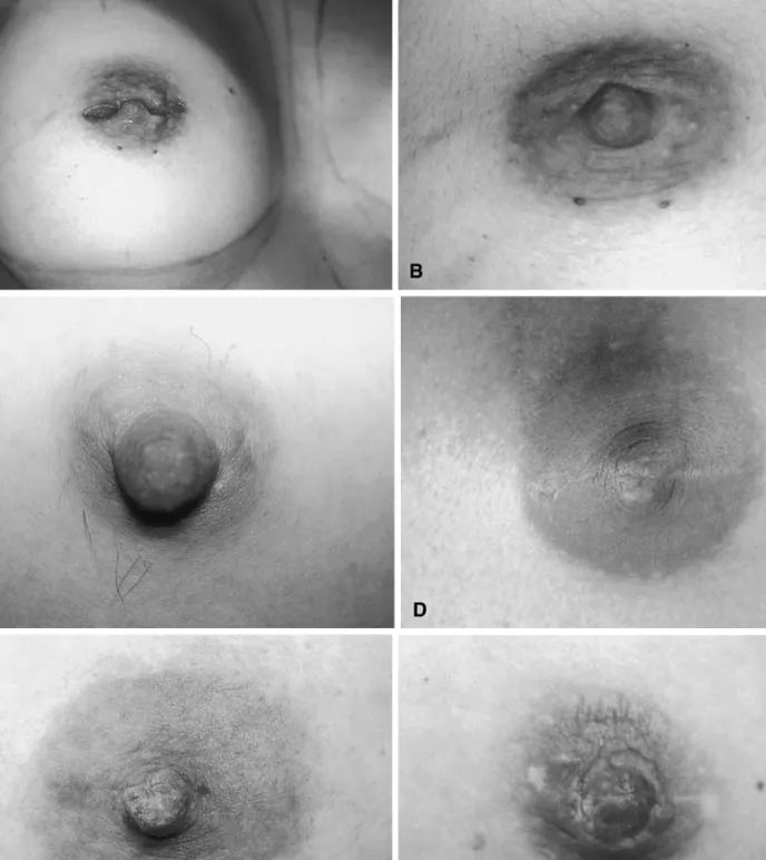
Fig. 1. Scars after a transareolar incision for breast augmentation. In general, the scar is interrupted in its horizontal course and therefore principally not well visible. (a) Intraoperative situs with transareolar incision. (b) Same right nipple–areola complex of a in detail: excellent scar and pigmentation. (c) A further example of a perfect scar. (d) Example of a ‘‘noticeable’’ scar. (e) Example of a ‘‘noticeable’’ distortion of the nipple. In this case, the incision could have been made at the lower base of the nipple. (f) Example of the only ‘‘obvious’’ rated scar with pigment changes that could be easily tattooed.
Among the greatest psychological benefits of breast augmentation seems to be the increased self-esteem occasioned by the improved breast contour, which leads to heightened sensuality, despite the quantitative amount of impaired sensation [6]. Regarding the physiologic peculiarities, there are only very rare reports on breast sensation after breast augmentation. In 1976, Courtiss and Goldwyn [5] provided the first significant clinical study evaluating changes of sensa-tion after augmentasensa-tions. Using crude assessment by finger touch, pain perception (Vitapulp), and light pressure to test sensibility, they found marked differ-ences in sensory perception between the skin of the breast, the areola, and the nipple (the areola was the most sensitive part of the breast and the nipple the least). Two years postoperatively, 15% of their patients had a persistently decreased sensation in the NAC, and larger implants were associated with greater sensory loss. They found no difference based on insertion of the implant through an inframammary, transareolar, or periareolar incision at either location. Pitanguy [13] stated that areolar sensibility and muscle mobility were not altered in his cases of augmentation.
In a recent study [1] that measured sensitivity 6 months after retropectoral breast augmentation, 20% of the women felt that they had decreased sen-sation. Yet, the authors concluded that these feelings are of little scientific value and therefore negligible because they found no significant changes in meas-urements of pressure and vibration thresholds. If the authors had used the appropriate set of 20 monofil-aments for pressure measurement instead of only 6 monofilaments, they probably would also have detected the decrease in pressure sensation that we found. Other studies on breast augmentation have
found no change, have reported an improvement of sensation postoperatively, or, as in most cases, have not commented on this issue.
Therefore, the main knowledge of breast sensation and its sequelae after surgery derives from studies of breast reduction and ‘‘normal-breasted’’ women versus those with macromastia [20,22,24]. From these reports, it is known that there is a wide range of sensation thresholds even in normal breasts without surgery, and that not all sensational modalities in one breast are distributed homogeneously.
In our study, special attention was given to sensa-tion of the nipple because no other incision for aug-mentation traverses the nipple itself. Whereas the nipple is thought by some authors to have a poor sensation and a paucity of nerves supplying it [5, 18], others, including women, consider the nipple to be very sensitive [4]. In contrast to these assumptions, we sought to quantify the severity of sensory impairment after augmentation. In our women, the mean values of the pressure threshold were increased by 1 to 2 Semmes–Weinstein filaments. This is considered a mild to moderate loss of sensation. The mean vibra-tory threshold also was increased (to approximate the values among women with macromastia who have had no surgery [20]) (Table 2). As a result, 11% of our women were aware of impaired sensation and judged it to be decreased.
The parameter that correlated best with the sub-jective outcome was temperature sensation. It had returned to normal [24]. Whether other incisions or implants could lead to a smaller objective sensory compromise remains undetermined.
Scars proved to be barely visible in most of the cases, and areas of hypopigmentation were rare. Yet,
Table 2. The mean values, standard deviations, and ranges for pressure and vibration thresholds and temperature in 36 breastsa
Area Mean ± SD (n = 36) Range Reference 22 (n = 50) Reference 20 (n = 11) log10F
Pressure (log10F) Nipple 3.57 ± 0.56 2.44–3.87 2.85 ± 0.44 28.5 ± 7.4 4.08
Areola 3.75 ± 0.40 1.65–4.56 2.69 ± 0.44 31.6 ± 4.0 4.17 Body 3.31 ± 1.09 1.65–4.56 2.36 ± 0.45 23.1 ± 13.0 3.84 Macromastia (cup D) (n = 13) Vibration (lm) Nipple 4.3 ± 5.9 0.3–14.2 — 0.3 ± 0.2 7.6 ± 6.8 Areola 0.2 ± 1.6 0.1–0.3 — 0.1 ± 0.6 8.1 ± 7.1 Body 1.3 ± 1.1 0.1–3.7 — 0.4 ± 0.3 8.4 ± 1.1
Temperature (°C) Nipple (%) Areola (%) Body (%) Reference 24
44 100 74 94 (100/75/91)
0 100 79 96 (100/84/97)
SD, Standard deviation
a
The four measure points on the areola and the four coordinates of the breast body were averaged. For comparison, the mean values of normal-breasted women (References) are given, including the vibratory threshold values of women with macro-mastia but no surgery (cup D). In Reference 20, the normal pressure thresholds were indicated in g/mm2. Note that the mean values for the pressure threshold of the patients in this study were significantly higher (all p < 0.001) than those in reference 22, but lower (better) than those in reference 20 The values for vibratory threshold after augmentation equaled or were better than the values of women with macromastia but no surgery in reference 20
the transareolar incision certainly is limited to breasts without ptosis because nearly every incisional pattern of mastopexy requires a periareolar approach. The question of compromised lactational function cannot be answered conclusively from this small study nor from literature [9,25].
In conclusion, the transareolar approach for breast augmentation showed no increased rate of complica-tions, including capsular contraction. The scars were of good quality, without major pigmental changes. Perceived sensation was normal 2 years after aug-mentation in 89% of the women, but objective mean pressure and vibration sensation were moderately compromised in all parts of the breast. This discrep-ancy shows once more that when clinicians are in doubt, the subjective feelings of their patients are more important than objective ‘‘reproducible’’ meas-urements.
If patients are appropriately selected with respect to the limitations of the technique (i.e., limited im-plant size in relation to the diameter of the areola and augmentation of only breasts without ptosis), the transareolar access has its definite place among the different incisions used in breast augmentation.
References
1. Banbury J, Yetman R, Lucas A, Papay F, Graves K, Zins JE: Prospective analysis of the outcome of subpectoral breast augmentation: sensory changes, muscle function, and body image. Plast Reconstr Surg 113:701–707, 2004 2. Broadbent TR, Woolf RM: Augmentation
mammapl-asty. Plast Reconstr Surg 40:517–523, 1967
3. Craig RDP, Sykes PA: Nipple sensivity following re-duction mammaplasty. Br J Plast Surg 23:165–172, 1970 4. Cooper AP: On the anatomy of the breast. Longmann, Orme, Green, Brown and Longmanns, London, 1840 5. Courtiss EH, Goldwyn RM: Breast sensation before
and after plastic surgery. Plast Reconstr Surg 58:1–13, 1976
6. Courtiss EH, Webster RC, White MF: Selection of alternatives in augmentation mammaplasty. Plast Rec-onstr Surg 54:552–557, 1974
7. Garner JS: CDC guideline for prevention of surgical wound infections, 1985: supercedes guideline for pre-vention of surgical wound infection published in 1982. Infect Control 7:193–200, 1986
8. Griffiths CO Jr: The submuscular implantation in aug-mentation mammaplasty Trans Fourth Internat Congr
Plast andReconstr Surg.Medica Excerpta, Amsterdam, 1964
9. Hurst NM: Lactation after augmentation mammapl-asty. Obstet Gynecol 87:30–34, 1996
10. Jaspers JJP, Posma AN, van Immerseel AAH, Gitten-berger-de-Groot AC: The cutaneous innervation of the female breast and nipple–areola complex: implications for surgery. Br J Plast Surg 50:249–259, 1997
11. Johnson GW, Christ JE: The endoscopic breast aug-mentation: the transumbilical insertion of saline-filled breast implants. Plast Reconstr Surg 92:801–808, 1993 12. Jones FR, Tauras AP: A periareolar incision for aug-mentation mammaplasty. Plast Reconstr Surg 1:641– 644, 1973
13. Pitanguy I: Transareolar incision for breast augmen-tation. Aesth Plast Surg 2:363–372, 1978
14. Pitanguy I: Transareolar incision for gynecomastia. Plast Reconstr Surg 38:414–419, 1996
15. Pitanguy I, Carreirao SE, Garcia LC: Transareolar incision for augmentation mammaplasty. Rev Brasil Chir 63:301–306, 1973
16. Price CI, Eaves FF, Nahai F, Jones G, Bostwick J: Endoscopic transaxillary subpectoral breast augmen-tation. Plast Reconstr Surg 94:612–619, 1994
17. Regnault P: Breast ptosis definition and treatment. Clin Plast Surg 3:193–203, 1996
18. Sarhadi NS, Dunn JS, Soutar DS: An anatomical study of the nerve supply of the breast, including the nipple and areola. Br J Plast Surg 49:156–164, 1996
19. Semmes-Weinstein J, Ghent L, Teuber LH: Frequency of response to a series of pressure stimuli according to log force in somatosensory changes after penetrating brain wounds in manHarvard University Press, Cambridge, pp 60–62, 1960
20. Slezak S, Dellon LA: Quantitation of sensibility in gigantomastia and alteration following reduction mammaplasty. Plast Reconstr Surg 91:1265–1269, 1993 21. Strasser E: An objective grading system for the evalu-ation of cosmetic surgical results. Plast Reconstr Surg 104:2282–2285, 1999
22. Tayrich GV, Kuzbari R, Rigel S, Todoroff BP, Schn-eider B, Deutinger M: Normal cutaneous sensibility of the breast. Plast Reconstr Surg 102:701–704, 1998 23. Tebbetts JB: A surgical perspective from two decades
of breast augmentation: toward state of the art in 2001. Clin Plast Surg 28:425–434, 2001
24. Terzis JK, Vincent MP, Wilkins LM, Rutledge K, Deane LM: Breast sensibility: a neurophysiological appraisal in the normal breast. Ann Plast Surg 19:318– 322, 1987
25. Widdice L: Commentaries: the effects of breast reduc-tion and breast augmentareduc-tion surgery on lactareduc-tion: an annotated bibliography. J Hum Lact 9:161–167, 1993