LETTER TO THE EDITOR
High hyperdiploid acute lymphoblastic leukemia in adults
shows clonal heterogeneity and chromosomal instability
at diagnosis and during the course of the disease
Anna Talamo
&Alfio Marazzi
&Alicia Rovo
&Urs Schanz
&André Tichelli
&Yves Chalandon
&Martine Jotterand
Received: 25 July 2011 / Accepted: 22 August 2011 / Published online: 23 September 2011 # Springer-Verlag 2011
Dear Editor,
High hyperdiploidy with 51–67 chromosomes (HeH)
constitutes a large cytogenetic subset of B cell precursor
childhood acute lymphoblastic leukemia (ALL) [
1
]. It is
much less common in adult B cell precursor ALL where it
was reported in nearly 10% of patients for whom outcome
was improved compared to the other cytogenetic groups,
but not as favorable as in children [
2
,
3
]. It is rarely found
in T cell or mature B cell ALL.
Automated four-color interphase fluorescence in situ
hybridization (I-FISH) previously revealed a high level of
clonal aneuploidy heterogeneity in HeH ALL at presentation
[
4
]. Numerical chromosome instability (CIN) was supposed
to be at the origin of this heterogeneity. To assess the
presence of clonal heterogeneity and numerical CIN in adult
HeH ALL at diagnosis and during disease course, we
focused on a series of ten ALL patients selected according
to the presence of HeH by conventional cytogenetics, age,
and availability of material for four-color I-FISH
investiga-tion using probes specific to chromosomes 4, 6, 10, and 17
(Supplement
1
). Probes were chosen based on the frequent
gain of these chromosomes in HeH ALL and its prognostic
significance [
5
]. Patients were referred between 1995 and
2009 from the university hospitals of Basel, Zurich, Bern,
Lausanne and cantonal/regional hospitals of St-Gallen,
Aarau, Mendrisio, Bellinzona, and Genolier Clinic. Two
patients were enrolled in the SAKK ALL 33-86/90 [
6
] and
GRAALL 2005 clinical trials, respectively. Ethical approval
for this project was obtained in accordance with the
guidelines of the local Ethical Review Board.
Thirty-four samples were analyzed (presentation, 7;
hematologic remission, 19; relapse, 8); status of heterogeneity
and CIN level were determined (Table
1
, Supplement
2
).
Significant aneuploidies were identified based on cutoff
values defined according to the Poisson distribution, and
combinations of aneuploidies were considered relevant when
at least one aneuploidy was determined to be significant.
Average CIN was determined for all four chromosomes
together and then for each selected chromosome.
Approaches used and cutoff levels were reported in detail
previously [
4
,
7
].
Electronic supplementary material The online version of this article (doi:10.1007/s00277-011-1317-x) contains supplementary material, which is available to authorized users.
A. Talamo
Cancer Cytogenetics Unit, Medical Genetics Service,
University Hospital and University of Lausanne (CHUV-UNIL), Lausanne, Switzerland
A. Marazzi
Institute of Social and Preventive Medicine, University of Lausanne,
Lausanne, Switzerland A. Rovo
:
A. TichelliHematology Department, University Hospital of Basel, Basel, Switzerland
U. Schanz
Department of Hematology, University Hospital of Zurich, Zurich, Switzerland
Y. Chalandon
Hematology Division, University Hospital of Geneva (HUG), Geneva, Switzerland
M. Jotterand (*) Medical Genetics Service,
University Hospital and University of Lausanne (CHUV-UNIL), Av. Pierre Decker 2,
CH 1011 Lausanne, Switzerland e-mail: [email protected] Ann Hematol (2012) 91:793–796 DOI 10.1007/s00277-011-1317-x
T able 1 C lones involving chromos omes 4, 6, 10, and 17 at pre sentation and/or during the course of disease in ten patients with high hyperdiplo id acute lympho b last ic le ukemia, expr es se d as p er ce nt age s Patients Clone s express ed as % 1 a 2 a 3 a 4 a 56 7 8 9 a 10 Analysis 131/05 b 88/06 b 363/09 b 241/05 b 1086/07 b 601/02 c 261/06 c 331/06 c 1282/07 d 1 181/08 c 29/09 d 540/ 09 d 2385/95 b 1527/97 c 27/98 c 158/98 c 388/02 b 95/ 03 c Normal 18.4 51.0 9.8 53.8 20.7 44.2 86.4 54.6 87.0 42.7 98.6 99.4 24.2 21.7 74.2 96.8 49.6 77 .4 +4 8.6 3.8 – 9.0 13.0 5.3 4.8 14.3 3.5 3.3 –– 1.2 1 1.4 – 2.6 –– +6 – 2.6 6.2 4 28.3 9.3 –– 3.7 4.4 –– 17.2 5.4 4.4 –– – +10 15.8 2.2 8.6 –– 6.4 2.0 4.6 – 3.4 –– 15.0 3.0 9.4 – 45.8 19 .8 +17 5.4 2.6 7.6 –– 2.2 –– 3.5 2.6 – – – – ––– – +6, +17 –– 7.4 – ––– – – 2.6 – – – – ––– – +6, +10 – 4.0 20.2 –– 13.9 –– – 7.8 –– 31.2 14.3 9.2 – 1.2 – +4, +17 4.8 1.2 –– ––– – – 1.1 – – – – ––– – +4, +6 – 2.6 – 8.8 34.3 1.7 –– – 1.6 –– – 3.3 ––– – +4, +10 16.8 3.6 –– – 1.4 5.6 24.3 – 2.8 –– – 3.7 ––– – +10, +17 9.2 1.2 10.8 – ––– – – 2.0 –– – – – – 1.2 – +4, +10, +17 14.0 2.4 –– ––– – – 2.1 – – – – ––– – +4, +6, +10 – 5.2 –– – 2.1 –– – 5.4 –– – 13.4 ––– – +6, +10, +17 – 1.8 23.2 – ––– – – 6.4 –– 1.0 – ––– – +4, +6, + 17 – 1.6 –– ––– – – 1.9 – – – – ––– – +4, +6, +10, +17 – 7.0 1.0 – ––– – – 6.0 – – – – ––– – +4, +4 – 1.2 – 8.6 – 1.5 –– – – – – – 1.8 ––– – +4, +4, +6 –– – 13.6 ––– – – – –– – – – – – – +4, +4, +6, +10 – 1.4 –– ––– – – – –– – 2.8 ––– – +6, +6 – ––– ––– – – – –– 1.4 – ––– – +6, +6, +10 – ––– ––– – – – –– 3.0 1.6 ––– – +4, +6, +6, +10 – ––– ––– – – – –– – 1.4 ––– – +4, +6, +10, +10 – ––– ––– – – – –– – 2.5 ––– – +6, +10, +10 – ––– – 2.7 –– – – – – – 2.1 ––– – +6, +10, +17, +17 –– 1.2 – ––– – – – –– – – – – – – +4, +10, +17, +17 1.2 ––– ––– – – – –– – – – – – – Others e 5.8 4.6 4.0 2.2 3.7 9.4 1.2 2.3 2.3 3.9 1.4 0.6 5.8 1 1.5 2.8 0.6 2.2 2 .8 T otal of abnorm al clone s 21 (13 ) 29 (13 ) 24 (15 ) 13 (8) 18 (15) 44 (34) 6 (3) 8 (5) 19 (16 ) 40 (25) 7 (7) 3 (3) 20 (13) 56 (43 ) 12 (9) 2 (1) 8 (5) 7 (6) V alues in parentheses are numbers of very small abnormal clones (<1%). Clones already pres ent in the previous analysis (<1%) are italicized. Study pa tients are further detailed in Supplement 1 . Four -color I-FISH using centromeric probes specific for chromosomes 4 (p-4n1/4, kindly provided by Prof. Mariano Rocchi, University of Bari, Italy), 6, 10, and 17 (D6Z1, D10Z1, and D17Z1, respectively; American T ype Culture Collection, Manassas, V A) was performed as for merly described [ 4 ] on 34 bone marrow samples, 5 of which were reported in T alamo et al. [ 7 ]. Probes were direc tly labeled by nick translation with four dif ferent fluorochromes (Cy3, Cy3.5, DEAC, and FITC). Automated four -color I-FISH anal ysis was realized with the scanning system Metafer 4/MetaCyte (MetaSystems, Altlussheim, Germany) using a motorized epifluorescence microscope (AxioImager Z1; Zeiss, Feldbach, Germany) equipped with a ×40 objective (Zeiss). In each sample, a minimum of 500 interphase nuclei were scored, except in one case (331/06) in which only 350 nuclei were available for classification aPatients in part reported by T alamo et al. [ 7 ] (12)
bPresentation cRelapse dHematologic
(bone marrow) complete remission eCumulated percentage of very small clones (<1%) 794 Ann Hematol (2012) 91:793–796
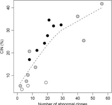
High levels of clonal heterogeneity were observed at
diagnosis and during disease course, at relapse particularly.
Clones detected at presentation generally reappeared at
relapse, mostly accompanied by newly generated ones
(Supplement
3
). Whereas the mean total number of
abnormal clones did not clearly differ between diagnostic
and relapse samples, the range of their variation did, being
much larger at relapse (Table
1
). Despite the small number
of patients, a significant correlation was observed between
number of abnormal clones and CIN, suggesting that the
higher the instability, the larger the number of abnormal
clones (Fig.
1
).
Data let surmise that the dynamic process at the origin
of HeH ALL is a complex one leading to coexistence of
sub-clones with different combinations of aneuploidy,
whose heterogeneity and variation result from a simultaneous
chromosome gain mechanism driven by underlying
chromosome instability and whose evolution will depend
on natural selection and acquisition of additional genetic
abnormalities [
1
,
8
].
Given the poor outcome associated with CIN in solid
tumors and myelodysplastic syndromes [
9
,
10
], the nature
and extent of clonal heterogeneity at diagnosis may be of
prognostic significance in HeH ALL. This question would
merit to be investigated in a large cohort of HeH ALL
patients.
Acknowledgments We are indebted to Dr. Mario Bargetzi (Cantonal Hospital, Aarau), Professor Michel Duchosal (University Hospital, Lausanne), Dr. Valérie Frossard (Valais Hospital, Sion), Dr. Jeroen Goede (University Hospital, Zurich), Dr. Urs Hess (Cantonal Hospital, Sankt-Gallen), Dr. Volker Kirchner (Genolier Clinic, Genolier), Dr. Leda Leoncini-Franscini (Oncology Institute of Southern Switzer-land, Bellinzona), Professor Thomas Pabst (University Hospital, Bern), Dr. Olivia Pagani (Regional Hospital, Mendrisio), and Professor Olivier Spertini (University Hospital, Lausanne) for referring patient samples and for providing clinical data. We are grateful to Dr. Joelle Tchinda (Oncology Diagnostics Laboratories, University Children’s Hospital, Zurich) for providing cytogenetic results from two analyses performed in her laboratory. We thank all the collaborators of the Cancer Cytogenetics Unit (Medical Genetics Service, University Hospital and University of Lausanne, Lausanne) and are indebted to Aurélie Diliberto for her technical assistance. We are grateful to Professor Jacques S. Beckmann (Medical Genetics Service, Department of Medical Genetics, University Hospital and University of Lausanne, Lausanne) and to Dr. Guy van Melle (Institute of Social and Preventive Medicine, University of Lausanne, Lausanne). We thank the GRAALL and SAKK cooperative groups for allowing us to analyze the samples of the patient who was included in the GRAALL 2005 study. We are indebted to the Mach Gaensslen Stiftung (Schweiz) for the award to Professor Martine Jotterand.
References
1. Paulsson K, Johansson B (2009) High hyperdiploid childhood acute lymphoblastic leukemia. Genes Chromosomes Cancer 48 (8):637–660. doi:10.1002/gcc.20671
2. Pullarkat V, Slovak ML, Kopecky KJ, Forman SJ, Appelbaum FR (2008) Impact of cytogenetics on the outcome of adult acute lymphoblastic leukemia: results of Southwest Oncology Group 9400 study. Blood 111(5):2563–2572. doi: 10.1182/blood-2007-10-116186
3. Moorman AV, Chilton L, Wilkinson J, Ensor HM, Bown N, Proctor SJ (2010) A population-based cytogenetic study of adults with acute lymphoblastic leukemia. Blood 115(2):206–214. doi:10.1182/blood-2009-07-232124
4. Blandin AT, Muhlematter D, Bougeon S, Gogniat C, Porter S, Beyer V, Parlier V, Beckmann JS, van Melle G, Jotterand M (2008) Automated four-color interphase fluorescence in situ hybridization approach for the simultaneous detection of specific aneuploidies of diagnostic and prognostic significance in high hyperdiploid acute lymphoblastic leukemia. Cancer Genet Cytogenet 186(2):69–77. doi:10.1016/j.cancergencyto. 2008.06.008
5. Sutcliffe MJ, Shuster JJ, Sather HN, Camitta BM, Pullen J, Schultz KR, Borowitz MJ, Gaynon PS, Carroll AJ, Heerema NA (2005) High concordance from independent studies by the Children’s Cancer Group (CCG) and Pediatric Oncology Group (POG) associating favorable prognosis with combined trisomies 4, 10, and 17 in children with NCI Standard-Risk B-precursor Acute Lymphoblastic Leukemia: a Children’s Oncology Group (COG) initiative. Leukemia 19(5):734–740. doi:10.1038/sj. leu.2403673
6. Wernli M, Tichelli A, von Fliedner V, Brun del Re G, Chapuis B, Fey MF, Fopp M, Gmur J, Grob JP, Jacky E et al (1994) Intensive induction/consolidation therapy without maintenance in adult acute lymphoblastic leukaemia: a pilot assessment. Working Party on Leukaemia of the Swiss Group for Epidemiologic and Clinical Cancer Research (SAKK). Br J Haematol 87(1):39–43
Fig. 1 Number of abnormal clones and CIN values (%) in 18 bone marrow samples from 10 patients with high hyperdiploidy acute lymphoblastic leukemia at disease presentation (black points), hema-tological complete remission (white points) and relapse (gray points). The correlation between number of abnormal clones and CIN level was measured using the Spearman correlation coefficient. There is an increasing curvilinear trend, as suggested by the broken line (a nonparametric smooth). The Spearman coefficient (0.89) was highly significant (p<10-6).
7. Talamo A, Chalandon Y, Marazzi A, Jotterand M (2010) Clonal heterogeneity and chromosomal instability at disease presentation in high hyperdiploid acute lymphoblastic leukemia. Cancer Genet Cytogenet 203(2):209–214. doi:10.1016/j.cancergencyto.2010.09.005
8. Greaves M (2009) Darwin and evolutionary tales in leukemia. The Ham-Wasserman Lecture. Hematology Am Soc Hematol Educ Program:3–12. doi:10.1182/asheducation-2009.1.3
9. Nakamura H, Saji H, Idiris A, Kawasaki N, Hosaka M, Ogata A, Saijo T, Kato H (2003) Chromosomal instability detected by
fluorescence in situ hybridization in surgical specimens of non-small cell lung cancer is associated with poor survival. Clin Cancer Res 9(6):2294–2299
10. Heilig CE, Loffler H, Mahlknecht U, Janssen JW, Ho AD, Jauch A, Kramer A (2010) Chromosomal instability corre-lates with poor outcome in patients with myelodysplastic syndromes irrespectively of the cytogenetic risk group. J Cell Mol Med 14(4):895–902. doi:10.1111/j.1582-4934. 2009.00905.x