Received: 13 June 2003 Revised: 31 December 2003 Accepted: 8 January 2004 Published online: 11 March 2004 © Springer-Verlag 2004
Abstract The purpose of this study
was to evaluate the monitoring and
diagnostic potential of MRI in fetal
lung development and disease using
lung volume and signal intensity
changes through gestation.
Thirty-five healthy fetuses (22–42 weeks)
were examined on a 1.5- T MR
system using sagittal T2w
single-shot fast specho imaging (TR
in-definite, TE 90 ms, slice
thick-ness/gap 3–5/0 mm, FOV 26–40 cm,
NEX 0.5). Fetal body and lung were
segmented manually and volumes
calculated. Signal intensities (SI) of
fetal lung and three reference values
were measured on the section best
displaying the lung. Regions of
interests were defined by including
the maximal organ area possible.
The following SI ratios were
gener-ated: lung/liver, lung/amniotic fluid,
lung/muscle, liver/fluid and liver/
muscle. Volumes and ratios were
correlated with gestational age. Data
from seven fetuses with pulmonary
pathology were compared with these
normative values. Absolute lung
vol-ume varied from 12.3 to 143.5 cm
3in correlation with gestational age
(P<0.001); lung volume relative to
total body volume ranged from 1.6
to 5.0%, decreasing with gestational
age (P=0.001). All SI ratios
mea-sured were unrelated to gestational
age. Diagnoses in the seven
abnor-mal fetuses were hydrothorax (n=2),
congenital cystic adenomatoid
mal-formation (n=2), diaphragmatic
her-nia (n=2) and pulmonary
sequestra-tion (n=1); their absolute and relative
lung volumes were below normal
(P<0.001). The SI ratios did not
differ significantly from those in the
normal population. Normative MR
fetal lung volumes may have
impor-tant clinical applications in
confirm-ing and quantifyconfirm-ing intrauterine
pul-monary hypoplasia and in
comple-menting ultrasound in the planning
of fetal and post-natal surgery. No
clinical relevance was found for fetal
lung SI values.
Keywords MRI · Fetal lung ·
SSFSE · Pulmonary hypoplasia ·
Signal intensity
Thomas M. Keller
Annett Rake
Sven C. A. Michel
Burkhardt Seifert
Josef Wisser
Borut Marincek
Rahel A. Kubik-Huch
MR assessment of fetal lung development
using lung volumes and signal intensities
Introduction
As a critical determinant of post-natal survival, fetal lung
growth and maturity are a key parameter in prenatal
diagnostics, both for risk assessment in preterm birth and
for predicting outcome in fetal disorders associated with
pulmonary hypoplasia. Diagnosis of lung maturity
cur-rently relies on surfactant-related lipid levels in amniotic
fluid withdrawn by amniocentesis [1, 2]. Non-invasive
imaging is clearly preferable. Ultrasound reveals the vast
majority of clinically significant fetal anomalies [3].
However, accurate prediction of lung development and
maturity with ultrasound is difficult, even in experienced
hands.
The introduction of ultrafast sequences has rapidly
expanded the indications for fetal magnetic resonance
T. M. Keller · S. C. A. MichelB. Marincek · R. A. Kubik-Huch Institute of Diagnostic Radiology, University Hospital Zurich,
Rämistrasse 100, 8091 Zurich, Switzerland A. Rake · J. Wisser
Department of Obstetrics, University Hospital Zurich,
Rämistrasse 100, 8091 Zurich, Switzerland B. Seifert Department of Biostatistics, University of Zurich, Sumatrastrasse 30, 8006 Zurich, Switzerland Present address: R. A. Kubik-Huch (
✉
) Institute of Radiology,Kantonsspital Baden, 5404 Baden, Switzerland
e-mail: [email protected] Tel.: +41-56-4863803 Fax: +41-56-4863809
imaging (MRI) [4–10]. MRI is a valuable adjunct to
ultrasound in the prenatal diagnosis of fetal chest masses
[11–14]. Several groups have investigated fetal lung
volume as a predictor of hypoplasia using echo-planar
imaging [15, 16] or fast T2-weighted imaging [12, 17,
18]. More recently, using a single-shot fast spin-echo
(SSFSE) sequence, Ikeda et al. focused on signal
intensi-ty (SI), showing that the abnormal densiintensi-ty of hypoplastic
lung in one fetus generated a marked difference in SI
from its twin [19]. Kuwashima et al. confirmed this
find-ing usfind-ing half-Fourier acquisition sfind-ingle-shot turbo
spin-echo (HASTE) sequences: after 26 weeks of gestation,
low-intensity fetal lung on MRI indicated pulmonary
hypoplasia [20].
The aim of our own study was to further investigate
the potential of MRI in fetal pathophysiology and
prena-tal diagnostics of normal lung growth as well as
assess-ment of lung maturity by combining the two parameters
of fetal lung volume and SI measurement.
Materials and methods
Patient population and study design. Between January 2000 and October 2001, 35 pregnant women, aged 29.4±5.1 years (mean value ± standard deviation, range: 19–39) and gestational age 31.6±6.5 weeks (22–42) were recruited either at routine prenatal ultrasound at weeks 18–22 of gestation in the obstetric clinic or following clinically indicated MR pelvimetry performed in the 3rd trimester in our department. All had uncomplicated singleton pregnancies. Fetal malformation and growth retardation were ex-cluded by prenatal ultrasound, performed in all cases by an experi-enced investigator with a 4-MHz sector probe (128xP/10, Acuson, Mountain View, CA) or multifrequency probe (3.5–5.1 MHz; Siemens Elegra, Issaquah, WA). The study protocol had been ap-proved by our institutional review board, and written informed consent was provided by all participating women.
MRI. Fetal MRI was performed on a 1.5 T system (Signa Horizon LX and CV/i, General Electric Medical Systems, Milwaukee, WI). A sagittal localizing two-dimensional (2D) fast spoiled gradient-echo (FSPGR) sequence was performed (TR 150 ms, TE 1.4 ms, flip angle 60°, bandwidth 31.25 kHz, slice thickness/gap 7–10/0–3 mm, FOV 32–36 cm, matrix 256×160, NEX 1) using the
body coil, followed by T2-weighted SSFSE MRI (TR indefinite, TE 90 ms, bandwidth 31.25 kHz, slice thickness/gap 3–5/0 mm,
FOV 26–40 cm, matrix 256×256, NEX 0.5) sagittal to the fetus
encompassing the entire fetal body. The body coil was used in the 16 cases in which fetal MRI was performed after clinically indi-cated MR pelvimetry; a torso phased-array coil to optimize signal-to-noise ratio was used in the remaining cases. Using maternal breath-hold SSFSE MRI, each individual image was acquired separately with an acquisition time of 0.8 s and a 6-s interimage interval to avoid saturation effects, resulting in a total imaging time of about 5 min. No subject was premedicated.
Fetal lung SI and three reference SI values—fetal liver, amni-otic and/or fetal gastric fluid and maternal muscle—were mea-sured using an Advantage Workstation 3.1 (GE Medical Systems, 283 rue de la Minière, B.P. 34 78533 Buc Cedex, France) on the same section to avoid signal variation because of different section spin saturations and to minimize nearfield artifacts of the surface coil (measurement on the same section was considered to be as equidistant from the surface coil as possible). The following SI ratios were generated: lung/liver, lung/fluid, lung/muscle, liver/ fluid and liver/muscle. The section best displaying the lung with-out amniotic fluid motion artifacts was chosen. A region of inter-est (ROI) was then defined by including the maximal organ area possible, omitting the border zones to avoid partial volume effects and, in particular with the fetal lung, omitting the blood vessels. SI values were measured on one section per lung per fetus and the means taken for statistical analysis.
Fetal body, lung and liver volumes were calculated on either a high-end workstation (Unix; Silicon Graphics, Mountain View, CA) running segmentation and 3D modeling software (ProVision Version 3.0b; Algotec, Raanana, Israel) as previously described by our group [7] or an Advantage Workstation 3.1 with an integrated 3D reconstruction tool. The same investigator (TK) manually seg-mented the fetal body, lung and liver on each section in all fetuses. The post-processing software then created a 3D surface model and automatically calculated the volume of each 3D reconstruction (Fig. 1). Lung volumes were calculated relative to total body and liver volumes (%).
Statistical analysis. All analyses were performed in StatView 5.0.1 (SAS Institute, Inc., Campus Drive, Cary, NC, 27511). Continuous variables were presented as means and standard deviations. Spear-man’s rank correlation was used to analyze the development of SI ratio and relative lung volume with gestational age. Bonferroni’s correction was used to address to the problem of multiple compar-isons. Since five SI ratios were analyzed, a corresponding P-value <0.01 was considered statistically significant. Similarly, relations of relative lung volume are considered statistically significant for P<0.025.
Pulmonary diagnostics. For comparison with these normative data, and following institutional review board approval, we in-cluded retrospectively all the fetuses with known pulmonary
Fig. 1 3D MR reconstructions
of T2-weighted SSFSE sequences (TR indefinite/ TE 90 ms, slice thickness/gap 3–4/0 mm, FOV 32 cm, matrix 256×256) of fetal body and
lung in uncomplicated preg-nancies at different weeks of gestation (left 26; middle 34; right 36)
pathology and MR data sets examined at our institute and logged in our relational database (Oracle version 8: Oracle, Redwood Shores, CA, 94065). Out of 60 examinations for this indication, seven fetuses with pulmonary pathology could be identified aged 27.0±5.5 (22–36) weeks of mothers aged 31.3±6.4 (25–42) years. The same SSFSE sequence as described above was used. Volumes (in lung sequestrum and CCAM including only healthy lung, in hydrops fetalis including only lung without pleural effusions and in diaphragmatic hernias including only residual lung) and SI measures (in lung sequestrum, CCAM and diaphragmatic hernias including only healthy lung) were performed as described above. A multiple linear regression was used to compare age-adjusted values of volumes and SI between normal and pathological fetuses.
Results
Fetal structure and amniotic fluid SI measurement were
feasible in all cases. In four cases, maternal muscle SI
could only be measured on one side; in another case, in
the 3rd trimester, it could not be measured at all, since
the muscle was outside the FOV, so that the
correspond-ing ratios were unobtainable. 3D reconstruction and
volumetry of fetal body, lung and liver from the MR data
sets acquired with the T2-weighted SSFSE sequences
were feasible in all cases.
Mean sizes of the regions of interests were for the
lung 615 mm
2(144–1,567 mm
2), for the liver 706 mm
2(72–2,028 mm
2), for the fluid 687 mm
2(32–2,932 mm
2)
and for the maternal muscle 549 mm
2(101–1,103 mm
2).
Mean SI ratios were lung/liver 2.68±0.76 (1.60–4.43),
lung/fluid 0.66±0.13 (0.47–1.05), lung/muscle 5.13±1.28
(2.54–7.72), liver/fluid 0.26±0.07 (0.16–0.45) and liver/
muscle 2.08±0.84 (1.00–4.02). Lung/fluid and lung/
muscle ratios did not correlate with gestational age
(P=0.36 and P=0.016, respectively). The lung/liver ratio
increased, and liver/fluid and liver/muscle ratios
de-creased with gestational age (P<0.001 in all cases).
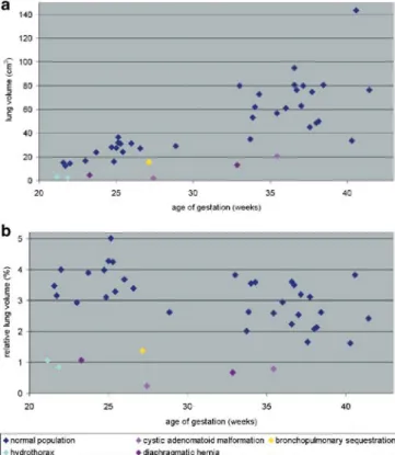
Lung volume ranged from 12.3 to 143.5 cm
3, liver
volume from 17.0 to 211.0 cm
3and fetal body volume
from 354 to 3,750 cm
3, all increasing with gestational
age (P<0.001). Relative lung/total body volume ranged
from 1.6 to 5.0%, liver/total body volume from 4.3 to
7.7% and lung/liver volume from 29.7 to 100.0%.
Rela-tive lung volumes decreased with gestational age (total
body: P=0.001 (Fig. 2); liver: P=0.001).
Pathologies in the seven abnormal fetuses were
hydrothorax, CCAM and diaphragmatic hernia (n=2
each) and lung sequestrum (n=1) (Table 1). Lung volume
ranged from 2.1 to 20.6 cm
3. Relative lung/total body
volume ranged from 0.2 to 1.4%. Absolute and relative
lung volumes were lower than in the age-adjusted
nor-mal population (P<0.001) (Fig. 2). The following SI
ratios did not differ significantly from those in the
nor-mal population: lung/liver (P=0.09), lung/fluid (P=0.38)
and liver/fluid (P=0.21); lung/muscle (P<0.001) and
liver/muscle (P=0.002) ratios were higher. Pregnancy
was terminated in the two cases of hydrops fetalis,
autopsy revealing arthrogryposis multiplex congenita in
one case, and no conclusive diagnosis in the other
(Fig. 3). In the case with a lung sequestrum (Fig. 4), the
coexistent pleural effusion was aspirated and a
thora-coamniotic shunt inserted during pregnancy, but the
in-fant died in the neonatal period. In the first CCAM case
(Fig. 5) the cyst was serially aspirated in pregnancy and
a thoracoamniotic shunt inserted, but death from
respira-Fig. 2 Correlation between fetal lung volume and gestational age
in normal fetuses and fetuses with pulmonary malformation. a Ab-solute lung volume increases with gestational age, while b lung volume relative to total body volume decreases, probably due to an increase in subcutaneous fat towards term. Absolute and relative lung volumes were smaller in malformed than in normal fetuses
Table 1 Clinical and MRI features in the fetuses with pulmonary
malformation
Case Gestational Diagnosis Relative fetal
age at MRI lung volume
(weeks) (% total body
volume) 1 22 Hydrops fetalis, hydrothorax 0.84 2 22 Hydrops fetalis, hydrothorax 0.87
3 24 Diaphragmatic hernia 1.06
4 28 CCAM 0.23
5 28 Lung sequestrum 1.39
6 33 Diaphragmatic hernia 0.67
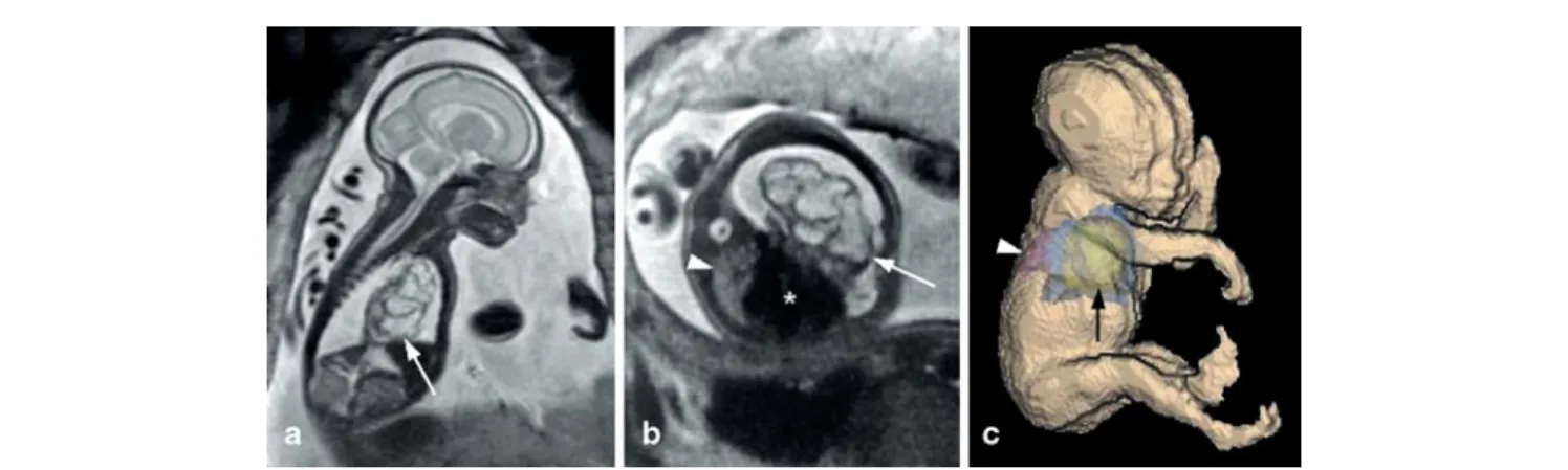
Fig. 3 Twenty-eight-week fetus with left lung CCAM. a Sagittal
and b axial T2-weighted SSFSE sequences (TR indefinite/TE 90 ms, slice thickness/gap 3–4/0 mm, FOV 32 cm, matrix 256×256), c 3D surface model. The CCAM (arrows) is displacing
the mediastinum and heart (asterisk) to the right, thus compressing
the right lung (arrowhead), resulting in a relative volume of only 0.2% (absolute volume, 2.1 cm3). Despite serial CCAM aspiration in pregnancy, death from respiratory failure occurred post-partum at 33 weeks of gestation
Fig. 4 Twenty-eight-week fetus with left lung sequestrum. a
Co-ronal T2-weighted SSFSE image (TR indefinite/TE 90 ms, slice thickness/gap 3–4/0 mm, FOV 32 cm, matrix 256×256). b 3D
re-construction. Arrowheads: pleural effusion and lung sequestrum (volume, 16.6 cm3). Arrows: normal right lung and rest of left lung (combined volume, 15.8 cm3; relative lung volume, 1.4%)
Fig. 5 Thirty-six-week fetus with CCAM. a Sagittal T2-weighted
SSFSE image (TR indefinite/TE 90 ms, slice thickness/gap 3–4/ 0 mm, FOV 32 cm, matrix 256×256) showing CCAM (arrows)
occupying left hemithorax. b 3D reconstruction of right lung (asterisk), relative volume 0.8% (arrow, CCAM)
tory failure occurred in the post-partum period, as in the
second CCAM case (Fig. 6). In one case with a
dia-phragmatic hernia scanned at 24 weeks, GA surgical
closure of the diaphragmatic defect was performed
post-partum; however, the fetus died a few weeks later of
pulmonary hypertension. The second case of a
diaphrag-matic hernia was associated with a cerebral
malforma-tion (Dandy Walker syndrome); the fetus died in the
neonatal period.
Discussion
As expected, absolute fetal lung volume increased with
gestational age in the normal population, confirming
pre-vious MRI studies [16, 17, 19]. However, fetal
volume-try used as we have previously described [7] showed a
decrease in relative lung volume towards term, probably
because of an increase in subcutaneous fat.
In contrast to earlier studies, fetal lung SI did not
change significantly with gestational age. However,
di-rect comparison is difficult if the differences in imaging
strategies and/or study populations are not considered
[15, 19, 20]. We found a highly significant change with
gestational age in the lung/liver SI ratio, but not in the
lung/fluid or lung/muscle ratios. It is true that using T2
*-weighted echo-planar sequences, Duncan et al. (1997)
also found a change in fetal liver SI with gestational
age, and suggested this might be due to a change in liver
iron concentration following a progressive decrease in
hepatic hemoglobin production [21]. As our study also
showed significant changes in the liver/fluid and liver/
muscle ratios, it is more likely that liver SI changes with
gestational age, which tends to invalidate Kuwashima
et al.’s interpretation of their results using fetal liver as
reference structure. On the evidence provided in the
present study, we believe that the liver is not a suitable
reference structure.
Our study included a small historical series of fetuses
with lung malformations. Ultrasound is the established
imaging modality in prenatal screening and malformation
diagnostics. However, its ability to assess fetal lung is
lim-ited [22]. Recent studies indicate that MRI may be a
valu-able adjunct, e.g., in identifying the origin of extensive
chest tumors [11, 13, 14] or in planning in utero
interven-tions, delivery, and immediate post-natal surgery [23–25].
As expected, lung volumes were smaller in the
mal-formed fetuses than in the normal population, confirming
previous reports [17, 26, 27]. As earlier investigators
stated [17], setting a relative lung volume threshold for
diagnosing pulmonary hypoplasia would require a very
high number of subjects. However, in our study the
lower limit of normal lung volume was 1.6%, against an
upper limit in the hypoplastic group of 1.4%, suggesting
that it should prove possible to create a percentile curve
based on a large study population to define pulmonary
hypoplasia.
We encountered technical challenges in the SI
analy-sis. In contrast to computed tomography, where tissue
density can be expressed as an absolute value in
Houns-field units, SI measures in MRI require comparison with
a reference structure. The ideal structure is one with
im-aging properties that remain unchanged during
pregnan-cy, in the vicinity of the fetal lung to avoid SI loss
be-cause of a difference in distance from the phased-array
coil. Conventional reference structures include fetal liver
and amniotic fluid [20, 21]. For the reasons already
stated, we believe that the liver is unsuitable for this
purpose, and we thus introduced maternal iliac muscle/
rectus abdominis as an additional reference structure.
Next to the technical challenges discussed above,
there are some other limitations to our study, one being
the small size of our normal fetus population.
Further-more, 3D reconstruction took several hours per fetus,
limiting not only the sample size as mentioned above,
but also the clinical feasibility of the method. Another
limitation is the relative rarity of pulmonary
malforma-tions, detected in only seven fetuses out of the total of 60
MRI scans performed for suspected fetal malformation.
Clinical outcome in these cases with pulmonary
hypo-plasia was poor: termination in two cases, and death
from respiratory failure in the remainder.
In conclusion, in accordance with previous studies
[16, 17, 19], we believe that normative MRI data for
fe-tal lung volume may have important clinical applications
in confirming and quantifying fetal pulmonary
hypopla-sia, a relatively rare but potentially lethal malformation.
MRI is likely to gain an increasing role as a
complemen-tary imaging method in diagnosing fetal lung
abnormali-ties and planning fetal and post-natal surgery. In contrast
to earlier studies [15, 19, 20], our study found no place
for SI measurement in assessing fetal lung development.
Acknowledgments We thank Heike Fischer for volunteer
recruit-ment, Nino Teodorovic, RT, and Dr. phil. Dani Nanz for technical assistance. We are grateful to Professor Dr. Renate Huch and Pro-fessor Dr. Rabih Chaoui for critical discussion of the study proto-col. This work was partly supported by a grant from the EMDO Foundation, Switzerland.
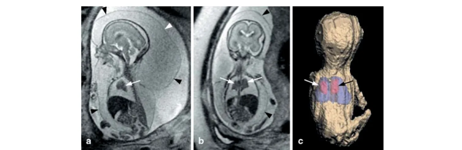
Fig. 6 Twenty-two-week fetus with hydrops fetalis. a Sagittal and b coronal T2-weighted SSFSE images (TR indefinite/TE 90 ms,
slice thickness/gap 3–4/0 mm, FOV 32 cm, matrix 256×256)
showing massive subcutaneous edema (arrowheads) and marked pleural effusions with secondary pulmonary compression
(ar-rows). c 3D surface model showing lung (arrows), volume 2.3 cm3 and relative volume of 0.9% (body volume was calculated by ex-cluding the subcutaneous edema). Autopsy at 24 weeks confirmed hydrops fetalis with subcutaneous edema and pleural effusions, with no concomitant malformations
References
1. Fenton BW, Lin CS, Seydel F, Macedonia C (1998) Lecithin can be detected by volume-selected proton MR spectroscopy using a 1.5 T whole body scanner: a potentially non-inva-sive method for the prenatal assess-ment of fetal lung maturity. Prenat Diagn 18:1263–1266
2. Horger EO, Finch H, Vincent VA (2001) A single physician’s experience with 4,600 genetic amniocenteses. Am J Obstet Gynecol 185:279–288 3. Levine D (2001) Ultrasound versus magnetic resonance imaging in fetal evaluation. Top Magn Reson Imaging 12:25–38
4. Levine D, Barnes PD, Edelman RR (1999) Obstetric MR imaging. Radiology 211:609–617 5. Kubik-Huch RA, Huisman TA,
Wisser J, Gottstein-Aalame N, Debatin JF, Seifert B, Ladd ME, Stallmach T, Marincek B (2000) Ultrafast MR imaging of the fetus. Am J Roentgenol 174:1599–1606 6. Coakley FV (2001) Role of magnetic
resonance imaging in fetal surgery. Top Magn Reson Imaging 12:39–51 7. Kubik-Huch RA, Wildermuth S,
Cettuzzi L, Rake A, Seifert B, Chaoui R, Marincek B (2001) Fetus and uteroplacental unit: fast MR imag-ing with three-dimensional reconstruc-tion and volumetry—feasibility study. Radiology 219:567–573
8. Breysem L, Bosmans H, Dymarkowski S, Schoubroeck DV, Witters I, Deprest J, Demaerel P, Vanbeckevoort D, Vanhole C, Casaer P, Smet M (2003) The value of fast MR imaging as an adjunct to ultrasound in prenatal diag-nosis. Eur Radiol 13:1538–1548 9. Huisman TA, Wisser J, Martin E,
Kubik-Huch R, Marincek B (2002) Fetal magnetic resonance imaging of the central nervous system: a pictorial essay. Eur Radiol 12:1952–1961 10. Ertl-Wagner B, Lienemann A, Strauss
A, Reiser MF (2002) Fetal magnetic resonance imaging: indications, tech-nique, anatomical considerations and a review of fetal abnormalities. Eur Radiol 12:1931–1940
11. Hubbard AM, Adzick NS, Crombleholme TM, Coleman BG, Howell LJ, Haselgrove JC, Mahboubi S (1999) Congenital chest lesions: diagnosis and characterization with prenatal MR imaging. Radiology 212:43–48
12. Walsh DS, Hubbard AM, Olutoye OO, Howell LJ, Crombleholme TM, Flake AW, Johnson MP, Adzick NS (2000) Assessment of fetal lung volumes and liver herniation with magnetic resonance imaging in congenital diaphragmatic hernia. Am J Obstet Gynecol 183:1067–1069
13. Mahieu-Caputo D, Sonigo P, Dommergues M, Fournet JC, Thalabard JC, Abarca C, Benachi A, Brunelle F, Dumez Y (2001) Fetal lung volume measurement by magnetic resonance imaging in congenital diaphragmatic hernia. Br J Obstet Gynaecol 108:863–868
14. Hata N, Wada T, Chiba T, Tsutsumi Y, Okada Y, Dohi T (2003) Three-dimen-sional volume rendering of fetal MR images for the diagnosis of congenital cystic adenomatoid malformation. Acad Radiol 10:309–312
15. Duncan KR, Gowland PA, Freeman A, Moore R, Baker PN, Johnson IR (1999) The changes in magnetic resonance properties of the fetal lungs: a first result and a potential tool for the non-invasive in utero demonstration of fetal lung maturation. Br J Obstet Gynaecol 106:122–125
16. Duncan KR, Gowland PA, Moore RJ, Baker PN, Johnson IR (1999) Assess-ment of fetal lung growth in utero with echo-planar MR imaging. Radiology 210:197–200
17. Coakley FV, Lopoo JB, Lu Y, Hricak H, Albanese CT, Harrison MR, Filly RA (2000) Normal and hypoplas-tic fetal lungs: volumetric assessment with prenatal single-shot rapid acquisition with relaxation enhance-ment MR imaging. Radiology 216:107–111
18. Moore RJ, Strachan B, Tyler DJ, Baker PN, Gowland PA (2001) In vivo diffusion measurements as an indica-tion of fetal lung maturaindica-tion using echo planar imaging at 0.5 T. Magn Reson Med 45:247–253
19. Ikeda K, Hokuto I, Mori K, Hayashida S, Tokieda K, Tanigaki S, Tanaka M, Yuasa Y (2000) Intrauterine MRI with single-shot fast-spin echo imaging showed different signal intensities in hypoplastic lungs. J Perinat Med 28:151–154
20. Kuwashima S, Nishimura G, Iimura F, Kohno T, Watanabe H, Kohno A, Fujioka M (2001) Low-intensity fetal lungs on MRI may suggest the diagno-sis of pulmonary hypoplasia. Pediatr Radiol 31:669–672
21. Duncan KR, Baker PN, Gowland PA, Issa B, Moore R, Worthington B, Johnson IR (1997) Demonstration of changes in fetal liver erythropoiesis using echo-planar magnetic resonance imaging. Am J Physiol 273:965–967 22. Heling KS, Tennstedt C, Chaoui R,
Kalache KD, Hartung J, Bollmann R (2001) Reliability of prenatal sono-graphic lung biometry in the diagnosis of pulmonary hypoplasia. Prenat Diagn 21:649–657
23. Adzick NS, Harrison MR, Crombleholme TM, Flake AW, Howell LJ (1998) Fetal lung lesions: management and outcome. Am J Obstet Gynecol 179:884–889 24. Flake AW, Crombleholme TM,
Johnson MP, Howell LJ, Adzick NS (2000) Treatment of severe congenital diaphragmatic hernia by fetal tracheal occlusion: clinical experience with fifteen cases. Am J Obstet Gynecol 183:1059–1066
25. Hubbard AM (2001) Magnetic reso-nance imaging of fetal thoracic abnor-malities. Top Magn Reson Imaging 12:18–24
26. Rypens F, Metens T, Rocourt N, Sonigo P, Brunelle F, Quere MP, Guibaud L, Maugey-Laulom B, Durand C, Avni FE, Eurin D (2001) Fetal lung volume: estimation at MR imaging-initial results. Radiology 219:236–241
27. Paek BW, Coakley FV, Lu Y, Filly RA, Lopoo JB, Qayyum A, Harrison MR, Albanese CT (2001) Congenital dia-phragmatic hernia: prenatal evaluation with MR lung volumetry—preliminary experience. Radiology 220:63–67