Virchows Arch (2005) 447: 772–777 DOI 10.1007/s00428-005-0019-y
C A S E R E P O RT
Lorenzo Taminelli . Khalil Zaman . Carole Gengler . Nicolas Peloponissios . Hanifa Bouzourene .
Jean-Michel Coindre . Isabelle Hostein . Louis Guillou
Primary clear cell sarcoma of the ileum: an uncommon
and misleading site
Received: 27 February 2005 / Accepted: 23 May 2005 / Published online: 14 July 2005
# Springer-Verlag 2005
Abstract A clear cell sarcoma, arising primarily in the ileum of a 35-year-old man, is reported. Histologically, the neoplasm infiltrated the full thickness of the intestinal wall. It consisted of strands and sheets of round to spindle-shaped cells with clear to eosinophilic cytoplasm, vesic-ular nuclei and prominent nucleoli. Vascvesic-ular invasion was present at diagnosis. Tumour cells expressed S-100 protein, melan-A and tyrosinase. They were negative for HMB45, CD117, cytokeratins, epithelial membrane antigen, smooth muscle actin, desmin, CD31, CD34, chromogranin and syn-aptophysin. Reverse transcription–polymerase chain reac-tion analysis performed on paraffin-embedded tissue showed EWS–ATF1 fusion transcripts representative of the t(12;22) (q13;q12) clear cell sarcoma reciprocal trans-location. The patient, who developed liver metastases 2 months after diagnosis, died of disease at 15 months. This case demonstrates that the gastrointestinal tract is a poten-tial site for primary clear cell sarcoma of soft tissues, and, furthermore, that cytogenetics and/or molecular techniques play a central role in the diagnosis.
Keywords Clear cell sarcoma . Ileum .
Immunohistochemistry . RT-PCR . t(12;22) (q13;q12)
Introduction
Clear cell sarcoma (also known as malignant melanoma of soft tissues) is a rare tumour that typically occurs in the tendon sheaths and aponeuroses of distal extremities of adolescents and young adults [3, 6,10,11]. Involvement of the gastrointestinal tract is exceedingly rare and, fre-quently, a source of diagnostic problems. Six cases of di-gestive tract clear cell sarcoma were heretofore reported in the literature, most of them very recently [1,4,5,7,13,15], including one in the ileum [4]. A second ileal case is de-scribed below. It occurred in a 35-year-old man and caused the death of the patient 15 months after diagnosis.
Clinical history
A 35-year-old, HIV-negative man without any previous significant medical history was admitted in March 2003 for a 15-kg weight loss over a few months and symptoms of intestinal obstruction (i.e. colic pain, vomiting) without fever. CT scans showed a segmental thickening of the ileum, situated 80 cm above the ileo–caecal valve. Pre-operative diagnoses included Meckel diverticulum, polyps and a malignant neoplasm. At laparotomy, a single circum-ferential, stenotic lesion of the ileum was found. There was neither other detectable tumour in the abdominal cavity nor any liver metastases. The ileal mass was resected along with 20 cm of ileum and one regional mesenteric lymph node. A pathologic diagnosis of unclassified non-epithelial malignant neoplasm was rendered. The mesenteric lymph node was devoid of tumour deposits, but the ascitic fluid, sampled at the time of surgery, contained clusters of ma-lignant cells. A lymphoma, a GIST, a neuroendocrine neo-plasm were excluded on the basis of immunohistochemical results. A melanoma (primary or metastatic) was strongly suspected owing to tumour cell reactivity for S-100 protein. L. Taminelli . C. Gengler . H. Bouzourene . L. Guillou
Department of Pathology, University Hospital, Lausanne, Switzerland
K. Zaman
Department of Oncology, University Hospital, Lausanne, Switzerland
N. Peloponissios
Department of Surgery, University Hospital, Lausanne, Switzerland
J.-M. Coindre . I. Hostein
Department of Pathology, Bergonié Institute, Bordeaux, France
L. Guillou (*)
Institut Universitaire de Pathologie, Rue du Bugnon 25,
1011 Lausanne, Switzerland
e-mail: [email protected] Tel.: +41-21-3147216
Careful examination of the patient, including extensive ex-amination of skin, eyes, nasosinusal cavities, throat, testes and soft tissues failed to detect any other potential primary tumour. The metastatic work-up was negative, as was the PET scan. A colonoscopy showed several polyps in the ascending and sigmoid colon, which turned out to be ad-enomatous polyps with low-grade dysplasia at microscopic examination.
Two months post-operatively, several metastases were detected in the liver, along with multiple peritoneal im-plants and a malignant ascitis. The tumour regressed dramat-ically after six courses of a platinum-based chemotherapy, but a second look, performed in November 2003, showed that the malignant process extended microscopically to the whole peritoneal subserosa and to the omentum. Lymph node and liver biopsy specimens, taken during the second surgical procedure, were tumour-free. From then on, and despite various treatments, the tumour gradually progressed in the abdomen, leading to several episodes of intestinal obstruction. Patient’s death occurred 15 months after dis-ease onset, as a result of Candida albicans septicemia. No autopsy was performed.
Materials and methods
Tumour samples were fixed in 4% buffered formalin, em-bedded in paraffin and stained with hematoxylin and eosin and Fontana–Masson stains. Additional sections were stud-ied with the following antibodies: S-100 protein (Dako-patts, Glostrup, Denmark, pre-diluted), melan-A (Dako, diluted 1:50), HMB45 (Dako, diluted 1:50), tyrosinase (Ludwig Institute, Lausanne, Switzerland, diluted 1:50, gift from Dr. D. Rimoldi), cytokeratins (clone C-11, diluted 1:40 and clone AE1/AE3, diluted 1:200, Novocastra Lab-oratories Inc., Newcastle upon Tyne, UK), CD31 (Dako, diluted 1:40), CD34 (Immunotech Beckam Coulter, Inc., Fullerton, CA, USA, diluted 1:160), CD45 (Dako, diluted 1:50), CD3 (Novocastra, pre-diluted), CD20 (clone L26, Dako, pre-diluted), CD56 (clone 1B6, Novocastra, diluted 1:50), smooth muscle actin (clone 1A4, Sigma Aldrich, St Louis, MI, USA, diluted 1:20,000), myeloperoxidase (Dako, diluted 1:2,000), synaptophysin (Dako, diluted 1:25), chromogranin-A (Dako, diluted 1:1,000), somato-statin (Bachem Peninsula Laboratories, Inc., San Carlos, CA, USA, diluted 1:800), serotonin (clone 5HT-H209, Dako, diluted 1:50) and CD117 (c-kit, Dako, polyclonal diluted 1:200). Immunostaining was performed according to the avidin biotin complex (ABC) method. Tissue sec-tions were submitted to microwave oven heating prior to staining, except for CD117 staining. All steps were per-formed at room temperature, and diaminobenzidine was used as chromogen. Appropriate positive and negative con-trols were employed throughout.
Reverse transcription–polymerase chain reaction (RT-PCR) was used to detect the presence of EWS–ATF1 transcripts in paraffin-embedded tumour samples. RNA ex-traction and RT-PCR reaction were performed as previous-ly described [9]. Briefprevious-ly, PCR amplification was performed
in duplicate using a 96-well plate (Applied Biosystems, Foster City, CA, USA) with a 50μl final reaction mixture containing 300 nM each primer; 200 nM probe ATF1 or 50 nM probe beta2-microglobulin; 200 μM of an equal concentration of dATP, dCTP, dGTP, dTTP, dUTP; MgCl2 4 mM; 1.25 U Taq polymerase and 0.25 U Amperase UNG in a 1X Real-Time PCR Buffer containing a passive ref-erence (Rox fluorochrome). Thermal cycling conditions were 2 min at 50°C for amperase activation, 10 min at 95°C for Taq polymerase activation, then 50 cycles of two PCR steps consisting of 15 s at 95°C and 60°C for 1 min. All reactions were performed in the ABI Prism 5700 Sequence Detection System (Applied Biosystems).
Primers and probe sequences were chosen with Primer Express software (Applied Biosystems). Primers were pur-chased from Eurobio (Les Ulis, France) and probes and QPCR Core kit containing Real-Time PCR Buffer, dNTP, MgCl2, amperase UNG and Taq polymerase were pur-chased from Eurogentec (Herstal, Belgium). Probes and primer sequences were as follows: forward primer EWS, 5′-CAT GAG CAG AGG TGG GCG-3′; reverse primer ATF1, 5′-CCC CGT GTA TCT TCA GAA GAT AAG TC-3′; probe ATF1, 5′-6-FAM-AGG AGG ACG CGG TGG AAT GGG-TAMRA-3′; forward primer beta2-microglob-ulin, 5′-TGA CTT TGT CAC AGC CCA AGA TA-3′; reverse primer beta 2-microglobulin, 5′-AAT CCA AAT GCG GCA TCT TC-3′; probe beta 2-microglobulin, 5′-6-FAM-TGA TGC TGC TTA CAT GTC TCG ATC CCA-TAMRA-3′.
Results
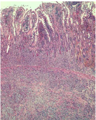
A 20-cm segment of ileum was resected, centered by a well-defined firm, circumferential mass, measuring 1.8 cm in maximal diameter. Cut section showed a whitish neo-plasm that ulcerated the overlying mucosa and infiltrated the whole thickness of the intestinal wall, extending into the subserosa (Fig.1.). The tumour cells, arranged in sheets and nests separated by fibrous septa of varying thickness, were polygonal, with a clear to eosinophilic cytoplasm. Tumour cell nuclei were oval, often vesicular with a single central prominent nucleolus (Fig.2.). Neither multinucle-ate giant cells nor melanin pigment were observed. Mitoses were numerous (up to 32 mitoses per ten high-power fields; one high-power field, 0.174 mm2). There was no necrosis, but vascular invasion was present. The mesenteric lymph node sampled was devoid of metastatic deposits. The peri-toneal and omental biopsies taken in November 2003 showed the same neoplastic proliferation, although tumour cells were very scarce, embedded in an abundant collag-enous matrix. These tumour cells could easily be over-looked in some samples.
Immunohistochemically, the tumour cells were diffusely positive for S-100 protein (Fig. 3.) and, focally, for ty-rosinase and melan-A. They were negative for HMB45, keratins, EMA, smooth muscle actin, desmin, chromogra-nin-A, synaptophysin, CD117, CD31, CD34, CD45, CD20, CD3 and myeloperoxidase. Ultrastructurally, the tumour
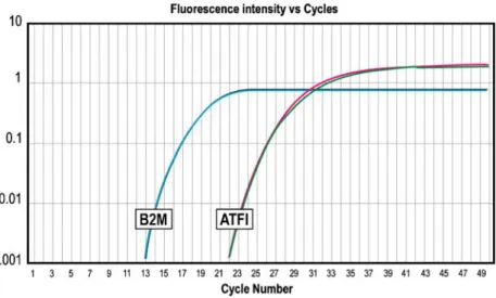
cells showed rounded to oval nuclei with smooth or indented contours, open chromatin and one or two prom-inent nucleoli. The cytoplasm was moderately abundant and contained few organelles as well as immature mela-nosomes. EWS–ATF1 fusion transcripts representative of the t(12;22) (q13;q12) clear cell sarcoma translocation were detected using real-time RT-PCR (Fig.4.). The fusion oc-curred between the exon 8 of EWS gene and the exon 4 of the ATF-1 gene. SYT-SSX1/2 fusion transcripts, represen-tative of the t(X;18) synovial sarcoma translocation, were not detected.
Discussion
Described in 1965 by FM Enzinger [6], clear cell sarcoma of tendons and aponeuroses is a peculiar type of sarcoma which shows features of melanocytic differentiation. Clin-ically, it presents as a slow-growing tumour (median size, 2–5 cm) that mainly affects young adults (20–40 years). These tumours may be present for years before coming to clinical attention. The distal extremities, particularly the feet, ankles, knees, wrists and hands are the most common sites of tumour development [3,6,10,11]. The lesions are almost always deep seated and intimately associated with tendons, aponeuroses and fascia.
Clear cell sarcoma is infrequent outside the limbs. Here-tofore, six cases of clear cell sarcoma of the digestive tract have been reported [1, 4, 5, 7, 13, 15]. The salient clin-icopathologic features of those cases, and the case under discussion, are summarized in Table1. Males seem to be predominantly affected, in contrast to soft tissue lesions, which are divided equally between the sexes. Median age is 37 years (mean, 40 years), and median tumour size is 4 cm. Clear cell sarcoma of soft tissue has a poor prognosis, de-spite various treatment regimens [3,6,10,11]. The tumour tends to metastasize to lungs but also to regional lymph nodes and bone. The 5-year survival rate is about 50–60%. Long term follow-up is mandatory because metastases can develop late in the course of the disease (10 years or more after diagnosis). Of the seven patients with a clear cell sarcoma of the gastrointestinal tract, two had metastatic disease at the time of diagnosis [13,15], and an additional four developed liver metastases 2 (current case) to 24 [4] months post-operatively. Only two patients were disease-free at last follow-up, 11 [1] and 18 [5] months after diagnosis. Follow-up of patients 4 and 6 of Table1have been updated recently (see Acknowledgements).
In the digestive tract, clear cell sarcoma has the same histology and immunoprofile as in the soft tissues. The tu-mour presents as bundles or nests of pale staining to eo-sinophilic spindle cells separated by dense fibrous tissue septa. Tumour cells have prominent vesicular nuclei with Fig. 2 Tumour cells are arranged in nests and fascicles separated by
collagenous septa. Nuclei are round to oval, vesicular, with prom-inent nucleoli. Mitotic figures are numerous. Hematoxylin and eosin stain ×400
Fig. 1 Diffuse infiltration of the mucosa and submucosa of the ileum by tumour cells. Hematoxylin and eosin stain ×50
Fig. 3 Strong reactivity of clear cell sarcoma cells for S-100 protein ×400
mostly a large, central, single nucleolus. Multinucleate giant tumour cells, a relatively common feature of clear cell sarcoma of soft tissues, seem to be rarer in the
gastroin-testinal tract (one of seven cases). Intracellular melanin accumulation is reported in half of the cases.
The tumour cells in clear cell sarcoma of soft tissues usu-ally express melanocytic makers, including S-100 protein, Fig. 4 Real-time PCR assay
demonstrating the presence of EWS–ATF1 transcripts. Graphs show fluorescence emission data during each PCR cycle. This fluorescence is directly related to the quantity of PCR products. ATF1, graph of amplification of EWS–ATF1 transcripts. B2M, graph of amplification of beta-2-microglobulin transcripts, used as internal control
Table 1 Clear cell sarcoma of the gastrointestinal tract Case Author
(reference) Age (years)
Sex Site Size (cm) Metastases at diagnosis Molecular confirmation Treatment Adequacy of surgical resection Outcome 1 Ekfors et al. [5]
38 M Duodenum 3 No No Truncal vagotomy,
pancreatico-duodenectomy, cholecystectomy
Complete AWNED at 18 months
2 Donner et al. [4] 37 M Ileum 6.5 No Yes (karyotype) Intestinal resection, along with tumour
Complete Liver mets at 24 and 46 months, AWD at 46 months 3 Fukuda et al. [7] 74 M Transverse colon 2 No Yes (RT–PCR on liver met) Partial colectomy+ regional lymphadenectomy
Complete Single liver met at 9 months (resected), AWD at 15 months 4 Pauwels
et al. [13]
30 M Stomach 4 Yes (peritoneum, regional lymph nodes) Yes (karyotype and FISH) Palliative partial gastric resection
Complete Pancreatic, liver and Douglas cul-de-sac mets at 4 months, AWD at 18 months 5 Zambrano
et al. [15]
15 F Jejunum 5 Yes (mesenteric lymph nodes) Yes (karyotype) Intestinal resection, along with tumour and enlarged lymph nodes + CT
Incomplete Tumour progression at 12 months, DOD at 16 months
6 Achten et al. [1]
57 M Jejunum 6.5 No Yes (FISH) Intestinal resection, along with tumour
Complete AWNED at 11 months
7 Current case 35 M Ileum 1.8 No Yes (RT-PCR) Intestinal resection, along with tumour
Complete Liver mets at 2 months, DOD at 15 months DOD Dead of disease, AWD Alive with disease, AWNED Alive with no evidence of disease, mets metastases, CT Chemotherapy, RT–PCR
HMB45, melan-A and the melanoma isoform of microph-thalmia transcription factor (MITF-M) [2]. In the gastroin-testinal tract, clear cell sarcoma cells consistently expressed vimentin and S-100 protein. HMB45 was expressed in three of seven cases only, and melan-A in two of four cases with available data. They were consistently negative for EMA, cytokeratins, muscle-specific actin, smooth muscle actin, desmin, CD34, chromogranin, synaptophysin, CD45 and CD117. Melanosomes at varying stages of development were visible ultrastructurally in all (6/6) cases with avail-able data.
The diagnosis of clear cell sarcoma is a challenging one, especially if the tumour occurs in an unexpected site such as the gastrointestinal tract. In our case, the diagnosis was considered only at the time of the peritoneal tumour growth 8 months after the primary tumour had been re-moved. A metastatic melanoma was initially suspected, but an extensive work-up failed to find any primary elsewhere. The other differential diagnoses, successively contemplat-ed and rulcontemplat-ed out on the basis of tumour immunoprofile, included a poorly-differentiated neuroendocrine carcinoma (tumour cells were negative for keratins, EMA, chromo-granin and synaptophysin), a malignant gastrointesti-nal stromal tumour (negativity for CD117 and CD34), a clear cell myomelanocytic tumour/PEComa (negativity for smooth muscle actin, strong positivity for S-100 protein), a paraganglioma (negativity for chromogranin and synapto-physin, diffuse positivity for S-100 protein), some sort of germ cell neoplasia (negativity for epithelial markers and placental alkaline phosphatase), a synovial sarcoma (negativity for epithelial markers, absence of detectable SYT-SSX fusion transcripts), a leiomyosarcoma (negativ-ity for muscle markers) and a lymphoma (negativ(negativ-ity for lymphoid markers). Although initially considered as im-probable, the other diagnostic hypotheses that could not be definitely ruled out included epithelioid malignant pe-ripheral nerve sheath tumour, sclerosing epithelioid fibro-sarcoma and clear cell fibro-sarcoma. Ruling out the diagnosis of malignant peripheral nerve sheath tumour was effec-tively difficult on account of the negativity of the HMB45 marker. Nevertheless, the focal positivity of the melan-A marker was a helpful indication of the melanocytic differ-entiation of the tumour. The detection of EWS–ATF1 fu-sion transcripts by RT-PCR finally demonstrated that this lesion was a clear cell sarcoma. The lack of primary tumour elsewhere strongly suggests that this tumour arose primarily in the ileum and was not a metastatic deposit from a soft tissue tumour, as has sometimes been reported [12].
The relationship between clear cell sarcoma and melano-ma has been controversial for a long time. Both neoplasms show morphologic features of melanocytic differentiation (melanin pigment), as well as immunohistochemical and ultrastructural features (melanosomes), and some authors refer to clear cell sarcoma as malignant melanoma of soft parts [3,5]. Clear cell sarcoma differs, however, from cuta-neous melanoma in several respects: it is almost always
deep-seated, associated with tendons, tendon sheaths and aponeuroses, seldom involves the epidermis and behaves differently, metastasizing predominantly to the lungs. In addition, clear cell sarcoma bears the t(12;22) (q13;q12) translocation which is specific for this tumour type and had never been identified in cutaneous melanoma [2,8]. This translocation, present in 75 to 90% of cases, fuses the acti-vating transcription factor 1 (ATF1) gene on chromosome 12 with the EWS gene on chromosome 22 [2,8].
Based on these differences, clear cell sarcoma was con-sidered heretofore as a clinicopathologic entity distinct from melanoma. However, recent gene expression profil-ing data would suggest that this is not really the case. While examining a series of sarcomas including clear cell sar-comas, Segal et al. [14] found that clear cell sarcomas do not‘cluster’ with other soft tissue sarcomas and may rather represent a distinct genomic subtype of melanoma. In ad-dition to genes of melanocytic differentiation, this tumour was also shown to express fibroblast growth factor recep-tor 1 (FGFR1), a tyrosine kinase receprecep-tor involved in angiogenesis and tumour cell growth and migration, sug-gesting that the use of tyrosine kinase inhibitors might have some value in the treatment of patients with un-controlled disease.
In conclusion, this case shows that clear cell sarcoma of soft tissues may occur as a primary neoplasm of the gas-trointestinal tract. It also underlines the pivotal role of mo-lecular techniques in the diagnostic approach when dealing with‘unusual’ locations.
Acknowledgements The authors thank Dr. P. Pauwels and Dr. R. Sciot for updating the follow-up of patients 4 and 6, respectively, and Dr S.Taylor for proofreading.
References
1. Achten R, Debiec-Rychter M, de Wever I, Sciot R (2005) An unusual case of clear cell sarcoma arising in the jejunum highlights the diagnostic value of molecular genetic techniques in establishing a correct diagnosis. Histopathology 46:472–474 2. Antonescu CR, Tschernyavsky SJ, Woodruff JM, Jungbluth AA, Brennan MF, Ladanyi M (2002) Molecular diagnosis of clear cell sarcoma. Detection of EWS–ATF1 and MITF-M transcripts and histopathological and ultrastructural analysis of 12 cases. J Mol Diagn 4:44–52
3. Deenik W, Mooi WJ, Rutgers EJ, Peterse JL, Hart AA, Kroon BB (1999) Clear cell sarcoma (malignant melanoma) of soft parts. A clinicopathologic study of 30 cases. Cancer 86:969– 975
4. Donner LR, Trompler RA, Dobin S (1998) Clear cell sarcoma of the ileum: the crucial role of cytogenetics for the diagnosis. Am J Surg Pathol 22:121–124
5. Ekfors TO, Kujari H, Isomaki M (1993) Clear cell sarcoma of tendons and aponeurosis (malignant melanoma of soft parts) in the duodenum: the first visceral case. Histopathology 22:255– 259
6. Enzinger FM (1965) Clear cell sarcoma of tendons and aponeuroses. An analysis of 21 cases. Cancer 18:1163–1174 7. Fukuda T, Kakihara T, Baba K, Yamaki T, Yamaguchi T,
Susuki T (2000) Clear cell sarcoma arising in the transverse colon. Pathol Int 50:412–416
8. Graadt van Roggen JF, Mooi WJ, Hogendoorn PCW (1998) Clear cell sarcoma of tendons and aponeuroses (malignant melanoma of soft parts) and cutaneous melanoma: exploring the histogenetic relationship between these two clinicopatho-logical entities. J Pathol 186:3–7
9. Hostein I, Andraud-Fregeville M, Guillou L, Terrier-Lacombe MJ, Deminiere C, Ranchere D, Lussan C, Longavenne E, Bui NB, Delattre O, Coindre JM (2004) Rhabdomyosarcoma: value of myogenin expression analysis and molecular testing in di-agnosing the alveolar subtype: an analysis of 109 paraffin-embedded specimens. Cancer 101:2817–2824
10. Lucas DR, Nascimento AG, Sim FH (1992) Clear cell sarcoma of soft tissues. Mayo Clinic experience with 35 cases. Am J Surg Pathol 16:1197–1204
11. Montgomery EA, Meis JM, Ramos AG, Martz KL (1993) Clear cell sarcoma of tendons and aponeuroses. A clinicopathologic study of 58 cases with analysis of prognostic factors. Int J Surg Pathol 1:89–100
12. Ohba Y, Suzuki H, Hiraga H, Ito T, Sawa H, Nagai M, Satoh SI, Iwaki H, Nagashima K (1999) Melanotic peritoneal sar-comatosis originating from clear cell sarcoma. Pathol Int 49: 653–657
13. Pauwels P, Debiec-Rychter, Sciot R, Vlasveld T, Den Butter B, Hagenmeijer A, Hogendoorn PCW (2002) Clear cell sarcoma of the stomach. Histopathology 41:526–530
14. Segal NH, Pavlidis P, Noble WS, Antonescu CR, Viale A, Wesley UV, Busam K, Gallardo H, DeSantis D, Brennan MF, Cordon-Cardo C, Wolchok JD, Houghton AN (2003) Classi-fication of clear-cell sarcoma as a subtype of melanoma by genomic profiling. J Clin Oncol 21:1775–1781
15. Zambrano E, Reyes-Mugica M, Franchi A, Rosai J (2003) An osteoclast-rich tumour of the gastrointestinal tract with features resembling clear cell sarcoma of soft parts: reports of 6 cases of a GIST simulator. Int J Surg Pathol 11:75–81