Introduction
Biomineralization of the ferrimagnetic iron oxide magnetite (Fe3O4) is known to occur in a wide variety of organisms (Kirschvink et al. 1985, Webb et al. 1990). Many of these studies have focused on how these organisms (magnetotactic bacteria, honey bees, birds and fish, for example) may use biogenic magnetite particles for geomagnetic field sensing – a behaviour known as magnetotaxis (for review see Kobayashi & Kirschvink 1995). Recently, however, attention has focused on the discovery and confirmation of the presence of biogenic magnetite (Fe3O4) and maghemite (gFe2O3) in the human brain (Kirschvink et al. 1992, Dobson et al. 1995, Dunn et al 1995, Kobayashi & Kirschvink 1995, Dobson & Grassi 1996). This discovery is espe-cially important in light of the fact that biogenic magnetite may provide plausible mechanisms linking
human health effects to environmental electro-magnetic field exposure (Kirschvink 1992, 1996, Kobayashi & Kirschvink 1995, Dobson & St. Pierre 1996).
Although considerable research has been done on bacteria and animals, much less is known about humans. In fact, the role of biogenic magnetite in the human central nervous system (CNS) is still not understood. Comparison of the magnetic properties of human brain tissue with those of the magneto-tactic bacteria, Magnetospirillum magnetomagneto-tacticum (MV-1) and the dissimilatory iron-reducing bacteria, Geobacter metallireducens (GS-15 – an anaerobic microorganism, non-magnetotactic and non-motile) has revealed that the material found in the human brain behaves in a similar fashion to the magnetite found in GS-15 (Dobson et al. 1995, Dunn et al. 1995, Moskowitz et al. 1990). Although this does not rule out the possibility of geomagnetic field sensing in humans (either at present or as an evolutionary relic), it does indicate that the biogenic magnetite found in the human brain is not efficiently config-ured for sensing using the same mechanism known to be employed by other organisms.
1 1 1 1 1 1
Analysis of magnetic material in the human heart, spleen and liver
P.P. Grassi-Schultheiss, F. Heller & J. Dobson
Swiss Federal Institute of Technology, ETH-Hönggerberg, Zürich, Switzerland and Department of Physics, Biophysics Programme, University of Western Australia, Perth, Australia
Received 26 March 1997; accepted for publication 11 June 1997
Isothermal remanent magnetization (IRM) acquisition and alternating field (A.F.) demagnetization analyses were performed on human heart, spleen and liver samples resected from cadavers. The magnetic proper-ties of the samples were measured both at 77K and at 273K. A.F. demagnetization was performed at 273K. Results from the analyses of the tissue indicate the presence of ferromagnetic, fine-grained, magnetically interacting particles which, due primarily to magnetic properties, are thought to be magnetite and/or maghemite. The presence of superparamagnetic particles can be inferred from the increase in saturation IRM values when measured at 77K compared with measurements at 273K and the decay of remanent magne-tization upon warming from 77K. The concentration of magnetic material (assuming it is magnetite or maghemite) in the samples varies from 13.7 ng g–1to 343 ng g–1, with the heart tissue generally having the highest concentration. The presence of magnetic material in these organs may have implications for the function of biogenic magnetite in the human body.
Keywords: heart, liver, maghemite, magnetite, spleen
Address for correspondence: J. Dobson, The University of Western Australia, Department of Physics, Biophysics Pro-gramme, Nedlands, Perth WA6907, Australia. Tel: (+61) 9 380 3181 or 2761; Fax: (+61) 9 380 1014; Email: jdobson@uniwa. uwa.edu.au
The purpose of this study is to investigate the mag-netic properties of human organs other than the brain – the heart (apex cordis dextra), spleen and liver – in an attempt to determine the extent of magnetite/ maghemite biomineralization in humans and to advance the understanding of its function. The human lungs were not taken into consideration in this study because of the possibility of exogenous mag-netic contaminants, which are easy to assimilate by breathing (Cohen 1973).
Methods
Tissue samples used in the study were resected from eight cadavers during routine autopsies. All samples were taken within 30 h after death and were not chemically fixed, allowing a more accurate assessment of the magnetic biominerals (Dobson & Grassi 1996). Contamination arte-facts resulting from airborne contamination, the use of surgical scalpels on the tissue, cauterization of blood vessels and formalin fixing were previously examined and controlled (Dobson & Grassi 1996).
The resected samples were immediately placed in vials which had been soaked in a 30% HCl solution for at least 24 h, and put directly in liquid nitrogen to preserve the chemistry. The samples were then placed in quartz glass holders for magnetic measurement. These holders were previously cleaned in 30% HCl for at least 24 h, and then rinsed with distilled water. The samples were packed in the holders with cellophane (found to be non-magnetic in separate analyses), to prevent movement during the measurements. All empty holders were measured without tissue prior to the tissue measurements so that their contri-bution to the total magnetization could be subtracted from the overall signal (as described in Dobson & Grassi 1996). Acquisition of IRM of the samples was measured both at 77K and at 273K. The samples were exposed to D.C. magnetic fields in stepwise increments up to one Tesla (T) at 77K using an Oxford Instruments water-cooled electro-magnet. After each step the remanent magnetization was measured on a 2G SQUID magnetometer. After the final 1T magnetization step, the samples were allowed to warm to 273K. The samples were then completely demagnetised, using an A.F. Schoensted Demagnetizer before being remagnetized in steps up to 1T at 273K. Following this, the samples were incrementally demagnetized and measured with the SQUID magnetometer in order to generate demagnetization curves.
The samples were weighed prior to measuring in order to calculate the mass concentration of magnetic material in the tissue. Tests to control for airborne contamination in the laboratory, and for the reproducibility of the measurements, were regularly carried out.
Results
IRM acquisition curves reveal the presence of low
coercivity magnetic material in all tissue samples measured (Figure 1). All samples reached magnetic saturation by 200 mT, which is consistent with the presence of magnetite and/or maghemite in the tissue.
Calculations of the concentration of magnetic material were performed assuming the material responsible for the magnetization of the tissue was biogenic magnetite. As maghemite has similar mag-netic properties, the calculated concentrations would be only slightly different if the presence of that mineral represented some fraction of the overall magnetization in the tissue (the saturation magneti-zation for magnetite is 476 Am–1 while that of
maghemite is 426 Am–1 – Merrill & McElhinney
1983). These calculations show that concentrations range from 14 ng g–1 to more than 300 ng g–1, with
the heart samples generally having the highest concentration (Table 1). With the exception of sample 96/268, the liver and spleen concentrations were broadly in the same range as human brain tissue. The heart samples, however, have concen-trations which are generally about five to ten times higher than brain tissue.
Alternating field demagnetization of IRM shows that the samples have R values (Wohlfahrt ratios – the ratio of saturation IRM demagnetized to the remanent coercive force value to the undemagne-tized saturation IRM) less than 0.5, indicating that the grains are magnetically interacting (Figure 2). The characteristics of these curves (R values and Median Coercivites) are similar to those reported from brain tissue samples (Kirschvink et al. 1992, Dunn et al. 1995, Dobson & Grassi 1996).
Comparison of IRM acquisition curves measured 1111 0111 0111 0111 0111 0111 111
Figure 1. Examples of IRM acquisition curves for heart, liver and spleen tissue measured at 77K. M is the remanent magnetization.
1 1 1 1 1 1
Table 1. Weight in grams, saturation remanent magne-tization (Jr–measured at 77K) and calculated magnetite/ maghemite concentrations for all heart, spleen and liver samples
Sample Weight (g) Jr (Am2kg–1) Concentration
(ng g–1) Heart 96/245 1.236 1.58E-05 343 96/268 0.768 4.70E-06 102 96/515 1.340 9.26E-06 201 96/549 0.570 1.13E-05 245 96/624 0.440 5.72E-06 124 96/674 0.193 7.78E-06 169 97/025 0.413 5.06E-06 110 Spleen 96/245 1.466 1.07E-06 23.3 96/268 1.493 1.42E-05 308 96/425 0.778 7.70E-07 16.7 96/515 1.129 1.69E-06 36.7 96/549 1.368 6.34E-07 13.7 96/614 0.783 3.88E-06 84.3 96/674 1.019 1.94E-06 42.2 Liver 96/245 1.695 2.40E-06 52.2 96/268 1.387 7.30E-06 158 96/425 0.961 1.54E-06 33.5 96/515 0.979 1.68E-06 36.5 96/549 1.160 1.70E-06 36.9 96/624 1.448 3.56E-06 77.4 96/674 1.112 5.06E-06 110 97/025 0.729 1.62E-06 35.2
Figure 2. IRM acquisition and AF demagnetization for heart sample 96/674 at 273K. The R value (see text for explanation) is 0.23 and the Median Coercivity is 32 mT. M is the remanent magnetization
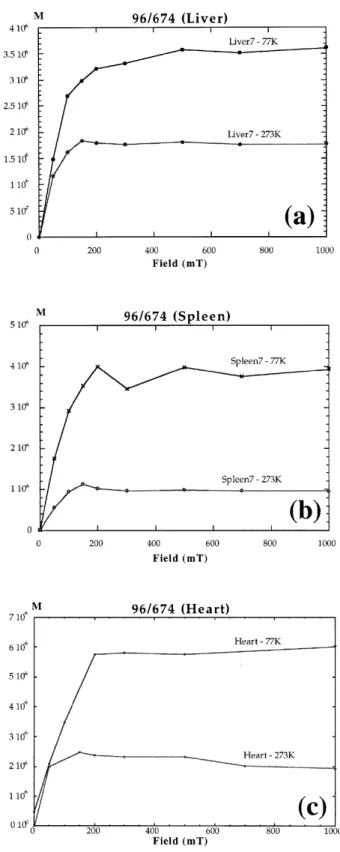
Figure 3. IRM acquisition curves for samples from: (a) liver; (b) spleen; and (c) heart measured at both 77K and 273K. M is the remanent magnetization
at 77K and 273K show that there is a difference in saturation remanent magnetization at the two temperatures (Figure 3). The significance of this will be discussed in the next section.
Discussion and conclusions
The IRM acquisition and AF demagnetization curves indicate that ferro or ferrimagnetic, fine-grained, magnetically interacting particles are present in varying concentrations in all tissue samples measured. The magnetic properties of the tissue are consistent with the material being biogenic magnetite and/or maghemite. The presence of these iron biominerals in the human brain and other organisms has been reported previously but they have not been observed in the human heart, spleen and liver.
Earlier magnetic investigations of these organs (particularly the spleen and liver) have revealed the presence of the iron biomineral hemosiderin (for review see St. Pierre et al. 1989). Although this material is thought to be antiferromagnetic (though this is still unresolved) and known to occur in several forms (especially in the liver and spleen), none of the forms would be likely to contribute to the magnetization of this tissue when measuring isothermal remanence – especially at 273K.
Mössbauer analysis of hemosiderin in the liver and spleen reveals that the maximum blocking temperature for this iron oxyhydroxide is 150K on a time scale of 10–8to 10–9seconds (St. Pierre et al.
1989). Even if it is antiferromagnetic with a defect moment, any hemosiderin present in the tissue therefore, would not contribute to the overall rema-nent magnetization, as even 77K would be above its blocking temperature at the time scale of the IRM measurements (several seconds per measurement). Previous Mössbauer investigations of these organs, however, would not have shown evidence of the presence of biogenic magnetite and/or maghemite as the concentrations are three to four orders of magnitude too low for resolution using this technique.
The presence of superparamagnetic particles in the tissue can be inferred from the increase in satu-ration IRM values when measured at 77K compared with measurements at 273K, as more superpara-magnetic grains will become superpara-magnetically blocked as energy available for thermal agitation is removed from the system. These particles are magnetic but of very small grain size and consequently have very short relaxation times. Magnetic relaxation time
is temperature dependent and will increase as the particles are cooled. As the relaxation time increases, the magnetization becomes stable on the time scales required for the measurement. This is an indication that there is a range of grain sizes present in the tissue.
Calculated concentrations of magnetic material in the tissue are generally consistent within each organ type, with heart tissue having the highest concen-trations. There was, however, one anomalous result – 96/268. It is not clear why the concentrations in this spleen and liver are so much higher than in the other samples. There was no observed pathology in either of the organs and all samples were handled in the same manner as our previous studies of brain tissue in order to avoid sources of contamination and artefacts (Dobson & Grassi 1996). It is therefore not likely that the magnetic material in these samples represents contamination due to airborne particles or surgical instruments.
Although all of the evidence presented here indi-cates that biogenic magnetite and/or maghemite likely is present in human organs other than the brain, in order to confirm these results it will be necessary in the future to extract and directly observe these particles using electron microscopy.
Recent studies indicate that exposure to electro-magnetic fields may have an influence on coronary heart disease in railway workers (Ptitsyna et al. 1996). As biogenic magnetite provides a possible mecha-nism for interactions of weak magnetic fields with the human brain (Kirschvink 1992, 1996, Dobson & St. Pierre 1996) it is important to examine its presence and understand its role in other human organs as well. Finding biogenic magnetite in organs other than the brain is a further indication that it may not have a role in geomagnetic field sensing in humans as this function would normally involve the central nervous system. It may represent another iron storage mech-anism for the body; however, at this point the role of magnetite in humans is still unknown.
Acknowledgements
We would like to thank Professor A. Aguzzi and Dr M. Klein from the Neuropathology Department of the University Hospital, Zürich for providing the tissue samples and Professors William Lowrie, Niels Kuster (both from the ETH Zurich), H. Gregor Wieser (University Hospital, Zürich) and Dr T. St Pierre for helpful discussions. We also thank Mr Beat Geyer and Mr Sandro Moser for technical assistance and Dr J. Webb for his helpful review. 1111 0111 0111 0111 0111 0111 111
References
Cohen D. 1973 Ferromagnetic contamination in the lungs and other organs of the human body. Science 180, 745–748.
Dobson J, Grassi PP. 1996 Magnetic properties of human hippocampal tissue: evaluation of artefact and contam-ination sources. Brain Res Bull 39, 255–259.
Dobson J, St. Pierre TG. 1996 Application of the ferro-magnetic transduction model to D.C. and pulsed magnetic fields: Effects on epileptogenic tissue & impli-cations for cellular phone safety. Biochem Biophys Res
Commun 227, 718–723.
Dobson J, Fuller M, Moser S, et al. 1995 Evocation of epileptiform activity by weak DC magnetic fields and magnetite biomineralization in the human brain. In: Baumgartner C, Deecke L, Stroink G, Williamson SJ, eds. Biomagnetism: Fundamental Research and Clinical
Applications. Amsterdam: Elsevier/ISO Press; 16–19.
Dunn JR, Fuller M, Zoeger J, et al. 1995 Magnetic material in the human hippocampus. Brain Res Bull 36, 149–153. Kirschvink JL. 1992 Comments on “Constraints on biolog-ical effects of weak extremely-low-frequency electro-magnetic fields”. Phys Rev A 46, 2178–2184.
Kirschvink JL. 1996 Microwave absorption by magnetite: A possible mechanism for coupling non-thermal levels of radiation to biological systems. Bioelectromag 17, 187–194.
Kirschvink JL, Jones DS, MacFadden BJ, eds. 1985
Magnetite Biomineralization and Magnetoreception in
Organisms: a New Biomagnetism. New York: R.P.
Plenum Publishing Corp.
Kirschvink JL, Kobayashi-Kirschvink A, Woodford BJ. 1992 Magnetite biomineralization in the human brain.
Proc Acad Sci USA 89, 7683–7687.
Kobayashi A, Kirschvink JL. 1995 Magnetoreception and electromagnetic field effects: Sensory perception of the geomagnetic field in animals and humans. In: Blank M, ed. ACS Advances in Chemistry Series No. 250,
Electromagnetic fields: Biological interactions and mech-anisms. American Chemical Society; 367–394.
Merrill RT, McElhinney MW. 1983. The Earth’s Magnetic
Field: It’s History, Origin and Planetary Perspective.
London: Academic Press, Inc.; 401 pp.
Moskowitz BM, Frankel RB, Bazylinski DA. 1990 Rockmagnetic criteria for the detection of biogenic magnetite. Earth Planet Sci Lett 120, 283–300.
Ptitsyna NG, et al. 1996 Coronary heart diseases: assess-ment of risk associated with work exposure to ultra-low-frequency magnetic fields. Bioelectromag 17(6), 436–444.
St Pierre TG, Webb J, Mann S. 1989 Ferritin and hemo-siderin: structural and magnetic studies of the iron core. In: Mann S, Webb J, Williams RJP, eds.
Biomineraliza-tion: Chemical and Biochemical Perspectives. Weinheim:
VCH Verlangsgesellschaft; 295–344.
Webb J, St. Pierre TG, Macey DJ, 1990 Iron biomineral-ization in invertebrates. In: Frankel RB, Blakemore R, eds. Iron Biominerals. New York: R.P. Plenum Publishing Corp; 193–220. 1 1 1 1 1 1