ORIGINAL PAPER
Factors Influencing the efficacy of intra-articular steroid
injections in patients with juvenile idiopathic arthritis
Peter Marti&Luciano Molinari&Isabel B. Bolt&
Reinhard Seger&Rotraud K. Saurenmann
Received: 18 February 2007 / Accepted: 16 May 2007 / Published online: 12 June 2007 # Springer-Verlag 2007
Abstract A retrospective chart review was performed of all patients with juvenile idiopathic arthritis (JIA) followed at our clinic who had an intra-articular steroid injection between 1 January 1997 and 31 December 2001. The aim of the study was to evaluate the outcome of intra-articular steroid injections (iaS) and determine prognostic factors. During the study period, 202 iaS were performed in 60 patients, of whom 37 had oligoarticular JIA, 15 had polyarticular, rheumatoid factor-negative JIA and four each had systemic and enthesitis-related JIA. The median duration of remission was 23.1 months (range: 0–69 months). At last follow-up, 103 joints (51%) of 47 patients were still in remission after a median follow-up time of 28 months (range: 1–69 months). For the total cohort, the remission was longer for wrist and finger joints [risk ratio (RR): 0.2], with concomitant treatment with methotrexate (RR: 0.28) and for enthesitis-related arthritis (RR: 0.34). For the group of knee joints, remission was longer with concomitant treatment with methotrexate (RR: 0.37), with triamcinolone hexacetonide (RR: 0.77) and with general anaesthesia for the procedure (RR: 0.56). Mild side effects were observed in 45 iaS (22.3%), and skin atrophy occurred at the injection site in 2% of injections, but no major adverse event occurred in our cohort. In conclusion, iaS is a safe procedure with a median duration of remission of 23.1 months. The remission was longer in the joints of the upper extremity, with concomitant treatment with methotrexate
and when the injection was performed under general anaesthesia.
Keywords Intra-articular steroid injection . Juvenile idiopathic arthritis . Prognosis . Side effects . Treatment
Abbreviations
ANA anti-nuclear antibody
HLA B27 human leucocyte antigen B27 iaS intra-articular steroid injection JIA juvenile idiopathic arthritis NSAR non-steroidal anti-rheumatic drugs RR risk ratio
TNFα tumour necrosis factor alpha
Introduction
Juvenile idiopathic arthritis (JIA) is the most common rheumatic disease of childhood, with a prevalence between 7 and 400 per 100,000 children [10]. Seven subgroups of JIA have been defined by the International League of Associations for Rheumatology (ILAR) [15], with oligoar-ticular JIA being the most common. Recent studies on the long-term outcome of affected children show that JIA will often extend into adulthood [6, 11, 13] and may cause severe joint destruction if the arthritis is uncontrolled for longer periods of time [13]. New drugs for the treatment of arthritis, especially the group of TNFα-blockers, are increasingly used to treat JIA and have significantly improved the quality of life of affected children [9, 18]. However, the higher number of treatment options increases the need to assess their respective effectiveness and safety
P. Marti
:
L. Molinari:
I. B. Bolt:
R. Seger:
R. K. SaurenmannDepartment of Paediatrics, University Children’s Hospital,
Zurich, Switzerland
R. K. Saurenmann (*)
Pediatric Rheumatology, University Children’s Hospital,
Steinwiesstr. 75, 8032 Zurich, Switzerland e-mail: [email protected]
in order to choose the best suited therapy for an individual patient.
Intra-articular corticosteroid injection (iaS) is a treatment option for patients with active arthritis in a small number of joints [7]. It is recommended as the treatment of choice for oligoarticular JIA but may also be used in cases where only a few joints remain actively inflamed during a treatment with systemic disease-modifying drugs such as methotrex-ate [4,14, 19]. The duration of remission after iaS varies widely [1,2,14]. The peak incidence of oligoarticular JIA is at preschool age, and small children will usually need general anaesthesia for joint injections. Possible complica-tions occurring after iaS include infeccomplica-tions, local skin atrophy or hypopigmentation, intra- or periarticular calcifi-cations or avascular necrosis of the epiphyseal bone [8,12,
14]. A knowledge of factors influencing the prognosis of iaS will help to select adequate treatment options and improve counselling of parents and patients.
The aim of our study was to evaluate the efficacy and safety of intra-articular steroid injections performed at our centre and to examine factors influencing the outcome of this procedure in a retrospective manner.
Patients and methods
The Paediatric Rheumatology database at the University Children’s Hospital of Zurich was searched for patients with JIA who had undergone iaS between 1 January 1997 and 31 December 2001. The charts of all 67 patients (288 injections) found were reviewed. Seven patients were excluded: three patients with eight injections performed in outside clinics, and two patients (four injections) who were followed at an outside clinic after the injections had been performed at our institution. Two patients with severe systemic JIA with a total of 74 joints injected for temporary relief of their refractory polyarticular arthritis were excluded from the analysis because the high number of injections in these two patients with extremely resistant disease skewed the analysis. Of the remaining 60 patients, the following data were collected: date of birth, gender, date of diagnosis of JIA, JIA subtype, results of anti-nuclear antibody (ANA), human leucocyte antigen (HLA) B27 and rheumatoid factor (RF) testing, diagnosis of uveitis, date of steroid injection, location of the injected joint, medication and dose injected, concomitant medication at the time of injection, date of arthritis relapse in the injected joint (if applicable), type of anaesthesia used (if applicable), side effects, such as nausea/vomiting, paradoxical reaction to sedation, increased appetite, flushed cheeks, local infections or atrophy/hypopigmentation at the site of injection. The end point of study follow-up was 31 October 2002.
Triamcinolone hexacetonide is used at our clinic as the drug of choice for intra-articular steroid injections. Due to
production shortfalls, this drug was not always available during the study period, and triamcinolone acetonide was used as a replacement medication from 1 September 1997 until 31 January 1999 and from 1 March to 31 October 2000. The dose of triamcinolone acetonide used was double the amount (in milligrams) of triamcinolone hexacetonide. For children with a body weight of ≥40 kg, the dose of triamcinolone hexacetonide injected was 40 mg in big joints (knee, shoulder, hip), 20 mg in medium joints (wrist, elbow, ankle, subtalar) and 5 mg in finger and toe joints. Children with a body weight between 20 and 40 kg would receive 75% of these doses, and children with a body weight <20 kg would receive 50% of these doses. All injections were performed by the same paediatric rheuma-tologist (RKS). Injections into hips were performed with ultrasound guidance, while injections into subtalar and midfoot joints were performed using fluoroscopy to control for correct position of the needle. Standard clinical routine consisted of asking all patients and their parents to keep the injected joint as quiet as possible for a 24-h period after injection.
Remission of arthritis in a joint was defined as the absence of signs of inflammation, such as capsular (soft tissue) swelling, effusion or the combination of tenderness and pain on motion. A joint with presence of any of the aforementioned signs of inflammation was considered to be actively inflamed.
Statistical analysis was performed using JMP IN 5.1
programme (SAS Institute, Cary N.C.) . We used the chi-square test and survival analysis (Kaplan Meier) with log rank/Wilcoxon to assess for differences among the groups. Factors significantly associated with the duration of remission were then tested in a Cox Proportional Hazards regression model.
The study was approved by the Institutional Review Board.
Results
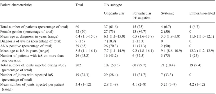
The 60 patients included in our study had 202 joint injections on 106 occasions (mean: 1.9 injections per occasion; range: 1–12). Table 1 presents the patients' characteristics and distribution of joints. No patients with psoriatic JIA or polyarticular, RF-positive JIA underwent joint injections during the study period, and no patient tested RF-positive. The distribution of joint types was highly significantly different among subtypes of JIA (chi-squarep<0.0001). For the duration of remission according to joint type, see Table2. Sixteen patients (26.7%) had only one joint injection, 19 patients (31.7%) had several joints injected at one occasion (mean: 2.3 joints; range: 2–12) and 25 patients (41.7%) had injections performed on several
occasions, with 49 repeated injections (24.3% of all injections) performed into a previously injected joint. In total, 153 different joints were injected, with 118 joints (77.1%) injected once, 25 joints (16.3%) injected twice, six joints (3.9%) injected three times and four joints (2.6%) injected four times.
One or more concomitant medications were used during 165 iaS (81.7%), including non-steroidal anti-rheumatic drugs (NSAR) (141 iaS; 69.8%), methotrexate (66 iaS;
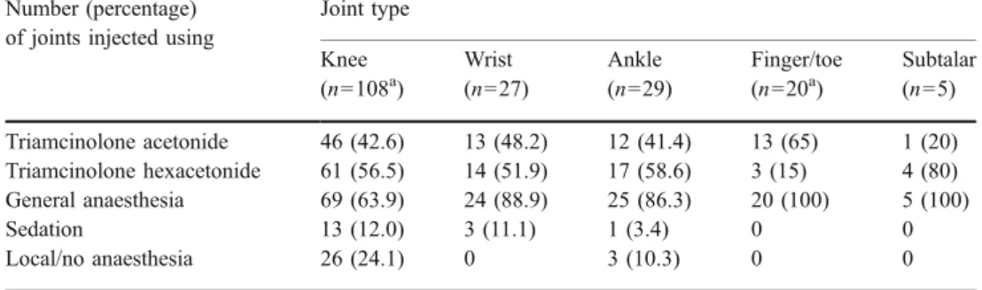
32.7%), systemic corticosteroids (16 iaS; 7.9%) and salazo-pyrine (one iaS). One hundred and fifty one (74.7%) iaS were performed under general anaesthesia, 18 (8.9%) in sedation with midazolam/nalbuphine, eight (4%) with local anaesthesia and 25 (12.4%) without anaesthesia. Table 3
shows how the distribution of joints injected under the dif-ferent types of anaesthesia differed significantly (chi-square p=0.014). Triamcinolone acetonide was used for 87/202 iaS (43.1%), triamcinolone hexacetonide for 108/202 (53.5%)
Table 1 Patients’ characteristics
Patient characteristics Total JIA subtype
Oligoarticular Polyarticular
RF negative
Systemic Enthesitis-related
Total number of patients (percentage of total) 60 37 (61.6) 15 (25) 4 (6.7) 4 (6.7)
Female gender (percentage of total) 42 (70) 27 (73) 13 (86.7) 2 (50) 0
Mean age at diagnosis in years (range) 6.4 (1.1–15.0) 6.1 (1.1–15.0) 6.5 (1.6–13.8) 3.0 (1.8–5.8) 11.6 (11.0–12.1)
Diagnosis of uveitis (percentage of total) 9 (15) 7 (18.9) 2 (13.3) 0 0
ANA positive (percentage of total) 39 (65) 26 (70.3) 11 (73.3) 2 (50) 0
Mean age at iaS in years (range) 8.5 (1.1–16.1) 7.7 (1.1–14.9) 9.2 (1.8–16.1) 9.4 (8.6–10.9) 12.3 (11.2–12.9)
Number of patients with iaS on more than one occasion
26 (43.3) 16 (43.2) 6 (37.5) 3 (75) 1 (25)
Total number of joints injected during study (percentage of total)
202 102 (50.5) 60 (29.7) 21 (10.4) 19 (9.4)
Number of joints with repeated iaS (percentage of total)
49 (24.3) 29 (28.4) 13 (21.7) 7 (33.3) 0
Mean number of joints injected per patient (range)
3.4 (1–12) 2.8 (1–9) 4.1 (2–8) 5.25 (3–7) 4.2 (1–12)
ANA, Anti-nuclear antibodies; RF, rheumatoid factor; iaS, intra-articular steroid injection; JIA, juvenile idiopathic arthritis
Table 2 Duration of remission according to joint type
Joint type Number
of joints injected
Number of joints with arthritis flare after iaS (percent)
Mean/median duration of remission until flare in months (range) Number of joints in ongoing remission at end of study (percent) Mean/median follow up time of joints with ongoing remission in months (range)
Risk ratio for relapse in the Cox regression model All joints 202 99 (49) 6.9 / 6.0 (0–30) 103 (51) 29.1/ 28.0 (1–69) Knee 108 55 (50.9) 8.0 / 6.0 (0–27) 53 (49.1) 27.2 /24.0 (1–69) 1.05 Ankle 29 19 (65.5) 4.5 / 3.0 (0–13) 10 (34.5) 18.2 / 14.0 (1–39) 1.88 Wrist 27 11 (40.7) 7.3 / 6.0 (0–24) 16 (59.3) 31.6 / 31.0 (13–52) 0.74 Finger/toe 20 4 (20) 8 / 5.5 (4–17) 16 (80) 40.3 / 46.0 (18–66) 0.2 Subtalar 5 4 (80) 3.5 / 1.5 (0–11) 1 (20) 13 2.58 Shoulder 4 2 (50) 18 / 18 (6–30) 2 (50) 25 /25 Elbow 3 1 (33.3) 2 2 (66.7) 39.5 / 39.5 (12–67) Hip 3 0 n.a. 3 (100) 28.7 / 13.0 (13–66) Midfoot 3 3 (100) 1 / 0 (0–3) 0 n.a. JIA subtype Oligoarticular 102 67 (65.7) 6.5 / 5 (0–27) 35 (34.3) 25.1 / 24 (1–69) Polyarticular RF negative 60 21 (35) 7.3 / 6 (2–24) 39 (65) 27.2 / 23 (1–66) Systemic 21 10 (47.6) 9.9 / 8 (0–30) 11 (52.4) 39.1 / 32 (25–67) Enthesitis-related 19 1 (5.3) 12 18 (94.7) 35.2 / 46 (11–46)
iaS and for 7 iaS the medication used was not recorded. Al-though the use of triamcinolone acetonide or triamcinolone hexacetonide was dependent only on availability of the drug, the distribution of joints injected with these two medications differed significantly (chi-squarep=0.036; Table3).
In a first analysis, the outcome of all iaS was evaluated using a survival (Kaplan-Meier) analysis. Table4 presents the factors tested and their respective association with the duration of remission.
In a second analysis, the outcome of the 108 knee joints was analysed for risk factors, as this was the largest ho-mogeneous group of joints injected in our cohort. Table 4
presents the factors significantly associated with a longer duration of remission in knee joints.
Minor side effects of the treatment were recorded for 26 patients and 45 injections (43.3 and 22.3%, respectively). These were related to anaesthesia in eight patients/15 injections
(13.3/7.4%) [six nausea/vomiting, three paradoxical reac-tion (agitareac-tion) following sedareac-tion]. Possible systemic ef-fects of corticosteroids were recorded in seven patients/16 injections (11.7/7.9%) (one flushed cheeks, three increased appetite, three mood changes) and local side effects to steroid injections were recorded in 12 patients/14 injections (20/6.9%) (14 skin atrophies combined with hypopigmen-tation at the injection site in 7/14). Ten of the 14 skin atrophies (71.4%) and five of the seven hypopigmentations (71.4%) observed were transient, with complete resolution occurring during the study period leaving a rate of 2% (4/ 202 injections) of local long-term side effects. The occurrence of skin atrophy or hypopigmentation did not correlate with joint type, age at diagnosis, age at injection or type of anaesthesia used. No major side effects, such as infections, skin necrosis or avascular necrosis related to the steroid injections, were observed in our cohort.
Table 4 Factors tested for association with duration of remission
Risk factor tested Significance of association with duration of remission
Total group Knee joint group
Kaplan Meier Cox regression Kaplan Meier Cox regression
Subtype of JIA p<0.0001* p=0.019*, RR 0.34 p=0.77
Concomitant treatment with methotrexate p<0.0001* p=0.0001*, RR 0.28 p=0.001* p=0.009*, RR 0.37
Larger number of joints injected at the same time p<0.0001* n.s. p=0.004* p=0.18, RR 0.84
Type of joint injected p=0.0003* p=0.014*, RR 0.2 n.a.
Joint of upper extremity p=0.0034* n.s. n.a.
Diagnosis of uveitis p=0.14 p=0.54
Negative ANA p=0.007* n.s. p=0.39
Presence of HLA B27 p=0.017* n.s.
Absence of skin atrophy at injection site p=0.02* n.s.
Time elapsed from diagnosis until injection p=0.12
Age at diagnosis p=0.19 p=0.54
Age at injection p=0.2 p=0.37
Gender p=0.44 p=0.25
Use of triamcinolone hexacetonide p=0.79 p=0.025* p=0.04*, RR 0.77
General anaesthesia p=0.9 p=0.01* p=0.049*, RR 0.56
Concomitant treatment with NSAR p=0.99
*Significant atp≤0.05
RR, Risk ratio; HLA B27, human leucocyte antigen B27; NSAR, non-steroidal anti-rheumatic drugs Table 3 Distribution of
medication and type of anaesthesia used
a
The triamcinolone type used was not recorded for one knee and four finger/toe joints
Number (percentage) of joints injected using
Joint type Knee (n=108a) Wrist (n=27) Ankle (n=29) Finger/toe (n=20a) Subtalar (n=5) Triamcinolone acetonide 46 (42.6) 13 (48.2) 12 (41.4) 13 (65) 1 (20) Triamcinolone hexacetonide 61 (56.5) 14 (51.9) 17 (58.6) 3 (15) 4 (80) General anaesthesia 69 (63.9) 24 (88.9) 25 (86.3) 20 (100) 5 (100) Sedation 13 (12.0) 3 (11.1) 1 (3.4) 0 0 Local/no anaesthesia 26 (24.1) 0 3 (10.3) 0 0
Discussion
Intra-articular steroid injections are commonly used for treatment of children with JIA and are considered the treatment of choice for oligoarticular joint involvement [4]. In our cohort iaS was a safe and very effective treatment with a median time of remission of 23.1 months.
The range of duration of remission after iaS varied widely in our cohort, but other investigators have had the same experience [2, 14, 16]. The current body of knowledge on factors influencing outcome is limited. In addition to the well-documented effect of the type of triamcinolone used [5,23], high erythrocyte sedimentation rate [16] and the subtype of arthritis [2] have been shown to be of importance. In our cohort, there was a significant difference in the duration of remission between the different joints, with a significantly longer remission in joints of the upper extremities, followed by the knees. The question that arises is whether weight-bearing may have an influence on the duration of remission in joints after iaS. Trauma- and/or stress-induced arthritis have been reported, especially in cases of psoriatic arthritis [17, 20]. However, the question of whether weight-bearing joints are at higher risk for relapse of arthritis or which other factors might be responsible for this result remains unclear. Breit et al. [2] found that the subtype of juvenile arthritis was more important than the type of joint injected, and they did not find a difference in the duration of remission between joints of the upper and lower extremities. It is difficult to compare our study with the study of Breit et al. as they used a scoring system to measure improvement of the arthritis rather than the duration of complete remission after injection as we did in the present study. The subtype of JIA was a highly significant risk factor in our total cohort. However, this factor was no longer significantly associated with the duration of remission when the analysis was restricted to the more homogeneous group of knee joints. This result is in agreement with the findings of other authors [5, 16]. Our results from the knee joint cohort suggest that the difference in outcome among subtypes of JIA found in our total cohort may reflect the highly significant difference in the distribution of joints injected in these subgroups. However, we can not exclude the possibility that the subtype of JIA may be of importance for the outcome of iaS in joints other than knees.
Concomitant use of methotrexate was the strongest predictive factor in our study and was highly significantly associated with a longer duration of remission both in the total cohort and in the subgroup of knee joints, whereas use of NSAR was not. Allen et al. [1] found that a higher dose of NSAR was significantly associated with a longer duration of remission after iaS, whereas most other authors do not comment on the influence of concomitant
medica-tion on the outcome of iaS [2, 5, 16]. We feel that our results confirm that iaS into residual joints during treatment with methotrexate is very effective and may be a viable alternative to TNFα-blocker use if only a few joints remain active with methotrexate alone. However, this hypothesis would have to be tested in a prospective study.
Several studies have shown a superior efficacy of triamcinolone hexacetonide compared to triamcinolone acetonide in patients with JIA [5, 23]. We found a significantly better efficacy of triamcinolone hexacetonide when we looked at the subgroup of knee joints but not for the total group. However, we must point out that our study is retrospective and was not designed to look for a difference in these two medications. Although the total number of joints injected with each type of triamcinolone was comparable, the joint types were significantly different among the groups. Our results confirm the findings of the superiority of triamcinolone hexacetonide over triamcino-lone acetonide for the knee joints, but in the total group this effect was probably confounded by the difference in response related to the type of joint injected.
The superiority of general anaesthesia over other types of pain reduction for the procedure was an unexpected result in the subgroup analysis of the knee joints. We were not able to assess the effects of anaesthesia type for other joints as the number of joints other than knees injected without general anaesthesia was too small for analysis. One might argue that this result only reflects differences in injection technique as it is technically easier to perform arthrocentesis in an anaesthetized child. However, the knee joint is not only the joint most commonly injected in our cohort, it is probably also the joint of the human body most easily accessible for injection. Although we cannot rule out differences in injection technique as a possible explanation for this unexpected result, other factors associated with the use of general anaesthesia for the injection must be considered. Several studies in adult patients with rheuma-toid arthritis were able to show that rest of the injected joint for 24–72 h improves the outcome of iaS in knee joints [3,
22] but not in wrists [21]. No similar study in children has been conducted to our knowledge. Although we generally advise parents and children to rest the injected joint for 24 h after the injection, we are not able to objectively con-trol the compliance to this advice. We therefore conclude that further studies are needed to assess the impact of general anaesthesia and bed rest on duration of remission after iaS.
The side effects of the procedure were mostly mild and transient, although they were not uncommon in our cohort. Most of these were related to side effects of the anaesthetic medication used or to transient systemic effects of the steroids injected. Hypopigmentation and skin atrophy at the injection site have been reported earlier [8, 14]. In
agreement with most other authors, we were able to demonstrate a high rate of complete resolution of these local corticosteroid side effects over time.
Our study is limited by a heterogeneous patient cohort and the variety of different joints injected in each patient. For some infrequently injected joints, factors influencing the outcome of the procedure could not be tested because of the small numbers, yet these joints may have influenced the overall result of our study. We attempted to compensate for this shortcoming with the analysis of the more homoge-neous group of knee joints.
In conclusion, we have shown that intra-articular corticosteroid injection is a safe and effective treatment for patients with JIA. It is especially efficacious in joints of the upper extremities and knees and in combination with methotrexate. Although side effects are quite common, they are usually mild and do not require interventions.
References
1. Allen RC, Gross KR, Laxer RM, Malleson PN, Beauchamp RD, Petty RE (1986) Intraarticular triamcinolone hexacetonide in the management of chronic arthritis in children. Arthritis Rheum
29:997–1001
2. Breit W, Frosch M, Meyer U, Heinecke A, Ganser G (2000) A subgroup-specific evaluation of the efficacy of intraarticular triamcinolone hexacetonide in juvenile chronic arthritis. J
Rheu-matol 27:2696–2702
3. Chakravarty K, Pharoah PD, Scott DG (1994) A randomized controlled study of post-injection rest following intra-articular
steroid therapy for knee synovitis. Br J Rheumatol 33:464–468
4. Cron RQ, Sharma S, Sherry DD (1999) Current treatment by United States and Canadian pediatric rheumatologists. J Rheuma-tol 26:2036–2038
5. Eberhard BA, Sison MC, Gottlieb BS, Ilowite NT (2004) Comparison of the intraarticular effectiveness of triamcinolone hexacetonide and triamcinolone acetonide in treatment of juvenile
rheumatoid arthritis. J Rheumatol 31:2507–2512
6. Foster HE, Marshall N, Myers A, Dunkley P, Griffiths ID (2003) Outcome in adults with juvenile idiopathic arthritis: a quality of
life study. Arthritis Rheum 48:767–775
7. Ilowite NT (2002) Current treatment of juvenile rheumatoid
arthritis. Pediatrics 109:109–115
8. Job-Deslandre C, Menkes CJ (1990) Complications of intra-articular injections of triamcinolone hexacetonide in chronic
arthritis in children. Clin Exp Rheumatol 8:413–416
9. Lovell DJ, Giannini EH, Reiff A, Jones OY, Schneider R, Olson JC, Stein LD, Gedalia A, Ilowite NT, Wallace CA, Lange M, Finck BK, Burge DJ (2003) Long-term efficacy and safety of etanercept in children with polyarticular-course juvenile rheuma-toid arthritis: interim results from an ongoing multicenter, open-label, extended-treatment trial. Arthritis Rheum 48:218–226 10. Manners PJ, Bower C (2002) Worldwide prevalence of juvenile
arthritis why does it vary so much? J Rheumatol 29:1520–1530 11. Narayanan K, Rajendran CP, Porkodi R, Shanmuganandan K
(2002) A follow-up study of juvenile rheumatoid arthritis into
adulthood. J Assoc Physicians India 50:1039–1041
12. Neidel J, Boehnke M, Kuster RM (2002) The efficacy and safety of intraarticular corticosteroid therapy for coxitis in juvenile
rheumatoid arthritis. Arthritis Rheum 46:1620–1628
13. Oen K, Malleson PN, Cabral DA, Rosenberg AM, Petty RE, Cheang M (2002) Disease course and outcome of juvenile rheumatoid arthritis in a multicenter cohort. J Rheumatol 29:
1989–1999
14. Padeh S, Passwell JH (1998) Intraarticular corticosteroid injection in the management of children with chronic arthritis. Arthritis
Rheum 41:1210–1214
15. Petty RE, Southwood TR, Baum J, Bhettay E, Glass DN, Manners P, Maldonado-Cocco J, Suarez-Almazor M, Orozco-Alcala J, Prieur AM (1998) Revision of the proposed classification criteria for juvenile idiopathic arthritis: Durban, 1997. J Rheumatol 25:1991–1994
16. Ravelli A, Manzoni SM, Viola S, Pistorio A, Ruperto N, Martini A (2001) Factors affecting the efficacy of intraarticular cortico-steroid injection of knees in juvenile idiopathic arthritis. J
Rheumatol 28:2100–2102
17. Sandorfi N, Freundlich B (1997) Psoriatic and seronegative inflammatory arthropathy associated with a traumatic onset: 4
cases and a review of the literature. J Rheumatol 24:187–192
18. Schneider R, Passo MH (2002) Juvenile rheumatoid arthritis.
Rheum Dis Clin North Am 28:503–530
19. Sherry DD, Stein LD, Reed AM, Schanberg LE, Kredich DW (1999) Prevention of leg length discrepancy in young children with pauciarticular juvenile rheumatoid arthritis by treatment with
intraarticular steroids. Arthritis Rheum 42:2330–2334
20. Wallace DJ (1994) Does stress or trauma cause or aggravate rheumatic disease? Baillieres Clin Rheumatol 8:149–159 21. Weitoft T, Ronnblom L (2003) Randomised controlled study of
postinjection immobilisation after intra-articular glucocorticoid
treatment for wrist synovitis. Ann Rheum Dis 62:1013–1015
22. Weitoft T, Larsson A, Saxne T, Ronnblom L (2005) Changes of cartilage and bone markers after intra-articular glucocorticoid treatment with and without postinjection rest in patients with
rheumatoid arthritis. Ann Rheum Dis 64:1750–1753
23. Zulian F, Martini G, Gobber D, Plebani M, Zacchello F, Manners P (2004) Triamcinolone acetonide and hexacetonide intra-articular treatment of symmetrical joints in juvenile idiopathic arthritis: a