ORIGINAL ARTICLE
Use of Imatinib in the Prevention of Heterotopic Ossification
Clément M. L. Werner, MD&Stefan M. Zimmermann, MD&Carola C. Würgler-Hauri, MD&Joseph M. Lane, MD&
Guido A. Wanner, MD&Hans-Peter Simmen, MD
Received: 21 February 2012/Accepted: 28 March 2013/Published online: 21 June 2013 * Hospital for Special Surgery 2013
Abstract Background: Heterotopic ossification (HO) is a common complication following orthopedic and trauma sur-gery, which may have substantial negative effects on the postoperative outcome. Angiogenesis appears to play a critical role in heterotopic ossification. One of the involved signaling molecules is platelet-derived growth factor (PDGF) which may be inhibited by imatinib. Questions/Purposes: Our goal was to prevent HO by pharmacologically interfering with the molecular signaling pathways involved in the developmental process. We hypothesized that by administering a proven inhibitor of PDGF expression, heterotopic bone formation may be prevented. Methods: The effect of imatinib on HO formation was studied in a murine model which reliably pro-duces islets of HO within the soft tissue following Achilles tenotomy. The control group underwent Achilles tenotomy only. The imatinib group received imatinib mesylate. After trial completion, the limbs were harvested and scanned by micro-CT. Heterotopic bone volume was then identified and quantified. Results: The mean volume of heterotopic bone formed in the control group was 0.976mm3 compared to 0.221 mm3in the imatinib group. The volume of HO in the
treatment group was reduced by 85% compared to the control group. Conclusions: The administration of imatinib was associated with a significantly reduced volume of HO. This may be due to the inhibitory effect of imatinib on the PDGF signaling pathway during development of HO. Clinical Relevance: The successful reduction of HO formation following imatinib administration has led to further insight concerning the pathogenesis of HO which in the future may lead to new clinical approaches towards the prevention of HO.
Keywords heterotopic ossification . prevention . imatinib mesylate . murine . PDGF
Introduction
Heterotopic ossification of muscles, tendons, and ligaments is a widely encountered problem throughout orthopedic and trauma surgery. Heterotopic ossification may result in joint contracture, ankylosis, pain, spasticity, swelling, fever, neurovascular compression, lymphedema, and pressure ulcers leading to significant disability [7]. It is defined as the forma-tion of mature lamellar bone in soft tissues outside of the skeletal periosteum following local trauma or operation [12]. Patients with high-grade ossification frequently necessitate reoperation thereby largely increasing the costs of treatment.
Pharmacological agents used to prevent the formation of heterotopic ossification (HO) include non-steroidal anti-in-flammatory drugs [18]. Low-dose perioperative radiation has also been shown to be very effective at preventing HO formation [18]. However, both prophylactic modalities have the drawback of both impeding bone remodeling and thus fracture healing as well as reducing the strengths of the interface between a porous-coated device and trabecular bone [8, 9]. Some surgeons even avoid the use of any HO prophylaxis in favor of fracture healing or implant integra-tion. In addition, all current therapeutical options to prevent heterotopic ossification merely decrease the incidence, but to this day cannot completely prevent their occurrence.
Electronic supplementary material The online version of this article (doi:10.1007/s11420-013-9335-y) contains supplementary material, which is available to authorized users.
C. M. L. Werner, MD
:
S. M. Zimmermann, MD:
C. C. Würgler-Hauri, MD
:
G. A. Wanner, MD:
H.-P. Simmen, MD Department of Traumatology, University Hospital,Zurich, Switzerland J. M. Lane, MD
Hospital for Special Surgery, 535 East 70th Street, New York, NY 10021 USA C. M. L. Werner, MD (*)
Klinik für Unfallchirurgie, UniversitätsSpital Zürich, Rämistrasse 100,
8091 Zürich, Switzerland e-mail: [email protected]
The exact mechanism leading to HO formation is not completely understood. During bone growth, development, and remodeling, angiogenesis (neovascularization) as well as osteogenesis are closely associated processes, sharing some essential mediators. The final event in endochondral ossification is the replacement of avascular (cartilage) tissue by highly vascularized bone. Angiogenic stimulators in-duced by hypoxic stress within the target tissue are a pre-requisite for the differentiation of stem cells to chondrocytes and subsequent heterotopic bone formation. The hypoxic tissue/cartilage template provides a target for capillary inva-sion and angiogenesis. Histological findings suggest that osteoblasts and osteoprogenitor cells always develop con-comitantly with endothelial cells in the newly formed blood vessels at sites where new bone is formed [1,3,23].
The Brooker grading scale of heterotopic ossification is based on the voluminal amount of ectopic bone. Although it is clear that severe heterotopic ossification is not incompatible with satisfactory functional results, several studies have shown that functional outcome in patients suffering from heterotopic ossification is dependent on the Brooker grade meaning that higher Brooker grades lead to significantly poorer outcome. Therefore, not only complete prevention—but a mere reduction in the amount of ectopic bone formation—may still suffice in order to significantly improve the functional outcome in patients with heterotopic ossification.
Attempts to prevent or treat aberrant bone formation have been restricted by the complexity and multiple causes of the disorder. New therapies are being devised to target the induc-tive molecules that may trigger the process, the progenitor cells, and local tissue environments conducive to osteogenesis. The modulation of factors thought to be associated with HO formation like hypoxia or neovascularization has only rarely been the target of investigations. Angiogenesis is required for endochondral bone formation. Although the exact mecha-nisms involved are not entirely clear, a possible explanation could be that new vessels are necessary to bring new stem cells into the target area where HO is about to be formed.
One of the signaling molecules involved in the process of angiogenesis is the platelet-derived growth factor (PDGF), which plays a key role in endochondral ossification. Imatinib mesylate has been shown to specifically inhibit PDGF (Glivec®, Novartis, Switzerland) [19]. The short-term inhibition of angiogenesis as regulated by PDGF is the property of imatinib that may limit the development of heterotopic ossification particularly in soft tissues.
We have therefore created the hypothesis that inhibition of new blood vessel formation resulting from blockage of PDGF expression by imatinib may ultimately reduce or inhibit the production of heterotopic bone. With this in mind, the primary aim of this project was to investigate whether imatinib could reduce the incidence and volume of HO in a murine model of soft tissue trauma.
Materials and Methods
The study was approved by the local animal care and ethical committees. The model used [11] is a well established
murine model in which HO formation reliably occurs within the soft tissue of a mouse hind limb 10 weeks after soft tissue trauma by means of an Achilles tenotomy.
Genetically unmodified CD1 mice were selected for the study. Identification was carried out by markings on the tail. Anesthesia was carried out using isoflurane 5–2% in oxygen (flow rate, 400 ml/min), via nose cone. The anesthetized mice underwent bilateral midpoint Achilles tendon tenotomy through a posterior approach, and the skin was closed using nonabsorbable sutures (Figs. 1 and 2). The animals were randomly assigned to one of two groups: a control group (n010) and a treatment group (n010). Peri-operative analgesia was mainly achieved with paracetamol (Dafalgan syrup 3%, 200 mg/kg), for 1–3 days [6].
To reduce stress and possible pain postoperatively, the animals were checked several times a day according to a predefined protocol. If they presented signs of postoperative distress (apathy, shivering, no chow and water intake), the treatment with paracetamol was extended. A score sheet was developed for detection of discomfort.
The control group underwent Achilles tenotomy only. The treatment group additionally received 10 mg/kg imatinib p.o. daily. This corresponds to the lowest dosage used in mice in the literature [22] over a period of 6 weeks, followed by 4 weeks of rest and cage activity only.
At 10 weeks after surgery, the mice were euthanized and the limbs harvested. Radiographic workup for the presence of HO was carried out using micro-CT of the hind legs with a nominal resolution of 30μm (b-cube, Swiss Federal Insti-tute of Technology, Zurich, Switzerland) (Fig.3). First, 2D overview images were generated. Those images showed a series of slices through the specimen. Skeletal, as well as ectopic, bone was separated from the background by a fixed thresholding procedure. The heterotopic bone compartments were then manually identified and analyzed as a separate compartment. HO was defined as any bone within soft tissue with a density at least equivalent to that of spongy skeletal bone. Following this, a 3D image of each limb was created, and the mass of heterotopic bone could be identified and quantitatively assessed (Fig.4). In cases of multiple foci, the individual volumes were added together.
Fig. 1. An intraoperative caption of a skin incision performed on the mouse hind limb.
Statistical analysis was conducted in collaboration with the Division of Biostatistics at the Institute for Social and Preventive Medicine of the University of Zurich, Switzer-land. The analysis was performed using SPSS software (IBM, Chicago, IL). Bone volume was analyzed using de-scriptive statistics (ANOVA), and differences between the groups were identified using the Wilcoxon rank sum test. Data were given as heterotopic bone volume in cubic milli-meter, the level of significance set at p<0.05. The histolog-ical stainings were semiquantitatively assessed.
Results
All animals survived, and there were no severe side effects; paracetamol use and postoperative mobility were compara-ble in the two groups. Overall, heterotopic ossifications occurred in n035 limbs (87.5%), while in n05 limbs (12.5%), no heterotopic bone could be found (see Figs. 3
and4). Heterotopic bone did not form as a single continuous mass but rather in a multitude of single independent areas.
There was no difference in the occurrence of HO be-tween groups, but the volume of HO was reduced in the treatment group (p00.028) In the control group, heterotopic ossification was found in 90% of the limbs (n018) and in the treatment group in 85% of the limbs. The volume of HO in the control group ranged between 0.000 and 7.429 mm3with a median of 0.976 mm3(SD, 1.70 mm3). The range in the imatinib group was between 0.000 and 1.649 mm3with a median of 0.221 mm3(SD, 0.42 mm3). This corresponds to a reduction of the volume of HO by 85% and was statistically significant (p00.028, Fig.5). Differences could be found not only in the total volume of HO formed, but also in the number of independent HO areas (mean of 4.2 islets in the treatment group vs. mean of 6 islets in the control group; see Fig. 4). This finding, however, was not statistically significant (p00.31).
Discussion
This study was conducted to investigate whether imatinib, a substance which is known to specifically inhibit PDGF, is capable of preventing HO formation in a well established murine model, where a soft tissue trauma by means of Achilles tenotomy reliably leads to HO formation within
Fig. 2. An intraoperative caption of the performed Achilles tenotomy.
Fig. 3. A CT scan of a left mouse hind limb where heterotopic ossifications are marked with arrows.
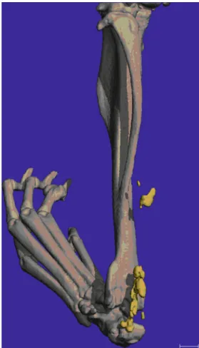
Fig. 4. 3D reconstruction of a mouse hind limb with heterotopic ossifications highlighted in yellow.
the soft tissue of the murine hind leg. If successful, future studies are planned to further analyze the involved signaling pathways to reassure that this reduction was in fact coupled with a decrease in PDGF-positive cells.
This study has its limitations. We were able to dem-onstrate the efficacy of imatinib in the prevention of HO formation in a soft tissue trauma model. The effect of PDGF suppression through imatinib on fracture healing (as it is very important with traumatic HO) has, howev-er, not been addressed and has yet to be examined. Future studies are to be conducted concerning this matter.
Imatinib may have also effectively decreased ectopic bone formation through other additional signaling pathways not addressed in this study. In addition, it is not known how imatinib affects wound healing or bony ingrowth with re-spect to arthroplasty. Here, too, further studies will need to be conducted in order to gain further insight. Lastly, it is not clear in how far these findings in a murine model are also applicable in humans.
Our fundamental hypothesis was that angiogenesis is a key factor in the development of heterotopic bone. Previous studies have proven that PDGF is one of many important
factors promoting this process, and imatinib in turn has been demonstrated to specifically inhibit PDGF expression.
An interesting finding was that although an impressing reduction in bone volume (85%, p00.028) could be ob-served in the group treated with imatinib, there was no complete prevention of the initiation of HO formation in the treatment group. A possible explanation for this could either be the relatively low dosage used in the trial or other—as mentioned above—additional signaling pathways involved. Future investigations will need to deal with potential side effects and lowest possible dosage of the drug. The actual dosage of 10 mg/kg daily p.o. approximately corresponds to the dosage used in humans to treat chronic myeloid leukemia. In mice, a tenfold dosage was initially proposed by the man-ufacturer (email communication with product manager at Novartis®, Switzerland).
The incomplete prevention of heterotopic ossification corresponds to clinical findings, where heterotopic ossifica-tion also may occur despite the use of current means of HO prophylaxis such as radiation or NSAR. A low-volume HO formation, however (Brooker grade 1 and 2), has showed to be of very little clinical significance in humans.
Morphologic and biochemical analysis of the heterotopic bone has shown an intense turnover and a high content of multiple growth factors (including PDGF), indicating a met-abolically active tissue [23] PDGF plays an important role in bone metabolism, especially in the bone healing process and reconstruction, acting as a stimulatory substance. The exact mechanism by which PDGF is related to altered bone for-mation is not yet completely understood. As stated earlier, a possible explanation could be that new vessels are necessary to bring in new stem cells into the target area where HO is about to be formed—and our hypothesis that PDGF inhibi-tion may ultimately also successfully inhibit heterotopic bone formation. Imatinib mesylate inhibits PDGF-controlled signaling pathways. This was shown in a study by Van Steensel et al. where imatinib mesylate successfully inhibited PDGF-BB-induced orbital fibroblast proliferation by blocking PDGF-receptor phosphorylation [19].
As previously stated, the effects of imatinib in a fracture healing model have yet to be investigated. Due to the com-plexity of the multiple signaling pathways involved in frac-ture healing, the exact influence and role of PDGF has not yet been definitively identified, and conflicting results have been found.
For now, promising emergent literature has already itified imatinib to be related to increased bone mineral den-sity and bone volume with long-time use though [2,4,5,10,
14,20,21]. These studies, however, deal with alterations of mature bone and may not exactly reflect the situation during callus formation and bone healing. One study [21] also mentions a positive effect of imatinib on bone density and volume in growing adolescent mice (using the same dosage as in the present one), and Ranly and Roussy demonstrated an inhibitory effect of PDGF on bone formation [16, 17]. These encouraging findings warrant further investigations involving imatinib as an agent to inhibit ectopic bone for-mation while at the same time possibly not impeding frac-ture healing. In contrast, Nash and colleagues have found a
Fig. 5. Graphic comparison between heterotopic bone volume (in cubic millimeter) in the control group and the treatment group.
stimulative effect of PDGF on the rate of fracture healing in rabbits[13].
We are aware that this new approach to limit HO forma-tion is the “beginning” of a study rather than a complete work. Nevertheless, following further research, PDGF sup-pression might represent a valuable future alternative in the prevention of heterotopic ossification.
Acknowledgments The study was supported by a grant of the Swiss Trauma Foundation.
Disclosures
Conflict of Interest: Clément ML.Werner, MD, Stefan M. Zimmermann, MD, Carola C. Würgler‐Hauri, MD, Joseph M. Lane, MD, Guido A. Wanner, MD, Hans‐Peter Simmen, MD have declared that they have no conflict of interest.
Human/Animal Rights: All institutional and national guidelines for the care and use of laboratory animals were followed.
Informed Consent N/A
Required Author Forms Disclosure forms provided by the authors are available with the online version of this article.
References
1. Alini M, Marriott A, Chen T, et al. A novel angiogenic molecule produced at the time of chondrocyte hypertrophy during endo-chondral bone formation. Dev Biol. 1996;176:124-132.
2. Berman E, Nicolaides M, Maki RG, et al. Altered bone and mineral metabolism in patients receiving imatinib mesylate. N Engl J Med. 2006;354:2006-2013.
3. Deckers M, Karperien M, van der Bent C, et al. Expression of vascular endothelial growth factors and their receptors during osteoblast differentiation. Endocrinology. 2000;141:1667-1674. 4. Dewar AL, Farrugia AN, Condina MR, et al. Imatinib as a potential
antiresorptive therapy for bone disease. Blood. 2006;107:4334-4337. 5. Fitter S, Dewar AL, Kostakis P, et al. Long-term imatinib therapy pro-motes bone formation in CML patients. Blood. 2008;111:2538-2547. 6. Flecknell PA. Laboratory Animal Anesthesia. London: Academic
Press (Elsevier); 1996.
7. Garland D. A clinical perspective on common forms of acquired heterotopic ossification. Clin Orthop Relat Res. 1991;13–29; 8. Goodman S, Ma T, Genovese M, et al. COX-2 selective inhibitors
and bone. Int J Immunopathol Pharmacol. 2003;16:201-205. 9. Goodman S, Ma T, Trindade M, et al. COX-2 selective NSAID
decreases bone ingrowth in vivo. J Orthop Res. 2002;20:1164-1169. 10. Jönsson S, Olsson B, Ohlsson C, et al. Increased cortical bone mineralization in imatinib treated patients with chronic myeloge-nous leukemia. Haematologica. 2008;93:1101-1103.
11. McClure J. The effect of diphosphonates on heterotopic ossifica-tion in regenerating Achilles tendon of the mouse. J Pathol. 1983;139:419-430.
12. Naraghi F, DeCoster T, Moneim M, et al. Heterotopic ossification. Orthopedics. 1996;19:145-151.
13. Nash TJ, Howlett CR, Martin C, et al. Effect of platelet-derived growth factor on tibial osteotomies in rabbits. Bone. 1994;15:203-208. 14. O'Sullivan S, Horne A, Wattie D, et al. Decreased bone turnover
despite persistent secondary hyperparathyroidism during prolonged treatment with imatinib. J Clin Endocrinol Metab. 2009;94:1131-1136.
15. Puzas JE, Miller MD, Rosier RN. Pathologic bone formation. Clin Orthop. 1989;245:269-281.
16. Ranly DM, McMillan J, Keller T, et al. Platelet-derived growth factor inhibits demineralized bone matrix-induced intramuscular cartilage and bone formation. A study of immunocompromised mice. J Bone Joint Surg Am. 2005;87:2052-2064.
17. Roussy Y, Bertrand Duchesne MP, Gagnon G. Activation of hu-man platelet-rich plasmas: effect on growth factors release, cell division and in vivo bone formation. Clin Oral Implants Res. 2007;18:639-648.
18. Saudan M, Saudan P, Perneger T, et al. Celecoxib versus ibuprofen in the prevention of heterotopic ossification following total hip replacement: a prospective randomised trial. J Bone Joint Surg Br. 2007;89:155-159.
19. van Steensel L, Paridaens D, Schrijver B, et al. Imatinib mesylate and AMN107 inhibit PDGF-signaling in orbital fibroblasts: a potential treatment for Graves' ophthalmopathy. Invest Ophthalmol Vis Sci. 2009;50:3091-3098.
20. Vandyke K, Dewar AL, Diamond P, et al. The tyrosine kinase inhibitor dasatinib dysregulates bone remodeling through inhibi-tion of osteoclasts in vivo. J Bone Miner Res. 2010;25:1759-1770. 21. Vandyke K, Fitter S, Dewar AL, et al. Dysregulation of bone
remodeling by imatinib mesylate. Blood. 2010;115:766-774. 22. Vuorinen K, Gao F, Oury TD, et al. Imatinib mesylate inhibits
fibrogenesis in asbestos-induced interstitial pneumonia. Exp Lung Res. 2007;33:357-373.
23. Yin M, Gentili C, Koyama E, et al. Antiangiogenic treatment delays chondrocyte maturation and bone formation during limb skeletogenesis. J Bone Miner Res. 2002;17:56-65.