Appl Microbiol Biotechnol (2005) 67: 234–239 DOI 10.1007/s00253-004-1804-2
A P P L I E D G E N E T I C S A N D M O L E C U L A R B I O T E C H N O L O G Y
Martin Braun . Inés García Rubio . Linda Thöny-Meyer
A heme tag for in vivo synthesis of artificial cytochromes
Received: 19 August 2004 / Revised: 27 September 2004 / Accepted: 19 October 2004 / Published online: 10 December 2004 # Springer-Verlag 2004
Abstract A genetic approach is described here that enables the specific covalent attachment of heme via a short C-terminal peptide tag to an otherwise non-heme-binding pro-tein. Covalent attachment of heme to the apo-protein is catalysed by the cytochrome c maturation system of Escher-ichia coli. While its original enzymatic activity is retained, the resulting heme-tagged protein is red, has peroxidase activity and is redox active. The presence or absence of a C-terminal histidine tag results in low-spin heme iron with six- or high-spin heme iron with five coordinate ligands, respectively. The heme tag can be used as a tool for the rational design of artificial c-type cytochromes and metalloenzymes, thereby overcoming previous limitations set by chemical approaches. Moreover, the tag allows direct visualisation of the red fusion protein during purification.
Introduction
The design of molecules that mimic the catalytic proper-ties of naturally occurring enzymes is challenging, and the ability to design and prepare them would be useful for a variety of applications in chemistry and biology. Construc-tion of artificial metalloenzymes, in particular ones with covalently attached metal cofactors, is desirable, because metalloenzymes catalyse chemical transformations with high selectivity and reactivity under mild conditions. Sev-eral reports on the semi-synthesis of rationally designed metalloenzymes exist. By and large, they rely on
knowl-edge of the three-dimensional structure of the template enzyme (Lu et al.2001; Qi et al.2001; Ohashi et al.2003). Heme is an iron-containing cofactor that catalyses a vari-ety of reactions, depending on its surrounding matrix (Frankenberg et al. 2003). In the cytochromes, the heme iron acts as a simple redox centre. In the globins, heme reversibly binds gases such as O2, CO or NO. In cytochrome
P450, O2is activated and cleaved to form water and an O
atom, which is then inserted into a substrate. Such activities are of interest for the design of enzymes with new catalytic properties. Various attempts have been made to construct artificial heme enzymes (Watanabe2002; Frankenberg et al.
2003). Here we describe a genetic approach which results in cross-linking heme to a short fusion tag in a protein of choice. The heme tag is used as a cassette for covalent heme attachment to the C-terminus of an otherwise non-metal-binding protein. We show that the resulting heme-protein fusion is red, has intrinsic peroxidase activity and is redox active.
Development of the heme tag is based on our knowl-edge of maturation of c-type cytochromes in bacteria. These proteins carry one or more heme molecules bound covalently to the amino acid sequence motif CXXCH. The two cysteines are added to the heme vinyl groups, re-sulting in two covalent thioether bonds per bound heme. The histidine residue serves as an axial ligand of the heme iron. Bacterial c-type cytochromes are synthesised as pre-cursor polypeptides with an N-terminal signal sequence for export of the polypeptide to the periplasm by the general protein secretion system (Sec) (Thöny-Meyer and Künzler
1997). Maturation of apo-cytochrome c (non-heme-bound) to holo-cytochrome c (heme-bound) takes place on the peri-plasmic side of the cytoperi-plasmic membrane. In Escherichia coli eight cytochrome c maturation (Ccm) proteins CcmA– CcmH are required specifically for this process. The cor-responding genes ccmA–ccmH are expressed only under anaerobic growth conditions (Grove et al. 1996; Thöny-Meyer et al.1996) when c-type cytochromes are used for respiratory electron transport. Previously, we constructed plasmid pEC86 (Arslan et al.1998), which expresses the ccmA–ccmH operon constitutively from a vector promoter M. Braun (*) . L. Thöny-Meyer
Institut für Mikrobiologie, ETH Hönggerberg, Wolfgang-Pauli-Str. 10, 8093 Zürich, Switzerland e-mail: [email protected] Tel.: +41-1-6323551 Fax: +41-1-6321148 I. G. Rubio
Laboratorium für Physikalische Chemie, Eidgenössische Technische Hochschule, Wolfgang-Pauli-Str. 10,
even under aerobic conditions. The recently characterised minimal sequence requirements for heme insertion into a cytochrome polypeptide (Braun and Thöny-Meyer 2004) allowed the development of a short heme tag. The coding sequence of this tag was fused in frame to the malE gene encoding the maltose-binding protein (MBP). The recom-binant fusion protein was tested for heme binding in the presence or absence of the ccm-plasmid pEC86.
Materials and methods
Strains, plasmids and growth conditions
E. coli strain DH5α (Hanahan1983) was used for cloning. MBP fusions were expressed in E. coli EC06 (ΔccmA-H) (Thöny-Meyer et al.1995) harbouring pEC86 (Arslan et al.
1998). Plasmid pMal-p (New England BioLabs) was used for the construction of MBP fusions under the control of the Ptacpromoter, which is repressed by the Lac repressor.
Final antibiotic concentrations were 200 μg ml−1for am-picillin and 10μg ml−1for chloramphenicol. For expression of proteins 0.9 l of Luria–Bertani broth were inoculated with 9 ml of an overnight culture of the appropriate strain at 30°C. Cultures were grown to an optical density of A600=0.8
and induced with 0.1 mM IPTG. Cells were harvested 17 h after induction by centrifugation at 3300 g.
Plasmid construction
Plasmids encoding MBP-heme tag fusions (Table 1) are derived from pMal-p. The ScaI/Eco52I DNA fragments encoding the microperoxidases of pMP126 and pMP259 (Braun and Thöny-Meyer2004) were cloned into the cor-responding sites of pMal-p, resulting in the out-of-frame pMP263 and pMP268 heme tag fusions. To obtain in-frame fusions, pMP263 and pMP268 were cut with SacI, blunt-ended with T4 DNA-polymerase (Roche Diagnostics) and religated, resulting in plasmids pMP292 and pMP295, res-pectively. This led to additional codons of the amino acid sequence NSRYPAA just before the heme-binding motif CLACH. The DNA encoding the hexa-histidine (His6) tag
of MP292 was removed by PCR amplification of the gene fusion, using the primer pair malE (5′-GGTCGTCAGACT GTCGATGAAGCC-3′) that anneals within the malE gene and Mini-cycA4 (5′-TAAGAATTCAGCCGATCGCGTG
GCAGGCCAGG-3′) encoding the C-terminal end of MP292 without a His6tag, and pMP292 as the template. The
PCR-product was digested with EcoRI and KpnI and cloned into the corresponding site of pMP292, resulting in pMP301. All constructs were confirmed by DNA sequencing with primer malE (Microsynth, Balgach, Switzerland).
Protein extraction and purification
Periplasmic proteins were extracted by resuspending the cell pellets derived from 0.9-l cultures in 5 ml 1 mg ml−1 polymyxin B sulphate (Sigma), 500 mM sucrose and 100 mM Tris-HCl, pH 8. The suspension was stirred for 60 min at 4°C and centrifuged at 20,000 g for 20 min. The supernatant contained the periplasmic proteins. His6-tagged
proteins were purified by Ni-NTA affinity chromatography (Qiagen). MBP (MalE) fusions were purified from peri-plasmic protein extracts by affinity chromato-graphy on amylose resin (New England BioLabs). Beads were washed twice with 10 bed volumes (BV) of 50 mM Tris-HCl, pH 8, and proteins were eluted with 2 BV of the same buffer containing 10 mM maltose and used for further character-isation. Alternatively, red protein was purified from 2 ml of periplasmic extract over a 1 ml Mono-Q column HR 5/5 on an Aekta (Amersham Biosciences, Freiburg, Germany) puri-fier. A 20-ml gradient from 0 M to 0.8 M NaCl in 20 mM Tris, pH 8 was run and 1-ml fractions were collected.
Analytical methods
For in-gel staining of proteins with covalently bound heme, samples were separated by SDS-PAGE as described by Laemmli (Laemmli1970). Polyacrylamide gels were fixed in 10% TCA for 10 min and then washed twice with water for 10 min. Heme-bound proteins were detected using their intrinsic peroxidase activity by immersion of the gels in a solution of 10 mg o-dianisidine (one tablet, Sigma) in 10 ml 0.05 M Na-citrate, pH 4.4, and 0.7% H2O2. Molecular
masses of purified peptides were determined by mass spec-troscopy on an Applied Biosystems Voyager-DE Elite using 2,5-sinapinic acid as matrix. Optical spectra were recorded on a Hitachi model U-3300 spectrophotometer and export-ed to a Microsoft Excel table for calculations of difference spectra and determination of absorption maxima. Con-centrations of purified proteins were determined with the
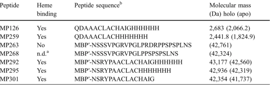
Table 1 Heme-tagged peptides and their properties
a n.d. Not determined b MBP Maltose-binding protein Peptide Heme binding
Peptide sequenceb Molecular mass
(Da) holo (apo)
MP126 Yes QDAAACLACHAIGHHHHHH 2,683 (2,066.2) MP259 Yes QDAAACLACHHHHHHH 2,441.8 (1,824.9) MP263 No MBP′-NSSSVPGRVPGLPRDRPPSPSPLNS (42,761) MP268 n.d.a MBP′-NSSSVPGRVPGLPPSPSPSLNS (42,324) MP292 Yes MBP′-NSRYPAACLACHAIGHHHHHH 43,177 (42,560) MP295 Yes MBP′-NSRYPAACLACHHHHHHH 42,936 (42,319) MP301 Yes MBP′-NSRYPAACLACHAIG 42,354 (41,737)
Bradford assay. Molar extinction coefficients at 278 nm and molecular weights were calculated using programs on the ExPASy Molecular Biology Server at http://www. expasy.ch.
For EPR measurements, a Bruker E500 spectrometer was used working in X-band (microwave frequency 9.5 GHz). The spectrometer was equipped with a Bruker ER 4122 super-high Q cavity and an Oxford continuous helium flow cryostat. The spectra were taken at 15 K. A micro-wave power of 0.06 mw and a modulation amplitude of 0.5 mT were used. Samples prepared for EPR spectroscopy were purified by amylose affinity chromatography. They were concentrated and prepared in a 50 mM Tris buffer, pH 8.5, containing 25% glycerol in order to obtain a good glass upon freezing the sample. The final concentration of the samples was 122 μM for holo-MP292 and 36 μM for holo-MP301.
Results
The MBP–AACLACHHHHHH fusion binds heme in the presence of the Ccm factors
The finding that the peptide QDAAACLACHHHHHH (MP251) can bind heme in the presence of the Ccm system (Braun and Thöny-Meyer 2004) raised the question of whether covalent heme attachment can also occur in larger proteins that normally do not contain this cofactor. We thus fused the majority of the coding sequences of MP126 and MP259 (Table1) to the periplasmic MBP, resulting in MP292 and MP295, respectively. Both proteins have a proline residue preceding the AACLACH motif that might provide a kink in the protein structure and facilitate maturation. These tagged polypeptides were expressed in E. coli EC06 (ΔccmA–ccmH) in the presence and absence of pEC86 (ccmA–ccmH). Periplasmic extracts were ana-lysed spectroscopically for the presence of c-type cyto-chromes. None of the proteins bound heme in the absence of pEC86. In the presence of pEC86, periplasmic extracts of both MP292 and MP295, but not of MP263 (out-of-frame fusion) were red. His6-tagged proteins expressed in
the presence of pEC86 were purified from periplasmic extracts via Ni-NTA affinity chromatography and resulted in red fractions with spectroscopic characteristics of c-type cytochromes. Alternatively, the proteins were purified by affinity chromatography over amylose resin, resulting in red fractions for both MP292 and MP295. This purifica-tion indicates the native folding of the MBP derivatives that retain the sugar-binding characteristics of the protein. Proteins were further analysed for covalent heme binding by heme staining after SDS-PAGE. In contrast to MP263, both MP292 and MP295 had heme-dependent peroxidase activity. For both, an additional heme-staining band with about a twofold molecular weight was found (Fig.1), as it is often seen for c-type cytochromes. The molecular weight of MP292 was further determined by mass spectroscopy. The main peak detected was 42558.6 m/z, which corre-sponds very well with the calculated mass of apo-MP292
(42560 Da). A peak corresponding to the expected mass of holo-MP292 (43177 Da) was also detected.
The His6tag is not required for heme binding
Our previous work on heme peptides had shown that a His6
tag is important for heme insertion (Braun and Thöny-Meyer2004). Hence, we wondered whether the His6tag is
also required for heme attachment in the MBP fusion. We thus constructed plasmid pMP301, which encodes MP292 lacking the His6tag (Table1). MP301 was expressed in the
presence of the Ccm system, purified by amylose affinity chromatography and analysed for heme binding by heme stain. The results were identical to those obtained for MP292 and MP295 (Fig.2d, f). We conclude that the His6
tag is not required for heme binding.
Heme-tagged maltose-binding protein has spectral characteristics of a c-type cytochrome
Cytochromes of the c-type such as horse heart cytochrome c have typicalα bands at 550 nm in the reduced state. To analyse the spectral characteristics of the fusion proteins, ascorbate (5-mM and 10-mM final concentrations) and di-thionite (5-mM final concentration) were used as reducing agents and added to samples of amylose-purified MP292, MP295 and MP301. As previously described for micro-peroxidases MP126 and MP259 (Braun and Thöny-Meyer
2004), none of the fusion proteins was reduced with the weak reducing agent ascorbate (E0¼ þ58 mV). Dithio-nite-reduced minus air-oxidised difference spectra of both MP292 and MP295 showed maxima in the range of 417– 418 nm for the Soret band, 522–523 nm for the β band and 550 nm for the α band, which are typical for c-type cytochromes (Fig.3). While the Soret peak of MP301 also shifted from 408 nm to 415 nm when dithionite was added, Fig. 1 Heme binding of MP292 and MP295. Samples were sep-arated by SDS-15% PAGE and stained for the presence of proteins with Coomassie (a) or for covalently bound heme by peroxidase activity stain (heme stain) (b). Each lane was loaded with 5 μg protein
its amplitude diminished, so that the difference spectrum mainly reflects the absorption of the oxidised form (Fig.3) with a minimum at 406 nm. Furthermore, no peaks were detected at the position of theβ or α band (523 nm or 550 nm, respectively). Heme ligands have previously been shown to influence the spectral behaviour in the 550-nm range (Arcovito et al.2001; Lee et al.2001). Upon addition of 300 mM imidazole to MP301, the dithionite-reduced
spectrum resembled that of the other heme His6-tagged
fusion proteins (Fig. 3), suggesting that imidazole can function as sixth ligand in the MBP-heme tag protein lacking the His6tag. In summary, the oxidised and reduced
states of all three fusion proteins have spectral character-istics typical for c-type cytochromes.
The His6tag serves as sixth ligand
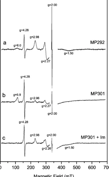
As the presence of imidazole significantly changed the optical spectra of MP301, we decided to characterise the heme ligands in more detail. EPR spectra of the two heme-binding fusion proteins, with and without exogenous imid-azole, were recorded. MP292 (Fig.4a) displays a characteristic low-spin ferric heme spectrum (spin, 1/2). The three fea-tures associated with the principal values of the anisotropic g-tensor are resolved (gz=2.98, gy=2.27, gx=1.50). These
values correspond typically to a bis-imidazole-coordinated heme centre, with approximately parallel axial ligands (Palmer
1983; Walker1999). In addition to the heme iron signal, two other lines were observed in the spectrum. The one at an effective g value of 4.28 is attributed to a non-heme ferric iron impurity in a highly rhombic environment (Pilbrow 1990). The region of the spectrum around g∼2 was omitted because of a strong signal from the cavity. Nevertheless, a signal at exactly g=2.00 was detected and assigned to a radical species. In spite of their high intensity in the spectra, the signals at g=4.28 and 2.00 are minority centres.
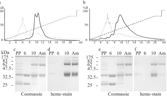
The EPR spectrum of MP301 (Fig. 4b) shows two different heme iron signals corresponding to two different species. The majority species has a signal at g ∼6 that corresponds to a typical high-spin ferric-heme iron (spin, Fig. 2 Purification of MP292 and MP301 from periplasmic extracts.
Periplasmic extracts of MP292 (a) and MP301 (b) were separated by anion exchange chromatography and absorption was monitored at 280 nm (..) and 410 nm (__). The gradient was from 0M to 0.8 M NaCl indicated as %B (- - -). Fractions of MP292 (c, d) and of MP301 (e, f) eluting after 6 ml (6) and 10 ml (10) were compared
with the periplasmic extracts (PP) and amylose affinity purified protein (Am) by SDS-12% PAGE. Gels were stained for peroxidase activity, showing the presence of covalently bound heme (d, f) and for the presence of proteins with Coomassie (c, e). Each lane was loaded with 5μg protein
Fig. 3 Spectral analysis of purified maltose-binding protein–heme tag fusions. Dithionite-reduced minus air-oxidised optical difference spectra are shown. +Im indicates the presence of 300 mM imidazole. The inset shows theα and β bands at higher magnification
5/2). This means that the axial coordination of the heme iron is weaker than in MP292, i.e. the iron is either five-coordinated or six-five-coordinated with a weak sixth ligand such as water (Palmer1983). Since the difference between the two proteins is the absence of the His6tag in MP301,
we conclude that the sixth ligand of iron in MP292 is probably a side chain from the His6tag.
The second species in Fig.4b also seems to correspond to a low-spin bis-histidine coordinated heme (gz=2.96,
gy=2.27, gx not resolved). Here, the sixth ligand could
either be of intra- or inter-molecular origin. If the latter were the case this signal would be related to dimers.
Figure4c displays the spectrum of sample MP301 after the addition of imidazole to the protein solution. The high-spin species is missing and has been transformed into a low-spin species with g values (gz=2.98, gy=2.26, gx=
1.50). The exogenous imidazole is probably coordinating the heme iron as the sixth ligand.
As can be seen in Fig.4a, there is also a small amount of high-spin signal in the spectrum of MP292 (small hump at g ∼ 6). This high-spin signal was completely removed upon addition of imidazole to the sample.
Enrichment of the heme-tagged protein by MonoQ ion exchange chromatography
One goal of this work was to create an optically detectable tag that facilitates protein purification. Periplasmic extracts containing heme-tagged proteins were separated over a MonoQ column, and absorption of fractions was mon-itored at 410 nm in addition to 280 nm. The absorption at 410 nm corresponds to heme absorption in the Soret region. While the main absorption at 280 nm for both ana-lysed fusion proteins MP292 and MP301 was in fraction no. 6, the absorption at 410 nm was maximal in fraction no. 10 (Fig. 2a, b). Optical spectra of these fractions were recorded to determine the ratio of A(410)/A(278). For MP292, fraction no. 6 had a ratio of 0.016 and fraction no. 10 of 0.228. This corresponds to a 3.7-fold enrichment of holo-protein from the initial periplasmic extracts (ratio = 0.061). In MP301 the enrichment of holo-protein in fraction no. 10 was 3.27-fold and the final ratio 0.17. For both MP292 and MP301 the ratios in fraction number 10 corresponded well with the ratios obtained after purification via amylose affinity chromatography (A(408)/A(278)=0.39 for MP292 and 0.16 for MP301). Under the same buffer conditions horse heart cytochrome c showed a ratio of 3.9. The calculated extinction coefficient at 280 nm for MP292 is 4.3-fold higher than that of cytochrome c. Taking these factors into consideration, purified MP292 contained approximately 40% and purified MP301 about 16% holo-protein. While fraction 10 still contains minor amounts of other contaminating proteins (Fig.2c, e), amylose affinity-purified protein was pure. Additionally, Western blot analysis with anti-MBP antibodies confirmed that the two major bands visible in fraction number 10 of MonoQ-purified samples indeed are MBP-fusions (data not shown).
Discussion
A C-terminal AACLACHAIG tag is recognised by the Ccm system for covalent attachment of heme to the MBP fusion, which leads to an artificial c-type cytochrome with per-oxidase and redox activities. Besides introducing these new characteristics, the initial feature of the MBP, i.e. the binding of amylose, is maintained.
The red colour of the fusion protein helps to fractionate the proteins during the initial steps of purification. In addition, protein-bound heme allows easy and sensitive detection by peroxidase activity. Transfer of MP292 to nitrocellulose membranes and subsequent detection by enhanced chemiluminescence (Feissner et al. 2003) re-sulted in detection of≥0.4 pmol of holo-protein (data not shown). This method is normally used as the last step in immunodetection protocols when antibodies linked to horse-Fig. 4 EPR spectroscopy. EPR spectra of MP292 (a), MP301 (b)
and MP301 after the addition of an excess of imidazole (c) are shown. The region around g ∼2 was omitted due to a strong baseline. All spectra were taken at 15 K
radish peroxidase are used. The covalent modification of a peptide with heme thus allows a one-step peroxidase detection in which antibodies are not needed. Recently a haemoglobin tag based on the bacterial haemoglobin of Vitreoscilla stercoraria has been developed (Park et al.
2003). This tag consists of 146 amino acids and binds heme non-covalently. By contrast, covalent binding as found in our heme tag enables detection of the tagged protein even under denaturing conditions during purification or after SDS-PAGE. The shortness of the tag also allows cloning by PCR, using primers that contain the coding sequence of the heme tag. In contrast to the haemoglobin tag, our system requires periplasmic targeting for heme attachment. This can be advantageous, as proteins over-produced in the bacterial periplasm have already a high degree of purity after the first steps of cell fractionation.
We have produced an artificial c-type cytochrome by the fusion of a heme tag to a periplasmic, non-cytochrome polypeptide that was produced in cells expressing the cytochrome c maturation system Ccm. The target protein obtained a new function, while the initial, maltose-binding characteristic was retained. The heme iron of many cyto-chromes in nature is either five- or six-coordinated, whereas heme-based sensors and enzymes are mostly five-coordi-nated with an open site to allow binding of small molecules such as O2 or CO (Lu et al. 2001). We found that in the
presence of a His6tag, the heme iron of the tagged fusion
protein was six-coordinated, but in the absence of the His6
tag it was five-coordinated. Previously, we could only speculate on the role of the His6tag in maturation of heme
peptides (Braun and Thöny-Meyer 2004). We now show that the His6 tag is not a general prerequisite for heme
incorporation when linked to CXXCH-tagged maltose-binding protein. However, when the tag is present it acts as sixth ligand to the heme iron.
In summary, covalent attachment of heme to a heme tag (with or without an additional His6 tag) fused to the
C-terminus of any periplasmically expressed protein can be a valuable tool for protein purification. The resulting red colour and the peroxidase activity are two characteristics worth considering when choosing an appropriate purifi-cation scheme for any protein of interest (Terpe 2003). Analogous to other chemically linked microperoxidase conjugates (Padeste et al. 2003), our tool also has the potential for a broad application, for example as biosensor or specific biocatalyst.
Acknowledgements We thank Olaf Christensen and Umesh Ahuja for experimental advice and helpful discussions and Arthur Schweiger for generous support. This work was supported by the Swiss National Foundation for Scientific Research.
References
Arcovito A, Gianni S, Brunori M, Travaglini-Allocatelli C, Bellelli A (2001) Fast coordination changes in cytochrome c do not necessarily imply folding. J Biol Chem 276:41073–41078
Arslan E, Schulz H, Zufferey R, Künzler P, Thöny-Meyer L (1998) Overproduction of the Bradyrhizobium japonicum c-type cytochrome subunits of the ccb3oxidase in Escherichia coli. Biochem Biophys Res Commun 251:744–747
Braun M, Thöny-Meyer L (2004) Biosynthesis of artificial micro-peroxidases by exploiting the secretion and cytochrome c mat-uration apparatuses of Escherichia coli. Proc Natl Acad Sci USA 101:12830–12835
Feissner R, Xiang Y, Kranz RG (2003) Chemiluminescent-based methods to detect subpicomole levels of c-type cytochromes. Anal Biochem 315:90–94
Frankenberg N, Moser J, Jahn D (2003) Bacterial heme biosynthesis and its biotechnological application. Appl Microbiol Biotech-nol 63:115–127
Grove J, Tanapongpipat S, Thomas G, Griffiths L, Crooke H, Cole J (1996) Escherichia coli K-12 genes essential for the synthesis of c-type cytochromes and a third nitrate reductase located in the periplasm. Mol Microbiol 19:467–481
Hanahan D (1983) Studies on transformation of Escherichia coli with plasmids. J Mol Biol 166:557–580
Laemmli UK (1970) Cleavage of structural proteins during the assembly of the head of bacteriophage T4. Nature 227:680–685 Lee JC, Gray HB, Winkler JR (2001) Cytochrome c’ folding triggered by electron transfer: fast and slow formation of four-helix bundles. Proc Natl Acad Sci USA 98:7760–7764 Lu Y, Berry SM, Pfister TD (2001) Engineering novel
metallopro-teins: design of metal-binding sites into native protein scaffolds. Chem Rev 101:3047–3080
Ohashi M, Koshiyama T, Ueno T, Yanase M, Fujii H, Watanabe Y (2003) Preparation of artificial metalloenzymes by insertion of chromium(III) Schiff base complexes into apomyoglobin mutants. Angew Chem Int Ed 42:1005–1008
Padeste C, Steiger B, Grubelnik A, Tiefenauer L (2003) Redox labelled avidin for enzyme sensor architectures. Biosens Bioelectron 19:239–247
Palmer G (1983) Electron paramagnetic resonance of hemeproteins. Iron porphyrins part II. Addison-Wesley, London
Park KW, Webster DA, Stark BC, Howard AJ, Kim KJ (2003) Fusion protein system designed to provide color to aid in the expression and purification of proteins in Escherichia coli. Plasmid 50:169–175
Pilbrow J (1990) Transition ion electron paramagnetic resonance. Clarendon, Oxford
Qi D, Tann C-M, Haring D, Distefano MD (2001) Generation of new enzymes via covalent modification of existing proteins. Chem Rev 101:3081–3111
Terpe K (2003) Overview of tag protein fusions: From molecular and biochemical fundamentals to commercial systems. Appl Microbiol Biotechnol 60:523–533
Thöny-Meyer L, Fischer F, Künzler P, Ritz D, Hennecke H (1995) Escherichia coli genes required for cytochrome c maturation. J Bacteriol 177:4321–4326
Thöny-Meyer L, Künzler P (1997) Translocation to the periplasm and signal sequence cleavage of preapocytochrome c depend on sec and lep, but not on the ccm gene products. Eur J Biochem 246:794–799
Thöny-Meyer L, Künzler P, Hennecke H (1996) Requirements for maturation of Bradyrhizobium japonicum cytochrome c550 in Escherichia coli. Eur J Biochem 235:754–761
Walker F (1999) Magnetic spectroscopic (EPR, ESEEM, Mössbauer, MCD and NMR) studies of low-spin ferriheme centers and their corresponding heme proteins. Coord Chem Rev 185–186:471– 534
Watanabe Y (2002) Construction of heme enzymes: Four approaches. Curr Opin Chem Biol 6:208–216