Differential regulation of TGF-b – induced, ALK-5– mediated VEGF
release by SMAD2/3 versus SMAD1/5/8 signaling in glioblastoma
Katharina Seystahl, Isabel Tritschler, Emese Szabo, Ghazaleh Tabatabai, and Michael Weller
Department of Neurology, University Hospital Zurich, Zurich, Switzerland
Corresponding Author: Katharina Seystahl, MD, Department of Neurology, University Hospital Zurich, Zurich Switzerland (katharina.seystahl@usz.ch).
Background. The transforming growth factor (TGF)-b and vascular endothelial growth factor (VEGF) pathways have a major role in the pathogenesis of glioblastoma, notably immunosuppression, migration, and angiogenesis, but their interactions have re-mained poorly understood.
Methods. We characterized TGF-b pathway activity in 9 long-term glioma cell lines (LTCs) and 4 glioma-initiating cell lines (GICs) in relation to constitutive and exogenous TGF-b-induced VEGF release. Results were validated using The Cancer Genome Atlas tran-scriptomics data.
Results. Glioma cells exhibit heterogeneous patterns of constitutive TGF-b pathway activation reflected by phosphorylation not only of SMAD2 and SMAD3 but also of SMAD1/5/8. Constitutive TGF-b pathway activity depends on the type I TGF-b receptor, ALK-5, and accounts for up to 69% of constitutive VEGF release, which is positively regulated by SMAD2/3 and negatively regulated by SMAD1/5/8 signaling in a cell line-specific manner. Exogenous TGF-b induces VEGF release in most cell lines in a SMAD- and ALK-5 – dependent manner. There is no correlation between the fold induction of VEGF secretion induced by TGF-b compared with hyp-oxia. The role of SMAD5 signaling is highly context and cell-line dependent with a VEGF inhibitory effect at low TGF-b and pSMAD2 levels and a stimulatory effect when TGF-b is abundant.
Conclusions. TGF-b regulates VEGF release by glioma cells in an ALK-5–dependent manner involving SMAD2, SMAD3, and SMAD1/ 5/8 signaling. This crosstalk between the TGF-b and VEGF pathways may open up new avenues of biomarker-driven exploratory clinical trials focusing on the microenvironment in glioblastoma.
Keywords: angiogenesis, glioblastoma, TGF-b, VEGF.
Transforming growth factor (TGF)-b, a cytokine with pleiotropic functions, plays a pivotal role in cancer biology and represents one of the key pathogenic factors in glioblastoma. Ligand bind-ing leads to the phosphorylation of SMAD family proteins that are involved in the regulation of gene transcription. Activation of the TGF-b/SMAD pathway correlates with poor prognosis in glioma patients.1Various anti –TGF-b strategies have been ex-plored in rodent glioma models2–5and clinical trials.6,7
TGF-b signaling is mediated via a heterodimeric receptor complex comprising type I and type II receptors.8Canonical
TGF-b signaling involves ligand binding to TGF-b receptor II (TGF-bRII), which associates with the type I receptor activin receptor-like kinase-5 (ALK-5) resulting in the phosphorylation of SMAD2 and SMAD3.9Pharmacological ALK-5 inhibition is an effective treatment strategy in rodent glioma models.4,5,10
The involvement of another type I receptor in TGF-b signaling,
the activin receptor-like kinase 1 (ALK-1), leading to the phos-phorylation of SMAD1/5/8, has been characterized in endothe-lial cells.11This alternative pathway may counteract the ALK-5/ pSMAD2 pathway and thereby balance TGF-b signaling.
A role for TGF-b in modulating vascular endothelial growth factor (VEGF) release has been proposed in several cell types. Both negative and positive regulation of VEGF by TGF-b has been described in endothelial cells.12,13In glioma cells, the ef-fect of TGF-b on the regulation of VEGF release also remains un-certain. For selected glioma cell lines, a time-dependent increase of VEGF release induced by exogenous TGF-b has been reported.14 Silencing of TGF-b1/2 or exposure to the
ALK-5 inhibitor, SD-208, led to reduced VEGF levels in the super-natant of LN-308 and LNT-229 glioma cells, indicating a role for TGF-b1/2in the regulation of constitutive VEGF release in vitro.15
However, pharmacological inhibition of ALK-5 by SB431542
Received 1 May 2014; accepted 27 July 2014
#The Author(s) 2014. Published by Oxford University Press on behalf of the Society for Neuro-Oncology. All rights reserved. For permissions, please e-mail: journals.permissions@oup.com.
Neuro-Oncology
Neuro-Oncology 17(2), 254–265, 2015 doi:10.1093/neuonc/nou218
Advance Access date 27 August 2014
inhibited the TGF-b – evoked VEGF release but had only minor effects on constitutive VEGF release in D270MG or D423MG cells.16These observations raise the possibility that VEGF is
not only regulated via pSMAD2/3 signaling but also involves other pathways in glioma cells.
Materials and Methods
Cell Culture
Nine long-term malignant glioma cell lines (LTCs),17 4
glioma-initiating cell lines (GICs),18,19and hCMEC/D320 (Supple-mentary material) were incubated under normoxia or in a hyp-oxia incubator (1% O2, 5% CO2, 378C).
Reagents
Recombinant TGF-b2(R&D Systems) was used for all
stimula-tion studies. SD-208 (Scios Inc.) inhibits ALK-5 at 0.048 mM in cell-free systems.4
Immunoblot Analyses
Whole cell lysates were subjected to SDS-PAGE under reducing conditions loading equal amounts of proteins (Supplementary material). Short interference RNA (siRNA)-mediated knockdown was applied to identify the specific band. For quantitative cor-relation analyses of baseline expression of total and phosphor-ylated SMAD proteins, band intensity was analyzed via densitometry using ImageJ software (open source). To com-pare the relative induction of phosphorylation after stimulation, we scored the responses into no (,20%), low (20%–50%), me-dium (.50%) or high induction of phosphorylation (.100% or no baseline phosphorylation).
Real-time Polymerase Chain Reaction
Gene expression was determined via real-time polymerase chain reaction (RT-PCR; see Supplementary material for proce-dure and primer sequences) using glycerinaldehyde-3-phosphate dehydrogenase (GAPDH) as a housekeeping gene with the DCTT-method for relative quantification.
RNA Interference
To silence gene expression, cells were transiently transfected using Metafectene Pro (Biontex) for LTCs and electroporation for GICs (Neon Transfection System, Invitrogen) and siRNA pools (80 – 120 nM final concentration), containing 4 selected siRNA duplexes, each with a modification pattern addressing off-target effects caused by both strands (ON-TARGETplus, SMARTpool, ON-TARGETplus Non-targeting siRNA Pool as a neg-ative control; Dharmacon).
Flow Cytometry
Signal intensity of flow cytometry analysis (Supplementary ma-terial) was calculated as the ratio of mean fluorescence of spe-cific versus isotype control antibody (spespe-cific fluorescence index).
Enzyme-linked Immunosorbent Assay
Supernatants (preparation as outlined in the Supplementary material) were analyzed by ELISA for VEGF levels (eBioscience) and TGF-b1/2(R&D Systems).
Luciferase Reporter Assay
The pGL3 SBE-4-Luc plasmid (B. Vogelstein) containing 4 copies of the SMAD-binding element (SBE) GTCTAGAC21was used for reporter gene assays (Supplementary material).
Results
TGF-b Pathway Activity in Human Glioma Cells
We first analyzed the expression levels of TGF-b receptors, their ligands, constitutive levels of total SMAD2-5, phosphorylated SMAD2, SMAD3 and SMAD1/5/8, and the TGF-b target gene plas-minogen activator inhibitor (PAI)-1. Correlation analyses of these parameters were performed either for all cell lines pooled or sep-arately for LTCs (Table1). A separate analysis for the GICs was omitted because of small sample size. We validated our results using Affymetrix gene expression data from the The Cancer Ge-nome Atlas (TCGA) (Supplementary material, Table S1).
TGF-bRII, ALK-5, and ALK-1 messenger RNA (mRNA) were differentially expressed with a trend towards lower mRNA ex-pression for all receptors in GICs. ALK-1 mRNA levels were lower than ALK-5 levels and more than 100 times lower in gli-oma than in endothelial cells (Fig.1A – C, left). The highest TGF-bRII protein levels were found for T98G cells, while 2 of 4 GICs (T-269, S-24) had TGF-bRII levels at the detection limit. The specific fluorescence index varied strongly for ALK-5 from 84 in LN-18 cells to 1.7 in LN-308 cells. ALK-1 protein was not detected at the cell surface in glioma cells using hCMEC as a positive control (Fig.1A – C, right). Overall, mRNA levels were not predictive of protein levels. TGF-b1and TGF-b2were
hetero-geneously expressed on mRNA and protein level (Fig.1D and E). TGF-b1mRNA correlated with secreted TGF-b1but not with
TGF-b2 mRNA or protein. TGF-b2 mRNA correlated with
TGF-bRII mRNA in our dataset as well as TCGA database (Table1; Supplementary material, Table S1).
pSMAD2 levels were highest in LN-428, LN-319, A172 and LN-308, less in LN-18, D247 and T98G cells, below detection limit in the other cell lines, and correlated with pSMAD3 levels. pSMAD1/5/8 was low in 2 of 4 GICs (Fig.1F). TGF-bRII and ALK-5 mRNA correlated with pSMAD2, and TGF-bRII surface expres-sion with pSMAD1/5/8 levels. Phosphorylation of SMAD proteins as the proximate readout of TGF-b activity correlated with li-gand expression as follows: pSMAD2 with TGF-b1and TGF-b2
mRNA; pSMAD3 and pSMAD1/5/8 with TGF-b2protein and
in-versely SMAD5 protein with the sum of TGF-b1and TGF-b2
pro-tein. PAI-1 mRNA levels did not correlate with TGF-b receptors, ligands (mRNA or protein), or downstream phosphorylation of SMAD (Fig.1G, Table1).
Endogenous TGF-b Promotes VEGF Expression and
Release in a Cell Line-specific Manner
VEGF expression on mRNA or protein level was similar in LTCs and GICs (Fig.2A and B). TGF-b1mRNA correlated with VEGF
Table 1. Correlation analyses for components of the TGF-b signaling pathway and vascular endothelial growth factor for all cell lines pooled (upper right) and long-term glioma cell lines separately (lower left) TGF-b1mRNA TGF-b2mRNA TGF-b1protein TGF-b2protein TGF-b1+ 2protein SMAD2protein pSMAD2 SMAD3 protein pSMAD3 SMAD5 protein
TGF-b1mRNA n/a r ¼ 0.43; P ¼ .14 r ¼ 0.62; *P ¼ .03 r ¼ 0.12; P ¼ .69 n/a r ¼ 0.13; P ¼ .68 r ¼ 0.58; *P ¼ .04 r ¼ 0.10; P ¼ .73 r ¼ 0.43; P ¼ .15 r ¼ 20.03; P ¼ .91
TGF-b2mRNA r ¼ 20.35; P ¼ .36 n/a r ¼ 0.04; P ¼ .90 r ¼ 0.15; P ¼ .62 n/a r ¼ 20.02; P ¼ .94 r ¼ 0.73; *P ¼ .004 r ¼ 20.13; P ¼ .67 r ¼ 0.37; P ¼ .21 r ¼ 0.31; P ¼ .31
TGF-b1protein r ¼ 0.58; P ¼ .11 r ¼ 20.23; P ¼ .55 n/a r ¼ 0.06; P ¼ .84 n/a r ¼ 20.30; P ¼ .32 r ¼ 0.25; P ¼ .40 r ¼ 0.13; P ¼ .67 r ¼ 0.34; P ¼ .24 r ¼ 20.45; P ¼ .13
TGF-b2protein r ¼ 0.23; P ¼ .55 r ¼ 20.17; P ¼ .68 r ¼ 0.45; P ¼ .23 n/a n/a r ¼ 0.44; P ¼ .13 r ¼ 0.36; P ¼ .23 r ¼ 20,16; P ¼ .61 r ¼ 0.59; *P ¼ .03 r ¼ 0.06; P ¼ 0.86
TGF-b1+2protein n/a n/a n/a n/a n/a r ¼ 20.16; P ¼ .60 r ¼ 0.09; P ¼ .76 r ¼ 20.13; P ¼ .68 r ¼ 0.52; P ¼ .07 *r ¼ 20.59; P ¼ .03
SMAD2 protein r ¼ 0.27; P ¼ .49 r ¼ 20.32; P ¼ .41 r ¼ 20.17; P ¼ .68 r ¼ 0.20; P ¼ .61 r ¼ 20.07; P ¼ .88 n/a r ¼ 0.29; P ¼ .36 r ¼ 20.03; P ¼ .93 r ¼ 0.45; P ¼ .13 r ¼ 0.30; P ¼ .32 pSMAD2 r ¼ 20.07; P ¼ .88 r ¼ 0.30; P ¼ .44 r ¼ 0.10; P ¼ .81 r ¼ 0.60; P ¼ .10 r ¼ 0.07; P ¼ .88 r ¼ 0.30; P ¼ 0.44 n/a r ¼ 20.27; P ¼ .37 r ¼ 0.62; *P ¼ .02 r ¼ 0.36; P ¼ .23 SMAD3 protein r ¼ 0.30; P ¼ .44 r ¼ 0.07; P ¼ .88 r ¼ 0.23; P ¼ .55 r ¼ 20.03; P ¼ .95 r ¼ 0.23; P ¼ .55 r ¼ 0.03; P ¼ .95 r ¼ 20.47; P ¼ .21 n/a r ¼ 20.55; P ¼ .05 r ¼ 20.09; P ¼ .76 pSMAD3 r ¼ 0.35; P ¼ .36 r ¼ 20.28; P ¼ .46 r ¼ 0.50; P ¼ .18 r ¼ 0.40; P ¼ .29 r ¼ 0.47; P ¼ .21 r ¼ 0.50; P ¼ .18 r ¼ 0.55; P ¼ .13 r ¼ 20.38; P ¼ .31 n/a r ¼ 0.03; P ¼ .91 SMAD5 protein r ¼ 20.77; *P ¼ .02 r ¼ 0.12; P ¼ .78 r ¼ 20.82; *P ¼ .01 r ¼ 20.43; P ¼ .25 r ¼ 20.70; *P ¼ .04 r ¼ 0.08; P ¼ 0.84 r ¼ 20.03; P ¼ .95 r ¼ 20.53; P ¼ .15 r ¼ 20.15; P ¼ .71 n/a
pSMAD1/5/8 r ¼ 0.17; P ¼ .68 r ¼ 20.43; P ¼ .25 r ¼ 0.08; P ¼ .84 r ¼ 0.33; P ¼ .39 r ¼ 0.33; P ¼ .39 r ¼ 0.53; P ¼ .15 r ¼ 20.13; P ¼ .74 r ¼ 0.63; P ¼ .08 r ¼ 20.07; P ¼ .88 r ¼ 20.30; P ¼ .44 SMAD4 protein r ¼ 20.08; P ¼ .84 r ¼ 0.77; *P ¼ .02 r ¼ 20.22; P ¼ .58 r ¼ 0.23; P ¼ .55 r ¼ 20.37; P ¼ .34 r ¼ 0.05; P ¼ .91 r ¼ 0.58; P ¼ .11 r ¼ 0.02; P ¼ .98 r ¼ 20.05; P ¼ .91 r ¼ 0; P ¼ 1 TGF-bRII mRNA r ¼ 20.03; P ¼ .95 r ¼ 0.50; P ¼ .18 r ¼ 20.20; P ¼ .61 r ¼ 0.22; P ¼ .58 r ¼ 20.28; P ¼ .46 r ¼ 20.38; P ¼ .31 r ¼ 0.27; P ¼ .49 r ¼ 0.03; P ¼ .95 r ¼ 20.50; P ¼ .18 r ¼ 20.17; P ¼ .68 TGF-bRII protein r ¼ 0.22; P ¼ .58 r ¼ 20.35; P ¼ .36 r ¼ 20.43; P ¼ .25 r ¼ 0.12; P ¼ .78 r ¼ 20.25; P ¼ .52 r ¼ 0.37; P ¼ 0.34 r ¼ 20.32; P ¼ .41 r ¼ 0.32; P ¼ .41 r ¼ 20.43; P ¼ .25 r ¼ 0.02; P ¼ .98 ALK-5 mRNA r ¼ 20.42; P ¼ .27 r ¼ 0.58; P ¼ .11 r ¼ 20.32; P ¼ .41 r ¼ 20.33; P ¼ .39 r ¼ 20.50; P ¼ .18 r ¼ 20.43; P ¼ .25 r ¼ 0.10; P ¼ .81 r ¼ 20.50; P ¼ .18 r ¼ 20.17; P ¼ .68 r ¼ 0.43; P ¼ .25 ALK-5 protein r ¼ 0.23; P ¼ .55 r ¼ 0.07; P ¼ .88 r ¼ 20.15; P ¼ .71 r ¼ 0.03; P ¼ .95 r ¼ 20.17; P ¼ .68 r ¼ 20.13; P ¼ .74 r ¼ 20.13; P ¼ .74 r ¼ 0.30; P ¼ .44 r ¼ 20.42; P ¼ .27 r ¼ 20.20; P ¼ .61 ALK-1 mRNA r ¼ 20.17; P ¼ .68 r ¼ 0.42; P ¼ .27 r ¼ 20.15; P ¼ .71 r ¼ 0.08; P ¼ .84 r ¼ 20.12; P ¼ .78 r ¼ 20.13; P ¼ .74 r ¼ 20.08; P ¼ .84 r ¼ 0.55; P ¼ .13 r ¼ 20.42; P ¼ .27 r ¼ 20.05; P ¼ .91 VEGF mRNA r ¼ 0.47; P ¼ .21 r ¼ 0.15; P ¼ .71 r ¼ 0.36; P ¼ .33 r ¼ 0.29; P ¼ .44 r ¼ 0.16; P ¼ .68 r ¼ 20.08; P ¼ .84 r ¼ 0.36; P ¼ .34 r ¼ 20.29; P ¼ .44 r ¼ 0.47; P ¼ .21 r ¼ 20.26; P ¼ .49 VEGF protein r ¼ 0; P ¼ 1 r ¼ 0.37; P ¼ .34 r ¼ 0.47; P ¼ .21 r ¼ 20.12; P ¼ .78 r ¼ 0.32; P ¼ .41 r ¼ 20.78; *P ¼ .02 r ¼ 20.37; P ¼ .34 r ¼ 0.40; P ¼ .29 r ¼ 20.35; P ¼ .36 r ¼ 20.42; P ¼ .27 PAI-1 mRNA r ¼ 0.18; P ¼ .64 r ¼ 0.62; P ¼ .09 r ¼ 0.13; P ¼ .74 r ¼ 0.07; P ¼ .88 r ¼ 20.03; P ¼ .95 r ¼ 20.07; P ¼ .88 r ¼ 20.03; P ¼ .95 r ¼ 0.67; P ¼ .06 r ¼ 20.30; P ¼ .44 r ¼ 20.43; P ¼ .25
pSMAD 1/5/8 SMAD4 protein TGF-bRII mRNA TGF-bRII protein ALK-5 mRNA ALK-5 protein ALK-1 mRNA VEGF mRNA VEGF protein PAI-1 mRNA TGF-b1mRNA r ¼ 0.20; P ¼ .51 r ¼ 0.07; P ¼ .83 r ¼ 0.41; P ¼ .17 r ¼ 0.35; P ¼ .24 r ¼ 0.31; P ¼ .31 r ¼ 0.41; P ¼ .17 r ¼ 0.33; P ¼ .27 r ¼ 0.55; *P ¼ .05 r ¼ 20.01; P ¼ .99 r ¼ 0.15; P ¼ .62 TGF-b2mRNA r ¼ 0.20; P ¼ .50 r ¼ 0.20; P ¼ .50 r ¼ 0.73; *P ¼ .005 r ¼ 0.17; P ¼ .58 r ¼ 0.80; *P ¼ .001 r ¼ 0.32; P ¼ .28 r ¼ 0.52; P ¼ .07 r ¼ 0.14; P ¼ .65 r ¼ 20.03; P ¼ .91 r ¼ 0.39; P ¼ .19 TGF-b1protein r ¼ 0; P ¼ . 1 r ¼ 20.04; P ¼ .90 r ¼ 20.04; P ¼ .90 r ¼ 20.04; P ¼ .89 r ¼ 20.06; P ¼ .84 r ¼ 20.06; P ¼ .86 r ¼ 0.09; P ¼ .76 r ¼ 0.60; *P ¼ .03 r ¼ 0.52; P ¼ .07 r ¼ 0.13; P ¼ .68 TGF-b2protein r ¼ 0.62; *P ¼ .02 r ¼ 0; P ¼ 1 r ¼ 0.44; P ¼ .20 r ¼ 0.38; P ¼ .13 r ¼ 0.02; P ¼ .95 r ¼ 0.02; P ¼ .94 r ¼ 20.02; P ¼ .94 r ¼ 20.24; P ¼ .43 r ¼ 20.43; P ¼ .14 r ¼ 0.06; P ¼ .85 TGF-b1+2protein r ¼ 0.35; P ¼ .24 r ¼ 20.20; P ¼ .51 r ¼ 0.07; P ¼ .82 r ¼ 0.27; P ¼ .37 r ¼ 20.17; P ¼ .58 r ¼ 20.09; P ¼ .78 r ¼ 20.05; P ¼ .87 r ¼ 0.28; P ¼ .35 r ¼ 0.36; P ¼ 0.23 r ¼ 0.25; P ¼ .42 SMAD2 protein r ¼ 0.55; P ¼ .05 r ¼ 20.08; P ¼ .80 r ¼ 20.03; P ¼ .93 r ¼ 0.24; P ¼ .44 r ¼ 20.03; P ¼ .91 r ¼ 20.07; P ¼ .82 r ¼ 20.09; P ¼ .76 r ¼ 20.37; P ¼ .21 r ¼ 20.81; *P ¼ .0007 r ¼ 20.10; P ¼ .75 pSMAD2 r ¼ 0.18; P ¼ .55 r ¼ 0.28; P ¼ .36 r ¼ 0.60; *P ¼ .03 r ¼ 0.10; P ¼ .74 r ¼ 0.60; *P ¼ .03 r ¼ 0.19; P ¼ .54 r ¼ 0.31; P ¼ .30 r ¼ 0.33; P ¼ .27 r ¼ 20.30; P ¼ .31 r ¼ 20.03; P ¼ .93 SMAD3 protein r ¼ 20.04; P ¼ .89 r ¼ 0.19; P ¼ .54 r ¼ 20.29; P ¼ .34 r ¼ 20.21; P ¼ .49 r ¼ 20.52; P ¼ .07 r ¼ 0.14; P ¼ .64 r ¼ 0.39; P ¼ .19 r ¼ 0.02; P ¼ .94 r ¼ 0.25; P ¼ .42 r ¼ 0.13; P ¼ .68 pSMAD3 r ¼ 0.50; P ¼ .08 r ¼ 20.19; P ¼ .54 r ¼ 0.35; P ¼ .24 r ¼ 0.26; P ¼ .39 r ¼ 0.43; P ¼ .14 r ¼ 20.06; P ¼ .86 r ¼ 20.09; P ¼ .76 r ¼ 0.08; P ¼ .79 r ¼ 20.36; P ¼ .23 r ¼ 0.09; P ¼ .76 SMAD5 protein r ¼ 0; P ¼ 1 r ¼ 0.31; P ¼ .31 r ¼ 0.19; P ¼ .54 r ¼ 20.02; P ¼ .96 r ¼ 0.43; P ¼ .14 r ¼ 20.13; P ¼ .67 r ¼ 0.27; P ¼ .36 r ¼ 20.12; P ¼ .69 r ¼ 20.64; *P ¼ .02 r ¼ 20.57; *P ¼ .04 pSMAD1/5/8 n/a r ¼ 20.23; P ¼ .45 r ¼ 0.38; P ¼ .20 r ¼ 0.71; *P ¼ .007 r ¼ 0.03; P ¼ .91 r ¼ 0.06; P ¼ .91 r ¼ 0.23; P ¼ .46 r ¼ 20.50; P ¼ .08 r ¼ 20.40; P ¼ .17 r ¼ 0.33; P ¼ .27 SMAD4 protein r ¼ 20.23; P ¼ .55 n/a r ¼ 0.25; P ¼ .40 r ¼ 20.02; P ¼ .96 r ¼ 20.10; P ¼ .75 r ¼ 22 0.04; P ¼ .89 r ¼ 0.53; P ¼ .06 r ¼ 0.48; P ¼ .10 r ¼ 20.11; P ¼ .72 r ¼ 20.13; P ¼ .68 TGF-bRII mRNA r ¼ 20.22; P ¼ .58 r ¼ 0.70; *P ¼ .04 n/a r ¼ 0.56; P ¼ .05 r ¼ 0.50; P ¼ .08 r ¼ 0.58; *P ¼ .04 r ¼ 0.45; P ¼ .12 r ¼ 0.10; P ¼ .75 r ¼ 20.23; P ¼ .46 r ¼ 0.23; P ¼ .46 TGF-bRII protein r ¼ 0.55; P ¼ .13 r ¼ 20.03; P ¼ .95 r ¼ 0.23; P ¼ .55 n/a r ¼ 0.09; P ¼ .76 r ¼ 0.35; P ¼ .25 r ¼ 0.37; P ¼ .22 r ¼ 20.09; P ¼ .75 r ¼ 20.27; P ¼ .36 r ¼ 0.29; P ¼ .33 ALK-5 mRNA r ¼ 20.73; *P ¼ .03 r ¼ 0.15; P ¼ .71 r ¼ 0.10; P ¼ .81 r ¼ 20.38; P ¼ .31 n/a r ¼ 0.05; P ¼ .87 r ¼ 0.06; P ¼ .84 r ¼ 0.03; P ¼ .92 r ¼ 20.10; P ¼ .75 r ¼ 0.15; P ¼ .62 ALK-5 protein r ¼ 0.03; P ¼ .95 r ¼ 0.45; P ¼ .23 r ¼ 0.72; *P ¼ .04 r ¼ 0.47; P ¼ .21 r ¼ 20.43; P ¼ .25 n/a r ¼ 0.40; P ¼ .17 r ¼ 0.23; P ¼ .45 r ¼ 20.02; P ¼ .94 r ¼ 0.19; P ¼ .53 ALK-1 mRNA r ¼ 0.18; P ¼ .64 r ¼ 0.60; P ¼ .10 r ¼ 0.52; P ¼ .16 r ¼ 0.22; P ¼ .58 r ¼ 20.33; P ¼ .39 r ¼ 0.70; *P ¼ .04 n/a r ¼ 0.32; P ¼ .28 r ¼ 0.03; P ¼ .94 r ¼ 0.20; P ¼ .52 VEGF mRNA r ¼ 20.56; P ¼ .12 r ¼ 0.46; P ¼ .21 r ¼ 0.30; P ¼ .44 r ¼ 20.24; P ¼ .52 r ¼ 0.04; P ¼ .91 r ¼ 0.35; P ¼ .36 r ¼ 0.13; P ¼ .74 n/a r ¼ 0.33; P ¼ .27 r ¼ 20.09; P ¼ .77 VEGF protein r ¼ 20.22; P ¼ .58 r ¼ 20.10; P ¼ .81 r ¼ 0.10; P ¼ .81 r ¼ 20.40; P ¼ .29 r ¼ 0.32; P ¼ .41 r ¼ 20.12; P ¼ .78 r ¼ 0.13; P ¼ .74 r ¼ 20.03; P ¼ .95 n/a r ¼ 0.46; P ¼ .11 PAI-1 mRNA r ¼ 0.20; P ¼ .61 r ¼ 0.42; P ¼ .27 r ¼ 0.25; P ¼ .52 r ¼ 0.07; P ¼ .88 r ¼ 0.17; P ¼ .68 r ¼ 0; P ¼ 1 r ¼ 0.35; P ¼ .36 r ¼ 20.08; P ¼ .84 r ¼ 0.53; P ¼ .15 n/a
Abbreviation: mRNA, messenger RNA.
Se
ys
tahl
et
al.:
TGF-b
-induced
VEGF
release
in
gliob
lasto
ma
256Fig. 1. TGF-b receptor expression and TGF-b pathway activity in human glioma cells. (A –C) Expression levels of TGF-b-RII (A), ALK-5 (B) and ALK-1 (C) on mRNA level assessed by RT-PCR (left) and protein level (specific fluorescence index) assessed by flow cytometry (middle) (A and B). Representative flow cytometric profiles are shown on the right. Data are expressed as mean and SEM of 2 – 4 independent experiments. (D) Expression of TGF-b1(squares) and TGF-b2(circles) mRNA assessed by RT-PCR. (E) Long-term glioma cell lines (LTCs) were serum starved for
24 hours and then exposed to serum-free medium for 24 hours. Glioma-initiating cell lines (GICs) were seeded, changed to fresh medium24 hours later, and supernatants were harvested 24 hours thereafter. TGF-b1(open bars) or TGF-b2(striped bars) released into the
supernatant were analyzed by ELISA (mean and SEM, n ¼ 2, in duplicates; n.d., below detection limit). (F) Whole cell lysates of LTCs (serum starved for24 h) or GICs were subjected to immunoblotting. Representative loading controls for GAPDH or b-actin were included. (G) PAI-1 mRNA expression assessed by RT-PCR.
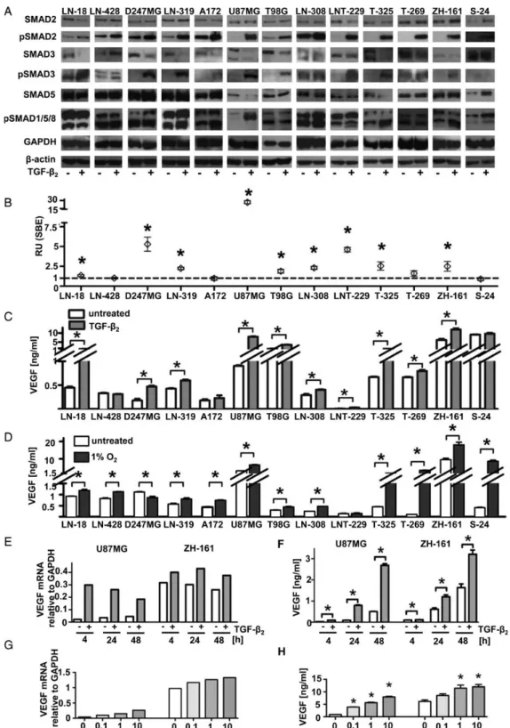
Fig. 2. Control of VEGF release by constitutive TGF-b pathway activity. (A) VEGF mRNA analyzed by RT-PCR and VEGF release into the supernatant (B) prepared as in Fig.1D and analyzed by ELISA (mean and SEM, n ¼ 5 in duplicates or triplicates) in long-term glioma cell lines (LTCs) and glioma-initiating cell lines (GICs). (C) Representative immunoblots (SMAD2, pSMAD2, SMAD3, pSMAD3, SMAD5, pSMAD1/5/8, GAPDH) of LN-308 cells after transfection with siRNA targeting TGF-bRII (72 h) or ALK-5 (48 h) or after pharmacologicial inhibition of ALK-5 by SD-208 (1 mM, 24 h, or respective controls (nontargeting siRNA or DMSO). (D) Representative immunoblots of LN-308 cells with siRNA targeting SMAD2 (48 h), SMAD3 (48 h), SMAD2, and SMAD3 (48 h), or SMAD5 (72 h) or nontargeting control. (E) Supernatants of LN-308 cells harvested 72 hours after transfection with siRNA targeting TGF-bRII were assessed for VEGF levels by ELISA. (F) Supernatants of U87MG, LN-308, and ZH-161 cells were analyzed for VEGF by ELISA after exposure to ALK-5 siRNA (left, 48 h post transfection for LN-308 and ZH-161, 72 h for U87MG) or SD-208 (right, 24 h, 1 mM). (G and H) Supernatants harvested after transfection with siRNA targeting SMAD2 (24 h for U87MG and ZH-161, 48 h for LN-308), SMAD3 (36 h for U87MG, 48 h for LN-308, 24 h for ZH-161), SMAD2/3 (36 h for U87MG, 48 h for LN-308, 24 h for ZH-161) (G), or SMAD5 (24 h for U87MG and ZH-161, 48 h for LN-308) (H). The representative experiments shown were performed in triplicate (Student t test, *P , .05).
Seystahl et al.: TGF-b-induced VEGF release in glioblastoma
mRNA, while total SMAD2 and total SMAD5 protein were in-versely correlated with VEGF release. TGF-b1 and TGF-b2
mRNA correlated with VEGF mRNA and also correlated inversely with SMAD2 mRNA in the TCGA database. However, neither TGF-b receptor mRNA nor protein in our cell line panel nor gene expression data of the TCGA nor pSMAD2/pSMAD3 nor pSMAD1/5/8 levels correlated with VEGF mRNA. PAI-1 mRNA correlated with VEGF mRNA in the TCGA but not in our dataset (Table1; Supplementary material, Table S1).
We chose LN-308 cells to study the regulation of constitutive VEGF release because of their high endogenous TGF-b expres-sion and high constitutive SMAD2 and SMAD3 phosphorylation. We added selected analyses for U87MG and ZH-161 cells. In LN-308, TGF-bRII silencing with an efficacy of 88% reduction assessed by RT-PCR (data not shown) reduced pSMAD2 and pSMAD3 levels and confirmed the involvement of TGF-b and TGF-bRII in their constitutive regulation. Interestingly, pSMAD1/5/8 levels were also reduced upon silencing of TGF-bRII. Pharmacological inhibition of the kinase activity of ALK-5 by the small molecule inhibitor SD-208 or silencing of ALK-5 also led to reduced pSMAD2 and pSMAD3 levels but did not affect constitutive pSMAD1/5/8 levels (Fig.2C). To investi-gate the contribution of the different SMAD signaling pathways to constitutive VEGF release, we established gene silencing of SMAD2, SMAD3, SMAD2/3 in combination, or SMAD5, which led to specific reductions of the corresponding target proteins. As expected, pSMAD2, pSMAD3, and pSMAD1/5/8 were also re-duced upon silencing of the respective SMAD proteins. In addi-tion, silencing of SMAD proteins did not affect only their own phosphorylation; instead, we observed reduced pSMAD3 upon silencing of SMAD2 in contrast with increased pSMAD2 upon si-lencing of SMAD3. pSMAD3 was increased upon sisi-lencing of SMAD5, while pSMAD2 was unaffected (Fig.2D).
Silencing of TGF-bRII reduced VEGF release in LN-308 cells (Fig.2E). Inhibition of ALK-5 by RNA interference or SD-208 also reduced VEGF release in U87MG and LN-308 cells, although the reduction was not significant in ZH-161 (Fig.2F). SMAD si-lencing had differential effects on VEGF release: SMAD2 or SMAD3 gene silencing reduced constitutive VEGF levels in U87MG and LN-308 but not in ZH-161 (Fig.2G). Even cosilenc-ing of SMAD2 and SMAD3 failed to affect VEGF release in ZH-161, but further reduced VEGF levels in LN-308 compared to the effect of silencing SMAD2 or SMAD3 alone. In contrast, silencing of SMAD5 increased the levels of constitutive VEGF re-lease in U87MG cells but had no effects in LN-308 or ZH-161 (Fig.2H).
Exogenous TGF-b Promotes VEGF Release in Human
Malignant Glioma Cells
We next assessed glioma cell response to exogenous TGF-b. All cell lines showed increased pSMAD2 and (except for S-24) in-creased pSMAD3, albeit to a different extent. The relative induc-tion of pSMAD2 correlated inversely with the endogenous pSMAD2 (r ¼ 20.66; P ¼ .01). pSMAD1/5/8 was increased in LN-319, A172, and U87MG LTCs as well as T-325, ZH-161, and S-24 GICs (Fig.3A). We next monitored the increase of TGF-b –dependent transcriptional activity using a SBE reporter plasmid.21TGF-b induced SBE reporter activity in all cell lines except in LN-428, A172, T-269, and S-24 cells. The highest
response was observed in U87MG (26-fold) (Fig.3B). Sufficient TGF-bRII surface expression was necessary for the induction of SBE reporter activity given the poor response in T-269 and S-24 cells, which exhibited TGF-bRII levels at the detection limit (Fig. 1A). In addition, high responsiveness correlated with pSMAD3 induction by TGF-b (r ¼ 0.62; P ¼ .02). Surprisingly, the reporter-nonresponsive cell lines LN-428 and A172 ex-pressed high levels of ALK-5, in contrast with the highly respon-sive cell line U87MG (Fig.1B).
VEGF levels increased after TGF-b treatment in all cell lines except LN-428, A172, and S-24. The most pronounced increase was seen in U87MG cells (9-fold) (Fig.3C). The response in U87MG was maintained but reduced in extent when those cells were switched to GIC culture conditions. In ZH-161, the re-sponse was further increased when switched to serum-containing medium for 2 passages. Under GIC conditions, basal VEGF synthesis was increased in U87MG, both on mRNA and protein levels, and in ZH-161 on protein level (Supplemen-tary material, Fig. S1). Cell lines not exhibiting TGF-b2–
depen-dent VEGF induction showed only minor induction of pSMAD2 and pSMAD3 by TGF-b2(Fig.3A). The induction of VEGF by
TGF-b correlated with the induction of pSMAD3 (r ¼ 0.59; P ¼ .04) and reporter responsiveness (r ¼ 0.71; P ¼ .006). To assess whether cell lines not increasing VEGF release in response to TGF-b were generally less responsive to transcriptional activa-tion of the VEGF gene, we exposed the same cell lines to
hyp-oxia (1% O2, 24 h) as a major driver of VEGF gene
transcription.22Hypoxia led to increased VEGF levels in almost
all cell lines including LN-428, A-172, and S-24, which did not increase VEGF release upon stimulation with TGF-b. Conversely, LNT-229 cells were unaffected, and D247MG even showed
re-duced VEGF release under hypoxia (Fig. 3D). Of note,
hypoxia-induced VEGF release was higher overall in GICs, ing from 1.9-fold (ZH-161) to 32-fold (T-269) than in LTCs, rang-ing from 1.3-fold (LN-18) to 2.1-fold (U87MG). Accordrang-ingly, we found only minor induction of HIF-1a protein by hypoxia in LNT-229, which increased VEGF release in response to TGF-b but not to hypoxia. Indeed, we detected higher HIF-1a protein levels in ZH-161 than in LNT-229 cells under normoxic condi-tions. Of note, treatment with TGF-b left HIF-1a protein unaf-fected, and there was no correlation between the magnitude of the VEGF response to TGF-b versus hypoxia (data not shown). We selected U87MG and ZH-161 cells to examine the time and concentration dependence of VEGF induction by TGF-b. On mRNA level, the highest increase of VEGF (14-fold for U87MG and 1.4-fold for ZH-161) was observed at 4 hours for U87MG and at 24 –48 hours for ZH-161 (Fig.3E). The induc-tion of VEGF release into the supernatant was highest at 9-fold at 24 hours in U87MG cells and 2-fold at 24 – 48 hours in ZH-161 cells (Fig.3F). Furthermore, the induction of VEGF was concentration dependent in U87MG and ZH-161 cells both on mRNA (Fig.3G) and protein level (Fig.3H).
Molecular Pathways Mediating TGF-b – evoked VEGF
Release in Glioma Cells
To explore the pathways mediating the stimulatory effect of ex-ogenous TGF-b on VEGF release, we combined the inhibition of ALK-5 by SD-208 or siRNA, or siRNA-mediated gene silencing of SMAD2, SMAD3 or SMAD5, with the addition of exogenous
Fig. 3. Stimulation of VEGF release by exogenous TGF-b. (A) TGF-b pathway inducibility was examined by assessing the induction of pSMAD2, pSMAD3, or pSMAD1/5/8 via immunoblot with or without stimulation with TGF-b2(10 ng/mL) for 24 hours after 24 hour serum starvation.
Shown are immunoblots of SMAD2, pSMAD2, SMAD3, pSMAD3, SMAD5, pSMAD1/5/8, and representative loading controls for GAPDH and b-actin. (B) Transcriptional activation by TGF-b was assessed by a SBE reporter assay. Shown are relative units of SBE reporter signal after stimulation with TGF-b2(10 ng/mL) for 24 hours normalized to untreated control. (C and D) The supernatants of glioma cell lines maintained
with or without 10 ng/mL TGF-b2for 24 hours (C) or with or without hypoxia (D) after a 24 hours of serum starvation were analyzed for VEGF
by ELISA. (E and F) VEGF was assessed after 4, 24 and 48 hour exposure to 10 ng/mL TGF-b2in U87MG or ZH-161 on mRNA level (E) or protein
level using cell culture supernatants as in A (F). (G and H) U87MG or ZH-161 cells were analyzed for VEGF after 24 hour stimulation with TGF-b2at 0,
0.1, 1 or 10 ng/mL on mRNA level (G) and in cell culture supernatants (H).
Seystahl et al.: TGF-b-induced VEGF release in glioblastoma
TGF-b2. As expected, inhibition of ALK-5 by SD-208 or RNA
inter-ference reduced TGF-b2–evoked SMAD2 and SMAD3
phosphor-ylation in U87MG and ZH-161 cells. Interestingly, the induction of SMAD1/5/8 phosphorylation was also abrogated by inhibition or silencing of ALK-5, suggesting a hitherto unrecognized TGF-b–dependent signal transduction pathway from ALK-5 to SMAD1/5/8 in glioma cells. Both pharmacological inhibition and silencing of ALK-5 interfered with the TGF-b2– mediated
in-crease of VEGF (Fig. 4A, right panel). Silencing of SMAD2, SMAD3 or SMAD5 in U87MG or ZH-161 cells led to specific re-ductions of the target protein and reduced pSMAD2, pSMAD3, and pSMAD1/5/8 levels, respectively (Fig.4B). Interestingly, TGF-b2– induced SMAD1/5/8 phosphorylation was further
in-creased in the case of SMAD2 or SMAD3 gene silencing in U87MG but not in ZH-161, consistent with substrate competi-tion of SMAD proteins for ALK-5 (Fig.4B). TGF-b2–evoked
tran-scriptional activity was reduced upon silencing of either SMAD2, SMAD3, or SMAD5 in U87MG. A similar trend was observed in ZH-161; however, the reduction was only significant upon si-lencing of SMAD3 alone or combined with SMAD2 (Fig.4C). In both cell lines, the TGF-b–evoked VEGF release was reduced
upon silencing of SMAD2 or SMAD3. Cosilencing of SMAD2 and SMAD3 had no superior effect over single silencing in ZH-161. Further, silencing of SMAD5 reduced TGF-b2– evoked VEGF
re-lease in U87MG, but the reduction was not significant in ZH-161 cells (Fig.4D).
Discussion
Targeting angiogenesis is one of the major current strategies for glioblastoma treatment, but enthusiasm for this approach has declined after the failure of several antiangiogenic agents in phase III trials.23–25Although the anti-VEGF antibody bevaci-zumab showed activity for recurrent glioblastoma in phase II trials,26,27no survival gain was achieved in 2 phase III trials
in newly diagnosed glioblastoma patients.28,29Further, various
attempts to improve antiangiogenic therapy by combination with cytotoxics were unsuccessful.30 Nevertheless, VEGF is still one of the most promising targets that interferes with the malignant phenotype of glioblastoma. While hypoxia is considered the key driver of VEGF release in glioblastoma, hypoxia-independent control of VEGF release has received little attention.
TGF-b, an important molecule shaping the microenviron-ment in glioblastoma, was characterized here as a positive reg-ulator of the VEGF pathway. We have provided a comprehensive analysis of the TGF-b pathway and its control of VEGF release in LTC and GIC models. Specifically, we analyzed the role of en-dogenous TGF-b as a model of autocrine signaling versus that of exogenous TGF-b, mimicking paracrine signals from the microenvironment. As a precondition for autocrine signal-ing, TGF-bRII and ALK-5 were widely expressed, whereas
ALK-1 was not (Fig. 1), which indicates that an ALK-1/
pSMAD1/5/8 signaling axis of VEGF release characterized in en-dothelial cells12does not operate in glioma cells. All LTCs
re-leased TGF-b1and TGF-b2, but TGF-b2was not detected in 2
of 4 GICs, confirming that therapeutic approaches targeting only one TGF-b isoform are insufficient. Most cell lines exhibited
phosphorylated SMAD2, SMAD3, or SMAD5, suggesting consti-tutive TGF-b pathway activation, confirmed by reduced pSMAD levels when TGF-bRII or ALK-5 expression were sup-pressed, or when ALK-5 was inhibited pharmacologically (Fig.2C). In nonendothelial cells, bone morphogenetic proteins (BMP) are considered as major drivers of SMAD1/5/8 phosphor-ylation; however, TGF-b – dependent phosphorylation of SMAD1/5/8 has been recently described (although not yet for glioma cells).31,32We found that pSMAD1/5/8 levels in cellular lysates correlated with TGF-bRII levels at the surface and that TGF-bRII gene silencing reduced pSMAD1/5/8 levels (Fig.2C). That inhibition of ALK-5 via SD-208 or ALK-5 gene silencing left pSMAD1/5/8 unaffected suggests that a type-I receptor other than ALK-5 mediates SMAD1/5/8 phosphorylation in re-sponse to TGF-b. Still, we cannot rule out that these effects are mediated via minor ALK-1 levels escaping detection on pro-tein level.
To study the effect of endogenous TGF-b signaling on consti-tutive VEGF release, we experimented with different compo-nents of the TGF-b pathway. TGF-bRII gene silencing reduced VEGF release in LN-308 cells, which confirmed a role for TGF-b in the regulation of VEGF release in this cell line that re-leases high levels of active TGF-b33,34(Fig.2E). Furthermore,
VEGF release required ALK-5 activity in LN-308 and U87MG cells but not in ZH-161 cells (Fig.2F). This might suggest a ro-bust TGF-b– independent VEGF secretion in GICs, which was po-tentially due to constitutive HIF expression at normoxic
conditions35 and was confirmed for ZH-161. SMAD2 and
SMAD3 are necessary for TGF-b – dependent regulation of VEGF in LN-308 and U87MG cells, but again ZH-161 was unaf-fected, even by the combined SMAD2/3 silencing that reduced VEGF most efficiently in LN-308 cells (Fig. 2G). The role of SMAD5 in the regulation of constitutive VEGF release is more complex. LN-308 and ZH-161 did not show major changes upon SMAD5 gene silencing, whereas U87MG showed increased VEGF secretion (Fig.2H). This can be explained by an indirect ef-fect of BMP-2 being expressed by U87MG36and destabilizing
HIF-1a in glioma cells.37Thus, depletion of SMAD5, resulting
in reduced BMP signaling, might increase the stability of HIF-1a and thereby promote VEGF release. Furthermore, a di-rect inhibitory effect of BMP-9 on VEGF via ALK-1/pSMAD1/5/8 signaling has been described in endothelial cells.38 Thus,
TGF-b contributes to constitutive VEGF release in glioma cells via a TGF-bRII/ALK-5/SMAD2/3 signaling pathway. The pSMAD1/5/8 signaling axis, which is driven by both TGF-b and BMP, might be indirectly involved in selected cell lines, possibly as a negative feedback mechanism.
TGF-b is secreted not only by glioma cells but also by the tumor microenvironment (eg, endothelial, immune, or micro-glial cells). Thus, we studied the effect of exogenous TGF-b on VEGF release. We observed a differential pattern of responsive-ness to TGF-b as determined by changes in SMAD phosphoryla-tion and reporter activity (Fig.3A and B). We found that SMAD1/ 5/8 is also phosphorylated via TGF-b and ALK-5 in some glioma cell lines and may contribute to enhanced VEGF release (Fig.3A, Fig.4A and D). Sufficient TGF-bRII cell surface expression and phosphorylation of SMAD2/3 are necessary, but not sufficient, for SBE activation (Fig.3B) and VEGF release (Fig.3C), suggest-ing the involvement of additional downstream effectors. That
Fig. 4. TGF-b –dependent increase of VEGF is mediated by ALK-5 and involves different SMAD signaling pathways. (A) U87MG or ZH-161 cells were treated with or without SD-208 (1 mM, 25 h) or (siRNA) targeting ALK-5 (72 h for U87MG, 48 h for ZH161) and 24 hours before harvesting with or without TGF-b2(10 ng/mL). Total SMAD2, pSMAD2, SMAD3, pSMAD3, SMAD5, pSMAD1/5/8, or GAPDH were analyzed by immunoblot. Supernatants
were analyzed for VEGF by ELISA. (B) Levels of total SMAD2, pSMAD2, SMAD3, pSMAD3, SMAD5, pSMAD1/5/8, or GAPDH were assessed by immunoblot after silencing of SMAD2 (24 h), SMAD3 (36 h for U87MG, 24 h for ZH-161), SMAD2 and SMAD3 (ZH-161, 24 h) or SMAD5 (24 h) with or without additional treatment with TGF-b2(10 ng/mL, 12 – 24 h). (C) Effect of silencing of SMAD2, SMAD3, or SMAD5, or respective
controls on transcriptional activation in U87MG or ZH-161 cells with or without treatment with TGF-b2(10 ng/mL, 12 h) was examined by SBE
reporter assay. Shown are relative units of SBE reporter signal normalized to untreated control-transfected samples. (D) Supernatants from cells treated as in B were assessed for VEGF by ELISA.
Seystahl et al.: TGF-b-induced VEGF release in glioblastoma
high constitutive levels of pSMAD2 negatively correlated with the inducibility of pSMAD2 by exogenous TGF-b suggests a sat-uration of the signaling pathway by autocrine TGF-b that leads to decreased sensitivity towards exogenous TGF-b. ALK-5 ex-pression was not predictive for sensitivity to exogenous TGF-b because cell lines with poor activation of the SBE reporter ex-pressed rather high levels of ALK-5 and vice versa. Posttran-scriptional processes (eg, involving deubiquitinating enzymes regulating TGF-b receptor stability39) probably modulate
sensi-tivity towards TGF-b.
Exogenous TGF-b induced VEGF release in most cell lines (Fig.3C). Cells unresponsive to TGF-b were still responsive to hypoxia with regard to VEGF induction, placing differential reg-ulation upstream of the VEGF promoter (Fig.3D). TGF-b–depen-dent SMAD3 phosphorylation and transcriptional SBE activation correlated with TGF-b–evoked VEGF release, although no such correlation was seen for pSMAD2 or the TGF-b target gene, PAI-1. A differential activation of the transcription factor Sp1, a major driver of VEGF gene expression in nonhypoxic condi-tions40via competing SMAD signaling pathways, might play a
role in this context. Of note, the VEGF promoter itself contains SBE sites, and promoter activity is increased by SMADs.41That
depletion of SMAD5 led to increased constitutive VEGF release (Fig.2H), but reduced TGF-b evoked VEGF release (Fig.4D) in U87MG cells might be best explained via the dual activation of pSMAD1/5/8 by TGF-b and BMP. While endogenous BMP sig-naling might inhibit constitutive VEGF expression, either indi-rectly via destabilization of HIF37or directly via BMP-9-ALK1 signaling as described for endothelial cells,38a relative
predom-inance of TGF-b signaling might occur in the pharmacological context of exogenous stimulation by TGF-b inducing VEGF both via pSMAD2/3 and pSMAD1/5/8 signaling (Fig.5).
In summary, we show that TGF-b regulation of VEGF release in glioma cells depends, in an ALK-5 – dependent manner, not only on SMAD2/3-dependent pathways but also on pSMAD1/ 5/8 signaling, suggesting that anti-TGF-b strategies may indi-rectly inhibit VEGF pathway activation in glioblastoma and that this mechanism accounts for some of the angiogenic ac-tivity attributed to TGF-b. Moreover, since we have recently il-lustrated how integrin inhibition downregulates TGF-b pathway activation in glioblastoma,42combinatorial therapeu-tic approaches targeting sequentially or parallel integrins TGF-b and VEGF based on preferential pathway activation may allow better treatment options for glioblastoma.
Supplementary Material
Supplementary material is available at Neuro-Oncology Journal online (http://neuro-oncology.oxfordjournals.org/).
Funding
Swiss Cancer League/Oncosuisse to MW and GT (KLS 2579-02-2010), University of Zurich to IT (Forschungskredit 54251101), Betty and David Koetser Foundation to KS.
Acknowledgments
We thank Pfizer (New York, NY) for providing PF-03446962.
Conflict of interest statement. KS (Roche: Advisory boards), GT (MSD, Roche: Advisory boards), MW (Bayer, Isarna, Merck Serono, MSD, Roche: Research grants; Isarna, Magforce, Merck Serono, MSD, and Roche: Lectures, advisory boards).
References
1. Bruna A, Darken RS, Rojo F, et al. High TGFb-Smad activity confers poor prognosis in glioma patients and promotes cell proliferation depending on the methylation of the PDGF-B gene. Cancer Cell. 2007;11(2):147–160.
2. Sta¨nder M, Naumann U, Dumitrescu L, et al. Decorin gene transfer-mediated suppression of TGF-b synthesis abrogates experimental malignant glioma growth in vivo. Gene Ther. 1998; 5(9):1187 –1194.
3. Friese MA, Wischhusen J, Wick W, et al. RNA interference targeting transforming growth factor-b enhances NKG2D-mediated antiglioma immune response, inhibits glioma cell migration and invasiveness, and abrogates tumorigenicity in vivo. Cancer Res. 2004;64(20):7596 –7603.
Fig. 5. TGF-b – dependent regulation of VEGF release in glioblastoma. Proposed model for the regulation of VEGF by TGF-b (straight arrows) and HIF (dotted arrows). TGF-b released from cells of the tumor microenvironment (eg, endothelial, immune, and microglial cells) signals via TGF-bRII and ALK-5 leading to phosphorylation of SMAD2, SMAD3, and SMAD1/5/8 and increased VEGF release. BMP-dependent pSMAD1/5/8 signaling (dashed arrows) destabilizes HIF and thereby indirectly reduces VEGF. TGF-b – dependent SMAD1/5/8 signaling requires, beyond TGF-bRII, a non-ALK-5-type-I-receptor (TGF-b-RI-x). Overall, VEGF release is modulated in a cell-specific and context-dependent manner, with a predominance of either hypoxia-, TGF-b – , or BMP-driven signaling for ZH-161 GICs exhibiting low endogenous TGF-b levels but stable endogenous HIF-1a, VEGF release is highly resistant to TGF-b pathway inhibition at baseline conditions but is sensitive when TGF-b is abundant. In contrast, LN-308 cells, characterized by high endogenous TGF-b and pSMAD2/3 levels, are highly sensitive for TGF-b pathway inhibition via ALK-5 and SMAD2/3 but not for inhibition of SMAD5 at baseline. In U87MG cells, the duality of pSMAD1/5/8 signaling as driven both by TGF-b and BMPs becomes apparent. SMAD5 at baseline conditions with low endogenous TGF-b negatively regulates VEGF, probably via BMP-driven destabilization of HIF, but positively regulates VEGF when TGF-b is abundant.
4. Uhl M, Aulwurm S, Wischhusen J, et al. SD-208, a novel transforming growth factor beta receptor I kinase inhibitor, inhibits growth and invasiveness and enhances immunogenicity of murine and human glioma cells in vitro and in vivo. Cancer Res. 2004;64(21):7954 – 7961.
5. Tran TT, Uhl M, Ma JY, et al. Inhibiting TGF-b signaling restores immune surveillance in the SMA-560 glioma model. Neuro Oncol. 2007;9(3):259–270.
6. Bogdahn U, Hau P, Stockhammer G, et al. Targeted therapy for high-grade glioma with the TGF-b2 inhibitor trabedersen: results of a randomized and controlled phase IIb study. Neuro Oncol. 2011;13(1):132 –142.
7. Carpentier A, Brandes A, Kesari S, et al. Safety interim data from a three-arm phase II study evaluating safety and pharmakokinetics of the oral transforming growth factor-beta (TGF-b) receptor I kinase inhibitor LY2157299 monohydrate in patients with glioblastoma at first progression [abstract 2061]. J Clin Oncol 2013;31:(suppl).
8. Groppe J, Hinck CS, Samavarchi-Tehrani P, et al. Cooperative assembly of TGF-b superfamily signaling complexes is mediated by two disparate mechanisms and distinct modes of receptor binding. Mol Cell. 2008;29(2):157 –168.
9. Kubiczkova L, Sedlarikova L, Hajek R, et al. TGF-b - an excellent servant but a bad master. J Transl Med. 2012;10:183.
10. Zhang M, Kleber S, Rohrich M, et al. Blockade of TGF-b signaling by the TGFbR-I kinase inhibitor LY2109761 enhances radiation response and prolongs survival in glioblastoma. Cancer Res. 2011;71(23):7155– 7167.
11. Goumans MJ, Valdimarsdottir G, Itoh S, et al. Activin receptor-like kinase (ALK)1 is an antagonistic mediator of lateral TGFb/ALK5 signaling. Mol Cell. 2003;12(4):817– 828.
12. Bostro¨m K, Zebboudj AF, Yao Y, et al. Matrix GLA protein stimulates VEGF expression through increased transforming growth factor-b1 activity in endothelial cells. J Biol Chem. 2004;279(51): 52904–52913.
13. Liu Z, Kobayashi K, van Dinther M, et al. VEGF and inhibitors of TGFb type-I receptor kinase synergistically promote blood-vessel formation by inducing alpha5-integrin expression. J Cell Sci. 2009;122(Pt 18):3294–3302.
14. Koochekpour S, Merzak A, Pilkington GJ. Vascular endothelial growth factor production is stimulated by gangliosides and TGF-b isoforms in human glioma cells in vitro. Cancer Lett. 1996; 102(1– 2):209– 215.
15. Tabatabai G, Herrmann C, von Kurthy G, et al. VEGF-dependent induction of CD62E on endothelial cells mediates glioma tropism of adult haematopoietic progenitor cells. Brain. 2008; 131(Pt 10):2579–2595.
16. Hjelmeland MD, Hjelmeland AB, Sathornsumetee S, et al. SB-431542, a small molecule transforming growth factor-b-receptor antagonist, inhibits human glioma cell line proliferation and motility. Mol Cancer Ther. 2004;3(6):737–745. 17. Weller M, Rieger J, Grimmel C, et al. Predicting chemoresistance in
human malignant glioma cells: the role of molecular genetic analyses. Int J Cancer. 1998;79(6):640–644.
18. Rieger J, Lemke D, Maurer G, et al. Enzastaurin-induced apoptosis in glioma cells is caspase-dependent and inhibited by BCL-XL. J Neurochem. 2008;106(6):2436 –2448.
19. Weiler M, Pfenning PN, Thiepold AL, et al. Suppression of proinvasive RGS4 by mTOR inhibition optimizes glioma treatment. Oncogene. 2013;32(9):1099– 1109.
20. Weksler BB, Subileau EA, Perriere N, et al. Blood-brain barrier-specific properties of a human adult brain endothelial cell line. FASEB J. 2005;19(13):1872–1874.
21. Zawel L, Dai JL, Buckhaults P, et al. Human Smad3 and Smad4 are sequence-specific transcription activators. Mol Cell. 1998;1(4): 611–617.
22. Forsythe JA, Jiang BH, Iyer NV, et al. Activation of vascular endothelial growth factor gene transcription by hypoxia-inducible factor 1. Mol Cell Biol. 1996;16(9):4604–4613. 23. Wick W, Puduvalli VK, Chamberlain MC, et al. Phase III study of
enzastaurin compared with lomustine in the treatment of recurrent intracranial glioblastoma. J Clin Oncol. 2010;28(7): 1168–1174.
24. Batchelor TT, Mulholland P, Neyns B, et al. Phase III randomized trial comparing the efficacy of cediranib as monotherapy, and in combination with lomustine, versus lomustine alone in patients with recurrent glioblastoma. J Clin Oncol. 2013;31(26): 3212– 3218.
25. Stupp R, Hegi M, Gorlia T, et al. Cilengitide combined with standard treatment for patients with newly diagnosed glioblastoma and methylated O6-methylguanine-DNA methyltransferase (MGMT) gene promoter: Key results of the multicenter, randomized, open-label, controlled, phase III CENTRIC study [abstract LBA2009]. J Clin Oncol 2013;31:(suppl).
26. Friedman HS, Prados MD, Wen PY, et al. Bevacizumab alone and in combination with irinotecan in recurrent glioblastoma. J Clin Oncol. 2009;27(28):4733–4740.
27. Kreisl TN, Kim L, Moore K, et al. Phase II trial of single-agent bevacizumab followed by bevacizumab plus irinotecan at tumor progression in recurrent glioblastoma. J Clin Oncol. 2009;27(5): 740– 745.
28. Gilbert MR, Dignam JJ, Armstrong TS, et al. A randomized trial of bevacizumab for newly diagnosed glioblastoma. N Engl J Med. 2014;370(8):699–708.
29. Chinot OL, Wick W, Mason W, et al. Bevacizumab plus radiotherapy-temozolomide for newly diagnosed glioblastoma. N Engl J Med. 2014;370(8):709– 722.
30. Reardon DA, Turner S, Peters KB, et al. A review of VEGF/ VEGFR-targeted therapeutics for recurrent glioblastoma. J Natl Compr Canc Netw. 2011;9(4):414 – 427.
31. Wakefield LM, Hill CS. Beyond TGFb: roles of other TGFb superfamily members in cancer. Nat Rev Cancer. 2013;13(5): 328– 341.
32. Wrighton KH, Lin X, Yu PB, et al. Transforming Growth Factor b Can Stimulate Smad1 Phosphorylation Independently of Bone Morphogenic Protein Receptors. J Biol Chem. 2009;284(15): 9755– 9763.
33. Fontana A, Hengartner H, de Tribolet N, et al. Glioblastoma cells release interleukin 1 and factors inhibiting interleukin 2-mediated effects. J Immunol. 1984;132(4):1837– 1844. 34. Leitlein J, Aulwurm S, Waltereit R, et al. Processing of
immunosuppressive pro-TGF-b1,2 by human glioblastoma cells involves cytoplasmic and secreted furin-like proteases. J Immunol. 2001;166(12):7238–7243.
35. Wei J, Wu A, Kong LY, et al. Hypoxia potentiates glioma-mediated immunosuppression. PLoS One. 2011;6(1):e16195.
36. Chattopadhyay N, T-Felt Hansen J, Godbole MM, et al. Transforming growth factor beta receptor family ligands inhibit hepatocyte growth factor synthesis and secretion from
Seystahl et al.: TGF-b-induced VEGF release in glioblastoma
astrocytoma cells. Brain Res Mol Brain Res. 2004;121(1 – 2): 146– 150.
37. Pistollato F, Rampazzo E, Abbadi S, et al. Molecular mechanisms of HIF-1a modulation induced by oxygen tension and BMP2 in glioblastoma derived cells. PLoS One. 2009;4(7):e6206.
38. Shao ES, Lin L, Yao Y, et al. Expression of vascular endothelial growth factor is coordinately regulated by the activin-like kinase receptors 1 and 5 in endothelial cells. Blood. 2009;114(10): 2197– 2206.
39. Eichhorn PJ, Rodon L, Gonzalez-Junca A, et al. USP15 stabilizes TGF-b receptor I and promotes oncogenesis through the
activation of TGF-beta signaling in glioblastoma. Nat Med. 2012; 18(3):429 – 435.
40. Page`s G, Pouyssegur J. Transcriptional regulation of the Vascular Endothelial Growth Factor gene--a concert of activating factors. Cardiovasc Res. 2005;65(3):564–573.
41. Sanchez-Elsner T, Botella LM, Velasco B, et al. Synergistic cooperation between hypoxia and transforming growth factor-beta pathways on human vascular endothelial growth factor gene expression. J Biol Chem. 2001;276(42):38527–38535. 42. Roth P, Silginer M, Goodman SL, et al. Integrin control of the transforming growth factor-b pathway in glioblastoma. Brain. 2013;136(Pt 2):564– 576.