Safe, effective and durable epicardial left atrial appendage clip
occlusion in patients with atrial
fibrillation undergoing cardiac
surgery:
first long-term results from a prospective device trial
Maximilian Y. Emmert
a, Gilbert Puippe
b, Stephan Baumüller
b, Hatem Alkadhi
b, Ulf Landmesser
c,
Andre Plass
a, Dominique Bettex
d, Jacques Scherman
a, Jürg Grünenfelder
a, Michele Genoni
e,
Volkmar Falk
aand Sacha P. Salzberg
a,*
a
Clinic for Cardiovascular Surgery, University Hospital Zurich, Zurich, Switzerland
b Institute of Diagnostic and Interventional Radiology, University Hospital Zurich, Zurich, Switzerland c
Clinic for Cardiology, University Hospital Zurich, Zurich, Switzerland
d Institute for Anesthesiology, University Hospital Zurich, Zurich, Switzerland e
Clinic for Cardiac Surgery, Triemli Hospital Zurich, Zurich, Switzerland
* Corresponding author. Clinic for Cardiovascular Surgery, University Hospital Zurich, Rämistrasse 100, CH - 8091 Zurich, Switzerland. Tel: +41-44-2559362; fax: +41-44-2555047; e-mail: [email protected] (S.P. Salzberg).
Received 31 January 2013; received in revised form 13 March 2013; accepted 13 March 2013
Abstract
OBJECTIVES: Atrialfibrillation (AF) is a significant risk factor for embolic stroke originating from the left atrial appendage (LAA). This is
thefirst report of long-term safety and efficacy data on LAA closure using a novel epicardial LAA clip device in patients undergoing
cardiac surgery.
METHODS: Forty patients with AF were enrolled in this prospective ‘first-in-man’ trial. The inclusion criterion was elective cardiac
surgery in adult patients with AF for which a concomitant ablation procedure was planned. Intraoperative transoesophageal echocardi-ography (TEE) was used to exclude LAA thrombus at baseline and evaluate LAA perfusion after the procedure, while computed tomog-raphy (CT) was used for serial imagery workup at baseline, 3-, 12-, 24- and 36-month follow-up.
RESULTS: Early mortality was 10% due to non-device-related reasons, and thus 36 patients were included in the follow-up consisting of 1285 patient-days and mean duration of 3.5 ± 0.5 years. On CT, clips were found to be stable, showing no secondary dislocation 36 months after surgery. No intracardial thrombi were seen, none of the LAA was reperfused and in regard to LAA stump, none of the patients demon-strated a residual neck >1 cm. Apart from one unrelated transient ischaemic attack (TIA) that occurred 2 years after surgery in a patient with carotid plaque, no other strokes and/or neurological events demonstrated in any of the studied patients during follow-up.
CONCLUSION: This is thefirst prospective trial in which concomitant epicardial LAA occlusion using this novel epicardial LAA clip device is
100% effective, safe and durable in the long term. Closure of the LAA by epicardial clipping is applicable to all-comers regardless of LAA morphology. Minimal access epicardial LAA clip closure may become an interesting therapeutic option for patients in AF who are not amenable to anticoagulation and/or catheter closure. Further data are necessary to establish LAA occlusion as a true and viable therapy for stroke prevention.
CLINICAL TRIAL REGISTRATION: The trial is registered atwww.ClinicalTrials.gov, reference: NCT00567515.
Keywords:Left atrial appendage• Stroke • TIA • Atrial fibrillation • Epicardial clip occlusion • Anticoagulation
INTRODUCTION
Atrialfibrillation (AF) is a well-established risk factor for the
oc-currence of stroke, and the thrombo-embolic origin in AF is very
often related to the left atrial appendage (LAA) [1]. The standard
of care for medical management of patients with AF remains
oral anticoagulation [2,3], and current guidelines recommend
treatment of patients with a CHA2DS2-VASC >1 [2,3].
In this regard, recent data indicate that LAA closure may be less inferior to anticoagulation in patients with AF in whom oral
anticoagulation was deemed unsuitable [4]. Just recently, the
European Society of Cardiology (ESC) has recommended that if
patients are not amenable to anticoagulation (elevated
CHA2DS-VASC and/or HAS-BLED scores), LAA occlusion may be
warranted [2,3]. Percutaneous approaches to LAA closure have
set the basis for treating AF patients with contraindications to
anticoagulation (elevated CHA2DS-VASC and/or HAS-BLED
scores). In published series, anatomy-based selection for closure devices is necessary to accommodate individual anatomical varia-tions. These anatomical limitations do not apply to the epicardial
© The Author 2013. Published by Oxford University Press on behalf of the European Association for Cardio-Thoracic Surgery. All rights reserved. doi:10.1093/ejcts/ezt204 Advance Access publication 8 May 2013
approach; however, current surgical closure offers only suboptimal
results [5]. To the contrary, recent data indicate that the use of an
epicardial LAA clip device during elective open heart surgery is safe and provides excellent early outcomes at 3 months after
surgery [6,7]. However, so far, only short-term reports are
avail-able for epicardial LAA clip occlusion while the long-term efficacy
of this therapy concept remains to be elucidated.
In the present prospective ‘first-in-man’ clinical trial, for the
first time, we report long-term outcomes of epicardial LAA clip occlusion in patients undergoing elective open heart surgery.
MATERIALS AND METHODS
This prospective device trial being registered at www.
ClinicalTrials.gov(reference: NCT00567515), began in September 2007 with enrolment and was completed in July 2009 with a total of 40 patients with AF undergoing open heart surgery. While early outcome data for the initial 34 patients with regard to the feasibility of epicardial LAA clip placement and safety have been
described previously [7], in the present study, and for the first
time, we report long-term outcomes of epicardial LAA clip occlu-sion in patients undergoing elective open heart surgery.
The primary inclusion criterion was elective cardiac surgery in adult patients with AF for which an ablation procedure was planned. Exclusion criteria were reoperation, known thrombus in the LAA, patients from the intensive care unit, a history of peri-carditis, recent myocardial infarction (<90 days) and a known allergy to the devices component (Supplementary Table 1). Computed tomography (CT) was used preoperatively to assess anatomy of the left atrium, pulmonary veins and LAA. Intraoperative transoesophageal echocardiography (TEE) was used to rule out LAA thrombus at baseline (exclusion criteria) and to evaluate LAA perfusion at the end of the procedure. Patients underwent serial follow-up after 3 months, and then yearly up to 3 years after surgery. Clinical follow-up comprised clinical status, laboratory examination; electrocardiogram, chest
X-ray and CT workup [7]. After surgery, it was recommended to
cease anticoagulation after 3 months. If in sinus rhythm, then aspirin 100 mg/day, and if in AF 300 mg/day was recommended.
Device description
The LAA Clip System (Atriclip, Atricure, Dayton, OH, USA) con-sists of a self-closing, implantable clip, a disposable selection guide is used to choose between the four available clip sizes (35,
40, 45 and 50 mm). When closed, the clip applies uniform pres-sure over the length of the two parallel branches to enpres-sure con-sistent, reproducible and secure occlusion at the site of the basis
of the LAA as demonstrated in the pre-clinical work [8]. For the
initial trial, a reusable deployment tool was used. For market introduction, the deployment tool has since been redesigned and is a complete disposable set with the clip loaded within
(Fig.1).
Computed tomography protocol
All baseline and follow-up CT scans were performed according
to the previously published protocol [7] using a dual-source CT
scanner system with non-ionic contrast medium, standard scan-ning and image reconstruction parameters. In brief, LAA neck diameter was preoperatively measured on curved multiplanar reformations through the LAA. The LAA patency was visually analysed by two experienced independent readers in consensus.
Patency was defined as the presence of contrast material within
the entire LAA. Clip stability was assessed using two different measurements. In patients with contraindication to CT, such as impaired kidney function or refusal to undergo CT, serial chest
radiography was deemed sufficient to assess the stability of
implant. In all others, CT scan was used to precisely assess clip position and effect on surrounding structures.
This research was conducted in accordance with the ethical standards of the responsible committee on human experimenta-tion (instituexperimenta-tional and naexperimenta-tional) and with the Helsinki Declaraexperimenta-tion of 1975, as revised in 2000, and all patients gave informed written consent.
Statistical analysis
The Kruskal–Wallis test was used to test for significant changes
between the parameters average heart rate and heart rate vari-ability during scanning, as well as between the clip location (i.e. distances and angles), LA diameters and LV function parameters
across the CT examinations at different dates. AP-value of <0.05
was considered significant. Statistical analysis was performed
using commercially available software (SPSS, release 15.0, Chicago, IL, USA).
RESULTS
Mortality and complications
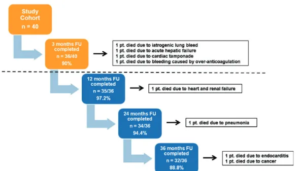
At total of 40 patients were included in this prospective device trial. While early mortality was 10% (4 of 40 patients) due to non-device-related reasons comprising iatrogenic lung bleed ( postoperative day 1), acute postoperative hepatic failure ( post-operative day 16), bleeding due to aortic tear at aortotomy suture line ( postoperative day 20) and out of hospital over
anticoagulation-related tamponade ( postoperative day 24) [7], a
total of 36 patients (36 of 40, 90% of the initial cohort) could be
successfully included in the follow-up assessments (Fig.2).
In the follow-up cohort (n = 36, 100%), mean follow-up
con-sisted of 1285 patient-days and mean duration of 3.5 ± 0.5 years. All 36 patients underwent follow-up at our institution and, during visits, electrocardiogram, laboratory workup and imaging Figure 1:LAA clip occlusion device.
AD UL T C ARDIA C
examinations were performed. During the 3-year follow-up period, 4 patients (4 of 36; 11.1%) died due to non-device-related
reasons including heart and renal failure (n = 1, 8 months
post-operatively), pneumonia (n = 1, 22 months postoperatively), mitral
valve endocarditis (n = 1, 28 months postoperatively) and
general-ized cancer (n = 1, 32 months postoperatively). Thus, 35 patients
completed 1 year follow-up (35 of 36, 97.2%), 34 patients the 2-year follow-up (34 of 36, 94.4%) and 32 patients the 3-year follow-up (32 of 36, 88.8%). None of these deaths was related to the device or study participation, as demonstrated by an inde-pendent Autopsy report and Data Safety Monitoring Board review. The occurrence of cardiac and non-cardiac complications
during follow-up is summarized in Table1.
Occurrence of stroke and neurological events
Only one transient ischaemic attack (TIA) occurred in one patient2 years after surgery (78-year old patient, CHA2DS-VASC = 4) in
the setting of documented sinus rhythm with only aspirin and
dis-continued statins (Table1). On CT and TEE, the LAA was occluded
and no intracavitary thrombi were seen. This event was thought to be due to carotid plaque, statin treatment and anticoagulation were reinstituted, and no further ischaemic event occurred. In all other patients during follow-up, no strokes, TIA or neurological events occurred (0 of 36, 0.0%).
Heart rhythm and the need for anticoagulation
At 3 years, 22 patients were in sinus rhythm (22 of 32, 68.8%) and of the 10 in AF (10 of 32, 31.2%), only 3 (3 of 10, 30.0%)were still being anticoagulated with warfarin (Fig.3). The mean
CHA2DS2-VASC Score of the entire study cohort (n = 40) was
3.7 ± 1.7 points.
Imaging
findings
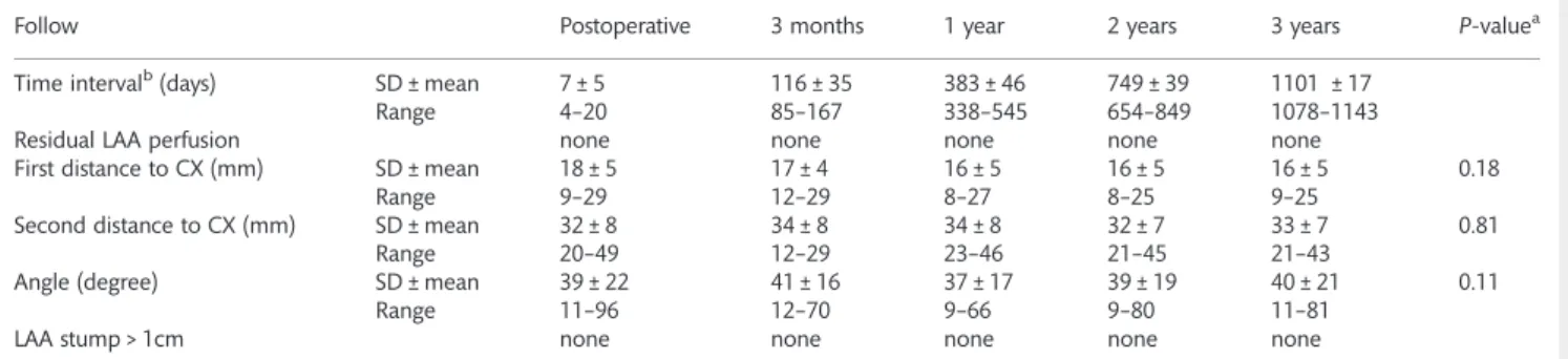
On CT, LAA clips were found to be stable, showing no secondary
dislocation (Table 2). Intracardial thrombi were not seen, and
Figure 2:Studyflow chart and follow-up. FU: follow-up.
Table 1: Mortality and major complications
Summary of mortality and adverse events Number of patients (n = 36) Overall mortality 4 (10.8%) Device-related mortality 0 (0%) Stroke 0 (0%)
Transient ischaemic attack 1 (2.7%) Myocardial infarction 1 (2.7%) Heart failure 1 (2.7%) Arrhythmia 1 (2.7%) Endocarditis 1 (2.7%) Renal failure 1 (2.7%) Pulmonary failure 0 (0%) Liver failure 1 (2.7%) Pneumonia 2 (5.2%) Malignancy 1 (2.7%)
Figure 3:Heart rhythm and anticoagulation status 3 years after clip implant-ation. At 3 years following clip implantation, 22 patients were in sinus rhythm (22 of 32, 68.8%) and of the 10 patients being in atrialfibrillation, 7 patients did not require the continuation of anticoagulation treatment (7 of 32, 21.9%), while 3 patients (3 of 32, 9.3%) were still being anticoagulated with warfarin.
none of the LAAs were reperfused. LAA occlusion was total and
complete in all patients (32 of 32, 100%) (Figs4and5). In regard
to LAA stump, none of the patients demonstrated a residual neck of >1 cm. Importantly, the results of imaging follow-up were con-sistent after 3 years follow-up in all patients. On chest radiography, the stability of the implant was also clearly documented.
DISCUSSION
Herein we report thefirst long-term outcome data on epicardial
LAA clip application during open heart surgery. In addition to
being 100% effective and safe in the short term [6, 7,8], the
results of this prospective device trial demonstrate the durable Figure 4:Computed tomography (CT) before and at 3-year follow-up after clip implantation. Exemplary CT before clip placement (A and C) and after clip implant-ation depicting the clip in stable position and fully excluding the left atrial appendage at a 3-year follow-up (B and D).
Table 2: Computed tomography findings at twelve-, twenty-four, and thirty-six month follow-up after LAA clip surgery
Follow Postoperative 3 months 1 year 2 years 3 years P-valuea
Time intervalb(days) SD ± mean 7 ± 5 116 ± 35 383 ± 46 749 ± 39 1101 ± 17
Range 4–20 85–167 338–545 654–849 1078–1143
Residual LAA perfusion none none none none none
First distance to CX (mm) SD ± mean 18 ± 5 17 ± 4 16 ± 5 16 ± 5 16 ± 5 0.18
Range 9–29 12–29 8–27 8–25 9–25
Second distance to CX (mm) SD ± mean 32 ± 8 34 ± 8 34 ± 8 32 ± 7 33 ± 7 0.81
Range 20–49 12–29 23–46 21–45 21–43
Angle (degree) SD ± mean 39 ± 22 41 ± 16 37 ± 17 39 ± 19 40 ± 21 0.11
Range 11–96 12–70 9–66 9–80 11–81
LAA stump > 1cm none none none none none
aFriedman’s 2-way paired ANOVA. The level of significance is 0.05. bTime Interval displayed as time between surgery and follow up.
LAA: left atrial appendage; CX: circumflex artery.
AD UL T C ARDIA C
occlusion of the LAA for over 3 years with excellent clinical out-comes. Importantly, during follow-up, no strokes occurred and anticoagulation could be often discontinued, most often decided by general practitioners and referring cardiologists. Moreover, our data indicate that successful closure of the LAA by epicardial clipping is applicable regardless of LAA morphology. Overall, no device-related complications occurred, the device performed as
it was designed to and fulfilled its purpose of totally excluding
the LAA from circulation.
LAA occlusion is performed to decrease the incidence of stroke and anticoagulation-related bleeding, and it appears that these complications did not occur in our series. When compared
with the CHA2DS-VASC score, we have seen a very low rate of
thrombo-embolic complications in our series. Considering a
mean CHA2DS-VASC score of close to 4, the expected stroke
rate would have been well above 10% [9].
It is obvious that the underlying cause of stroke in this
popula-tion is multifactorial, and not only related to the LAA [10]. That is
why we believe that sufficient anti platelet aggregation and also
statin treatment, when indicated, must be instituted in these patients when not in sinus rhythm. However, it is important to
note that the CHA2DS-VASC Score does not take into account
LAA perfusion. This score was created on patients in AF with LAA. Therefore, we believe that it is time for it to be revised, or at least used with precaution, as patients with occluded LAAs
will present with a decreased risk profile in the setting of
non-valvular AF. With this in mind, it appears that despite lack of data, closure of the LAA has become a well-accepted therapeutic option for patients not amenable for anticoagulation with AF.
Only one large trial allows the conclusion that effective LAA
closure provides stroke prevention [4], even though closure was
far from complete in this series. For precisely this purpose we
have initiated the creation of a web-based registry (www.laacr.
org) to collect long-term follow-up data on all patients
undergo-ing a surgical LAA closure.
While there are several methods to close the LAA, it is obvious that the limitation of surgical LAA occlusion is the invasiveness of the procedure, however effective it might be. For patients under-going heart surgery, it is our belief that LAA resection or clipping should therefore be performed in the setting of AF or even
without AF, but with significant risk factors. This is not only meant
for long-term protection but also in regard to the peri-operative
risk of thrombo-embolic complications [11]. Only effective LAA
therapies providing complete and durable occlusion should be
used, as incomplete occlusion will worsen the risk profile and
subject these patients to increased risk [5]. In their recent study,
Kanderianet al. [5] assessed 137 patients who underwent surgical
closure and demonstrated that there is a high occurrence of un-successful surgical LAA closure. On the other hand, the results for thoracoscopic ablations for stand-alone surgical treatment of AF,
in which the LAA is removed by stapling, are excellent [12]. Thus,
whatever option is chosen to address the LAA, documented
com-plete LAA occlusion is essential [5,7] and must be achieved.
In addition, also endocardial approaches to closing the LAA are currently being used. As surgeons, we believe that the high
anatomical variability [13] may make a more tailored approach
necessary. Currently, very scarce data are available on these new
approaches; nevertheless, the Protect AF trial [4] set the path for
Figure 5:In vivo monitoring of clip safety and stability using serial computed tomography (CT). CT image series of an exemplary patient before (A), at 3 months (B), 12 months (C) and at 36 months (D) after implantation indicating a stable clip position and durable left atrial appendage occlusion early postoperatively as well as throughout the entire follow-up period.
this new therapeutic option. Currently, and when compared with our study, in none of the published series a 100% effective-ness is reported for immediate LAA occlusion, and this is a major concern as these patients with contraindications to oral
anticoagulation will then require just this [14,15].
Another argument in favour of surgical closure is the electro-physiological role the LAA plays, and in particular in AF
recur-rences after left-sided catheter ablation for AF [16], in regard to
this specific issue, we have demonstrated complete electrical
iso-lation after LAA clip placement [17]. Therefore, it appears that
the epicardial approach of LAA removal by staplers or LAA
clip-ping may also provide adjunctive electrophysiological benefits.
Limitations
This was a surgical trial designed to evaluate this new device as a concomitant therapy option in patients undergoing cardiac surgery. The real potential for impact comes in the setting of the treatment for lone AF even in a stroke-prevention scenario, where stand-alone LAA therapies might effectively impact medicine.
CONCLUSION
This prospective device trial demonstrates, for thefirst time, the
long-term safety, durability and efficacy of epicardial LAA clip
oc-clusion using a novel epicardial clip device during cardiac surgery. In this series, no strokes occurred during follow-up, and anticoa-gulation could often be discontinued. The results show that suc-cessful epicardial LAA clip occlusion is applicable to all-comers regardless of LAA morphology. Minimally invasive epicardial LAA clip closure may represent an interesting therapeutic strategy for patients in AF who are not amenable to anticoagulation. Further studies are necessary to establish LAA occlusion as a true and viable therapy strategy for stroke prevention.
SUPPLEMENTARY MATERIAL
Supplementary material is available atEJCTS online.
Funding
This research was funded by an unrestricted research grant from Atricure.
Conflict of interest: Sacha P. Salzberg is a consultant for Atricure
and has received speaker fees.
REFERENCES
[1] Frost L, Engholm G, Johnsen S, Moller H, Husted S. Incident stroke after discharge from the hospital with a diagnosis of atrialfibrillation. Am J Med 2000;108:36–40.
[2] Camm AJ, Kirchhof P, Lip GY, Schotten U, Savelieva I, Ernst S et al. Guidelines for the management of atrialfibrillation: the task force for the management of atrial fibrillation of the European Society of Cardiology (ESC). Eur Heart J 2010;31:2369–429.
[3] Camm AJ, Kirchhof P, Lip GY, Schotten U, Savelieva I, Ernst S et al. Guidelines for the management of atrialfibrillation: the Task Force for the Management of Atrial Fibrillation of the European Society of Cardiology (ESC). Europace 2010;12:1360–420.
[4] Holmes DR, Reddy VY, Turi ZG, Doshi SK, Sievert H, Buchbinder Met al. Percutaneous closure of the left atrial appendage versus warfarin therapy for prevention of stroke in patients with atrialfibrillation: a ran-domised non-inferiority trial. Lancet 2009;374:534–42.
[5] Kanderian AS, Gillinov AM, Pettersson GB, Blackstone E, Klein AL. Success of surgical left atrial appendage closure: assessment by transeso-phageal echocardiography. J Am Coll Cardiol 2008;52:924–9.
[6] Ailawadi G, Gerdisch MW, Harvey RL, Hooker RL, Damiano RJ Jr, Salamon Tet al. Exclusion of the left atrial appendage with a novel device: early results of a multicenter trial. J Thorac Cardiovasc Surg 2011; 142:1002–9, 1009 e1001.
[7] Salzberg SP, Plass A, Emmert MY, Desbiolles L, Alkadhi H, Grunenfelder J et al. Left atrial appendage clip occlusion: early clinical results. J Thorac Cardiovasc Surg 2010;139:1269–74.
[8] Salzberg SP, Gillinov AM, Anyanwu A, Castillo J, Filsoufi F, Adams DH. Surgical left atrial appendage occlusion: evaluation of a novel device with magnetic resonance imaging. Eur J Cardiothorac Surg 2008;34: 766–70.
[9] Gage BF, Waterman AD, Shannon W, Boechler M, Rich MW, Radford MJ. Validation of clinical classification schemes for predicting stroke: results from the national registry of atrialfibrillation. JAMA 2001;285: 2864–70.
[10] Tugcu A, Okajima K, Jin Z, Rundek T, Homma S, Sacco RLet al. Septal pouch in the left atrium and risk of ischemic stroke. JACC Cardiovasc Imaging 2010;3:1276–83.
[11] Ryan K, Norbert B, John C. Perioperative Management Routine left atrial appendage ligation during cardiac surgery may prevent postoperative atrial fibrillation–related cerebrovascular accident. J Thorac Cardiovasc Surg 2013;145:582–9.
[12] Krul SP, Driessen AH, Zwinderman AH, van Boven WJ, Wilde AA, de Bakker JMet al. Navigating the mini-maze: systematic review of the first results and progress of minimally-invasive surgery in the treatment of atrialfibrillation. Int J Cardiol 2013;166:132–40.
[13] Di Biase L, Santangeli P, Anselmino M, Mohanty P, Salvetti I, Gili Set al. Does the left atrial appendage morphology correlate with the risk of stroke in patients with atrial fibrillation? Results from a multicenter study. J Am Coll Cardiol 2012;60:531–8.
[14] Shetty R, Leitner JP, Zhang M. Percutaneous catheter-based left atrial ap-pendage ligation and management of periprocedural left atrial append-age perforation with the lariat suture delivery system. J Invasive Cardiol 2012;24:E289–93.
[15] Bartus K, Han FT, Bednarek J, Myc J, Kapelak B, Sadowski J et al. Percutaneous left atrial appendage suture ligation using the lariat device in patients with atrial fibrillation: initial clinical experience. J Am Coll Cardiol 2012; pii:S07351097(12)03035-5.
[16] Di Biase L, Burkhardt JD, Mohanty P, Sanchez J, Mohanty S, Horton R et al. Left atrial appendage: an underrecognized trigger site of atrial fib-rillation. Circulation 2010;122:109–18.
[17] Starck CT, Steffel J, Emmert MY, Plass A, Mahapatra S, Falk V et al. Epicardial left atrial appendage clip occlusion also provides the electrical isolation of the left atrial appendage. Interact CardioVasc Thorac Surg 2012;15:416–8. AD UL T C ARDIA C