Printed in Great Britain
Heteronuclear filters in two-dimensional
PH/HJ-NMR spectroscopy: combined use
with isotope labelling for studies of
macromolecular conformation and
intermolecular interactions
GOTTFRIED OTTING AND KURT WUTHRICH
Institut fur Molekularbiologie und Biophysik, Eidgenossische Technische Hochschule-Honggerberg, CH-8093 Zurich, Switzerland
1. INTRODUCTION 40
2. APPLICATIONS OF HETERONUCLEAR FILTERS IN ' H - N M R STUD IES OF BIOLOGICAL MACROMOLECULES 42
2.1 nX-filters 47 2.2 nX-half-filters 51
2.3 Heteronuclear half-filters and heteronuclear three-dimensional NMR 55 3. FUNDAMENTAL CONSIDERATIONS ON HETERONUCLEAR FILTERS AND
THEIR USE IN 2D [lH,lH]-NMR 57 3.1 Hardware requirements 57
3.2 Product operator description of heteronuclear filtering 57
3.3 Extension of 2D \}H,lH\-NMR Experiments with Heteronuclear Filters 58
3.3.1 Phase cycling in the [lH,lH]-COSY experiment 59 3.3.2 Phase cycling in the [lH,lH]-NOESY experiment 61
3.3.3 Two alternative priority lists for the phase cycling in zD \}H,l H]-NMR with heteronuclear filters 61
4. 2D ['H/Hl-NMR EXPERIMENTS WITH wX-FILTERS 63
4.1 Product operator description of X-filters in [*H,lH]-COSY and [lH,l H]-NOESY 63
4.2 Experimental examples of [lH,lH]-COSY and [lH,lH]-NOESY with X-filters 65
5. 2D ['H/HJ-NMR EXPERIMENTS WITH X-HA LF-FI LTERS 67 5.1 X-Half-filter elements 67
5.2 X-Half-filters in [lH,lH]-COSY and ['H^Hi-NOESY 70
5.2.1 \}H,xHl-COSY and [*H,lH]-NOESY with X(w2)-half-filter 70
5.2.2 Experimental examples 72
5.2.3 [lH,lH]-COSY and [lH,lH]-NOESY with X(u2)-halj'-filter and
40 G. Otting and K. Wiithrich
5.2.4 Experimental examples 75
5.2.5 ?H,lH\-COSY and ^H^-NOESYwith X^-half-filter 78 5.2.6 Experimental examples 79
5.2.7 [lH,lH]-COSY and ^H^Hl-NOESY with X&J-half-filter and heteronuclear decoupling 81
6. 2D ['H/Hj-NMR WITH MX-DOUBLE-HALF-FILTERS 82
6.1 ^H,XH\-COS Y and [lH,lH]-NOES Y with X^wJ-double-half-filter 82
6.2 Experimental examples 85
7. CROSS-TALK AS A LIMITING FACTOR IN THE USE OF HETERONUCLEAR
HALF-FILTERS 89
8. ACKNOWLEDGEMENTS 94 9. REFERENCES 94
I. INTRODUCTION
The use of heteronuclear filters enables the editing of complex lH nuclear magnetic resonance (NMR) spectra into simplified subspectra containing a lesser number of resonance lines, which are then more easily amenable to detailed spectral analysis. This editing is based on the creation of heteronuclear two-spin or multiple-spin coherence and discrimination between protons that do or do not participate in these heteronuclear coherences. In principle, heteronuclear editing can be used in conjunction with one-dimensional or multidimensional 'H-NMR experiments for studies of a wide variety of low-molecular-weight compounds or macromolecular systems, and is implicitely applied in a wide range of heteronuclear NMR experiments with proton detection (e.g. Bax et al. 1983; Griffey & Redfield, 1987). In the present article we shall focus on the use of heteronuclear filters in two-dimensional (2D) [1H,1H]-NMR experiments. The selection of the material covered was primarily motivated by its impact on the practice of protein structure determination in solution, and on NMR studies of intermolecular interactions with biological macromolecules. Section 2 surveys potential applications of heteronuclear filters in this area. The remainder of the article is devoted to an introduction of the theoretical principles used in heteronuclear filters, and to a detailed description of the experimental implementation of these measurements. In writing the review we tried to minimize redundancy with the recent article in Quarterly Review of Biophysics by Griffey & Redfield (1987) and to concentrate on experiments that were introduced during the period 1986-9.
The practical significance of the experimental procedures discussed in this review is best assessed within the general framework of the NMR method for structure determination of biological macromolecules in solution (Wuthrich, 1986, 1989). Generally, heteronuclear filters can help in important ways to extend the use of this methodology to more complex systems, including bigger molecules and multimolecular aggregates. Similar to heteronuclear three-dimensional (3D)
Tabl e i . Survey of the editing characteristics of heteronuclear filters in homonuclear 2D experiments Filte r X-filte r 2X-filte r XCw^-half-filter " X(w 2 )-half-filter d X(w j ,w 2 )-double -half-filter " coherence " H X HXX ' H X H X HX( Tl ), HX(T 2 ) Tuning " N o N o Ye s (r ) Ye s (T ) Ye s (TI,T 2 ) Subspectru m Differenc e Su m Differenc e Su m Differenc e Su m Differenc e Su m doubly-selecte d X^-filtered / X(w 2 )-selecte d X(Wj)-selected / X(w 2 )-filtere d doubly-filtere d Proton s observed ' w , H(X ) I andH H( X and X' ) I andH H(X )
I" I andH I andH H(X
) V H(X ) V <»* H'(X ) I' and H' H'( X and X' ) I' and H ' I and H' V and H H'(X ) I" H'( X o r X' ) H(X ) I' I'' ° Th e type s o f participatin g spin s ar e identifie d an d thei r numbe r indicate s th e orde r o f coherence . X an d X ' ar e heteronuclea r spin s of th e sam e isotope . H designate s proton s tha t interac t wit h X i n a wa y tha t i s recognize d b y th e filter used : wit h wX-filters , thes e ar e al l th e proton s wit h nonvanishin g coupling s Jf HX t o n differen t X-spins . Wit h X-half-filter s an d X-double-half-filter s thes e ar e usuall y onl y thos e proton s whic h ar e directl y bonde d t o X an d hav e larg e one-bon d couplin g constants . * T , T t an d T 2 ar e tunin g delay s i n th e half-filte r elements . c Th e informatio n i n parenthese s indicate s th e numbe r o f heteronuclea r spin s t o whic h th e observabl e proton s mus t b e coupled , an d specifie s whethe r th e proton s observabl e i n w x an d i n w 2 mus t b e couple d t o th e sam e heter o spins . I designate s al l th e proton s tha t ar e no t classifie d a s H an d ar e no t recognize d b y th e filter [se e footnot e («)] . H an d H' , o r I an d I' , respectively , ar e identica l spin s fo r th e diagona l peak s an d differen t spin s fo r th e cross-peaks . * I n a n analogou s fashio n t o th e X-half-filters , 2X-half-filter s an d highe r orde r half-filter s ca n b e devised . Al l thes e half-filte r element s ca n als o b e combine d i n experiment s wit h heteronuclea r double-half-filters . " Fo r a n optimall y tune d filter delay , wit h r = ^/ 2 Jnx.-f Fo r optimall y tune d filter delays , wit h rt = i/2j HX an d T2 = i/2j HX , wher e .7 H X an d J'BX ma y hav e identica l o r differen t values .
42 G. Otting and K. Wuthrich
NMR, which is also reviewed in this issue of Quarterly Reviews of Biophysics (Fesik & Zuiderweg, 1990), the potentialities of heteronuclear filters can be greatly enhanced by combination with suitable isotope labelling techniques. This includes, for example, biosynthetically directed fractional labelling (Senn et al. 1989; Neri et al. 1989) or selective labelling by residue-type, which both can efficiently be obtained for recombinant proteins. Relative to heteronuclear 3D NMR the use of 2D ['H/HJ-NMR with heteronuclear filters may for certain applications be a valid alternative, both for fundamental as well as for practical reasons, and there are other applications where heteronuclear filters will foreseeably be the most powerful approach, especially for studies of intermolecular interactions with biological macromolecules (Fesik et al. 1988; Senn et al. 19876).
2. APPLICATIONS OF HETERONUCLEAR FILTERS IN ' H - N M R STUDIES OF BIOLOGICAL MACROMOLECULES
Table 1 gives a survey of the heteronuclear filters that are discussed in this review. The names listed in the first column indicate the order of the filter and distinguish between filters and half-filters. The order n of a wX-filter or a w-X-half-filter indicates the number of heteronuclear spins, X, to which a proton must be coupled in order to be recognized by the filter. For example, X-filters select for protons with
a non-vanishing coupling constant JHX to a single heteronuclear spin, 2X-filters
for those with two non-vanishing coupling constants to two heteronuclear spins X and X'. Higher order filters can be devised, but they are hardly of practical interest. The practical significance of heteronuclear filtering can best be appreciated on the background of some facts on conventional 2D [ ' H / ^ - N M R spectra (Ernst et al. 1987; Wuthrich, 1986). The 2D p H / H J - N M R spectra of prime importance for work with biological macromolecules contain an array of diagonal peaks in the
2D frequency plane (Fig. 1 b), with wx = w2, which display the chemical shift
positions of the resonance lines and resemble the conventional iD spectrum (Fig.
1 a). In addition, there are a large number of cross-peaks with OJ1 + w2. These come
about by correlation of individual resonance lines in the i D 'H-NMR spectrum
along w2 (Fig. 1 a) with different individual lines in the same 'H-NMR spectrum
displayed along 0)x (not shown in Fig. 1). Depending on the experiment used, the
cross-peaks manifest different types of interactions between proton spins, and thus contain the desired information needed for the determination of the molecular structure (Wuthrich, 1986, 1989). For example, in 2D nuclear Overhauser enhancement spectroscopy (NOESY) the cross-peaks represent nuclear Overhauser effects (NOE), which indicate that the protons corresponding to the two correlated peaks are separated only by a short distance, say less than 50 A, arid in 2D correlated spectroscopy (COSY) the cross-peaks represent scalar spin-spin couplings, which indicate that the protons corresponding to the two correlated peaks are part of the same chemical structure and are separated usually by not more than three chemical bonds. Since biological macromolecules contain
H-(a)
8 7 6 5 4 3 2 1 0
fflj (ppm)
Fig. 1. 'H-NMR spectra of the 1:1 complex formed between the DNA-binding domain consisting of the 76 N-terminal residues of Salmonella phage P22 C2 repressor and a 16 base-pair operator DNA-duplex (D2O solution, complex concentration 1-5 mM, 25 mM phosphate buffer, 100 mM-KCl, pD = 6-o, T= 28 °C, proton frequency 500 MHz), (a) Conventional iD spectrum. (6) Conventional homonuclear ['H.'HJ-NOESY spectrum recorded with a mixing time of 100 ms.
NMR lines (Fig. ia), and since there are numerous NOEs and scalar
proton-proton interactions, both the diagonal and large parts of the (j)1-<j)i plane
in 2D [1H,1H]-NMR spectra of proteins and nucleic acids are crowded with
44 G. Otting and K. Wiithrich
overlap of resonance lines becomes one of the limiting factors governing the feasibility of a detailed NMR investigation. With the insertion of a heteronuclear filter a much simpler subspectrum can be obtained because the resonances of only part of the protons in the system studied will be observable, for example, only
those with non-vanishing scalar couplings to a 15N nucleus.
In the practice of heteronuclear filtering pairs of 2D [1H,'H]-NMR spectra are
recorded in an identical way except for the phase of a (7r/2)(X)-editing pulse. The desired subspectra are then obtained as linear combinations of these recordings. In experiments with «X-filters or nX-half-filters two data sets are combined into two subspectra. With wX-double-half-filters, where two independent heteronuclear wX-half-filters are applied in the two frequency dimensions, four data sets are combined into four different subspectra. Depending on the combination used, the actual function of the filter may be either to select the resonances of X-coupled protons for observation in the subspectrum, or to filter X-coupled protons out of the subspectrum. The columns 4, 5 and 6 of Table 1 list the information contained in the different subspectra obtained with these procedures. The concept of using the same data sets for obtaining the different subspectra ensures that no desirable proton magnetization needs to be discarded in the course of the experiment. Each individual subspectrum has a signal-to-noise ratio which approximates that of the conventional, unfiltered spectrum. Furthermore, since all subspectra are obtained from a single experimental setup, the resonance positions of corresponding signals in the different subspectra are identical.
There are important differences between wX-filters and nX-half-filters. While
a X-filter selects for X-coupled protons along both (o1 and w2, a X-half-filter selects
only in one frequency dimension, wx or w2, and all protons are observed in the
other direction. The selection along the two frequency axes by a X-filter is for pairs of protons with non-vanishing spin-spin couplings to the same heteronuclear spin. As a result, the X-filter will suppress all diagonal peaks with the exception of those corresponding to X-coupled protons, and all cross-peaks except those between pairs of protons which are both coupled to the same X spin. Table 2 affords a survey of the combinations of non-vanishing heteronuclear spin—spin couplings that are recognized by the X-filter and the 2X-filter. In contrast, with a X-half-filter a subspectrum may be obtained that contains only diagonal peaks from X-coupled protons, and cross-peaks correlating the X-coupled protons with all protons that interact with them, independent of whether or not these are also coupled to X (' interaction' stands here, for example, for proton-proton NOE in the case of NOESY, or for proton-proton scalar coupling in the case of COSY). Another crucial difference between heteronuclear filters and half-filters is that
nX-filters select for the presence of heteronuclear couplings ^x and the filter effect
is independent of the size of the coupling constants, whereas the nX-half-filters are
tuned for distinct values of ^ x (column 3 of Table 1). Table 3 lists the
trigonometric functions which govern the distribution of the signal intensities between the different subspectra obtained with X-half-filter elements.
Tabl e 2 . Combinations of heteronudear spin-spin couplings required for observation of diagonal peaks and cross-peaks in 2D \}H, l H\-NMR experiments with nX-filters Combination s o f heteronudea r couplin g constant s require d fo r proto n observatio n Filte r Subspectrum " Diagona l peak s Cross-peak s X-Filte r Differenc e J( l H,X ) + o JCH,X) # o and JCH'.X ) 4 = o 2X-filte r Differenc e JCH.X ) * o JCH.X ) # o and JCH.X' ) + o 0 >
s
cI
and JOH.X' ) + o" and J( l H',X) 4 = o and J^H'.X' ) 4 = o " f o_ 0 Onl y th e differenc e spectr a ar e listed . Th e su m spectr a contai n al l th e diagona l peak s an d cross-peak s see n i n th e correspondin g & § conventiona l 2 D [ 1 H, 1 H]-NM R experiment , an d effect s o f th e nX-filter s ar e onl y manifeste d b y alteration s o f th e fine structur e o f th e g ' signal s fro m X-couple d protons . '" " 6 X an d X ' mus t b e tw o differen t spin s o f th e isotop e X . a oI
Tabl e 3 . Trigonometric functions governing the distribution of the signal intensity of X-coupled protons between the different subspectra obtained in 2D ^H^Hl-NMR experiments with X-half-filter elements Relativ e signa l intensity " § a . Filte r Subspectru m Diagona l peak s Cross-peak s n Differenc e si n (7TJ HX T) X I si n (TT7 HX T ) X I Su m co s (W7 HX T ) x 1 co s (7TJ HX T) X I X(w 2 )-half-filte r Differenc e 1 x si n (nJ HX T) 1 x si n (TT7 HX T ) Su m 1 x co s (TT^JX 7") J X CO S WHX T) X(w 1> w2 )-double - X(w,)-X(w 2 si n (w? H x T i) si n (TT7 HX T2 ) si n {nj ax rx ) si n (jt3 Hx .J2 ) b half-filte r doubly-selecte d X(w x )-filtered / co s {TTJ HX TX ) si n (7r7 HX T2 ) CO S (WJ HX T, ) si n (njf H .x .T 2 ) 1 ' X(o» 2 )-selecte d X(Wj)-selected / si n (rr7 HX T, ) co s (TT7 HX T2 ) si n (7rJ HX T, ) co s (ny H .x .T2 ) l> X(w 2 )-filtere d X(W,)-X((O 2 ) - CO S (n3 HX T1 ) CO S (7r7 HX T2 ) CO S (77j HX T! ) CO S (7r7 H .x .T 2 )* doubly-filtere d 0 ^i x designate s heteronuclea r spin-spi n couplin g constants , r, TY an d T2 ar e th e filter delay s i n th e X half-filte r element s (Fig . 14) . Th e filter dela y i s optimall y tune d wit h T = i/2jf Hx . Th e first ter m an d th e secon d ter m i n th e expression s fo r th e signa l intensitie s accoun t fo r th e filter effect s alon g io 1 an d w 2 , respectively . t> X an d X ' ma y b e eithe r th e sam e o r tw o differen t spins . J HX an d .7 H .X - ma y hav e identica l o r differen t values .
applications for wX-filters and wX-half-filters. As we shall see, the selection of the filter type will in each instance largely depend on considerations of the filter characteristics listed in Tables 1—3.
2.1 nX-filters
X-filters, and even more pronouncedly higher order X-filters with n = 2, 3,..., impose very stringent restrictions on the peaks selected in the 2D ['H/HJ-NMR difference spectra (Table 2). As a result, 2D ['H/HJ-NMR spectra recorded with a X-filter usually contain only a very small number of cross peaks and are therefore well resolved. Since the selection criterion is 'hard' in that there is no tunable range of spin-spin coupling constants to be chosen (Table 1), X-filters are ideally suitable for work with systems where a large spread of the values of the heteronuclear coupling constants would prevent proper tuning of a filter delay. Quite generally this situation arises in the presence of dihedral angle-dependent vicinal (i.e. between nuclear spins separated by three covalent bonds) or longer-range spin-spin coupling constants (Karplus, 1959). An illustrative example is presented by metallothionein (Fig. 2).
Mammalian metallothioneins consist of a polypeptide chain with 61 or 62
residues, which binds 7 bivalent metal ions, for example, Cd2+. The 3D structure
(Fig. 2) consists of two domains containing three and four metal ions, respectively. The cadmium ions are coordinated to the sulphur atoms of 20 cysteinyl side chains contained in the metallothionein amino-acid sequence (Fig. 3). Rabbit metallothionein-2a was the first protein for which isotope labelling and
heteronuclear NMR experiments were used to complement 1H-NMR
measurements in the determination of the 3D-structure (Neuhaus et al. 1984; Frey et al. 1985; Arseniev et al. 1988). In the initial studies the ^ - N M R lines of the metal-bound Cys were identified by a comparison of two protein preparations
containing, respectively, the NMR-inactive isotope 112Cd (I = o), and the
NMR-observable isotope 113Cd (/ = f) (Neuhaus et al. 1984). With the use of a X-filter
and a 2X-filter (X = 113Cd) the 'H-NMR lines of Cys bound to one or two 113Cd2+
ions could be identified directly in [113Cd7]-metallothionein, without the need for
preparation of the [112Cd7]-protein.
The values of the coupling constants 8Jf(H',118Cd) and 4J(Ha,113Cd) are
dependent on the dihedral angles x2> or %x and x2> respectively (Fig. 3). In rat
metallothionein-2, values of the 1H-U 3Cd coupling constants were found to range
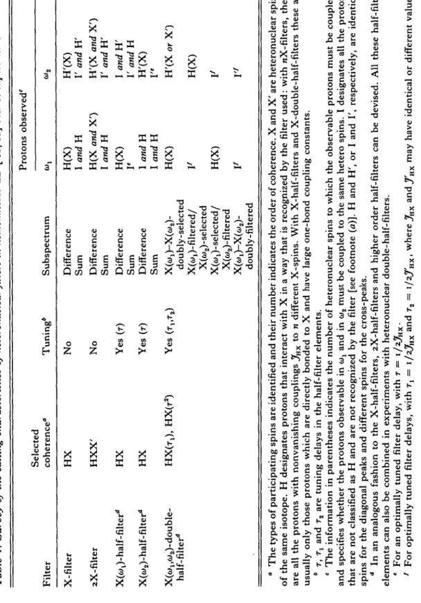
from o to 76 Hz. Fig. 4 shows a comparison of a conventional [1H,1H]-COSY
spectrum of rat metallothionein with the difference spectrum obtained from
pH^HJ-COSY with 113Cd-filter. The spectral region shown contains the diagonal
peaks of the Ca-protons and the cross-peaks between the Ca-protons (along w2)
and the C-protons (along (Oy). The difference spectrum (Fig. 46) contains only
diagonal peaks from protons coupled to 113Cd, and cross peaks between pairs of
protons which are both coupled to the same U3Cd ion. It is seen that both the
diagonal resonances of C-protons and cross peaks with C-protons can routinely
48 G. Otting and K. Wiithrich K31 'A61 (b) Ml Ml K30
Fig. 2. Stereoview of the solution of rat metallothionein-2 determined by NMR. The positions of the polypeptide backbone atoms N, Ca and C , and the side-chain heavy atoms of the 20 cysteinyl residues are connected by straight lines, and the cadmium ions are represented as dotted spheres of radius 0-9 A. (a) a-domain, (6) yff-domain. The first and last residues of each domain are identified by the one-letter amino-acid code and the sequence number (from Schultze et al. 1988).
Fig. 3. Cysteine sulphur-to-113Cd coordinative bond, x1 a nd X2 a r e m^ two dihedral angles that define the conformation of the side chain. In metallothionein 8 of the 20 cysteinyl residues are bound to two Cd'J+ ions.
- 1-5
40
Fig. 4. (a) Conventional phase-sensitive ['H^HJ-COSY spectrum, and (6) difference spectrum from ['H.'HJ-COSY with U3Cd-filter of rat [U3Cd7]-metallothionein-2 (D2O, protein
concentration 10 mM, 20 mM Tris buffer pD 7-0, 50 mM-KCl, 25 °C, :H frequency 360 MHz). The spectral region (Wj = ri-5"5 ppm, w2 = 3-9-5'5 ppm) is displayed, which contains the cross-peaks between C*-protons and C^-protons. In (b) the resonances originating from U3Cd-bound cysteines are framed and identified with the sequence positions. The diagonal peak at 475 ppm corresponds to the incompletely suppressed solvent resonance; the diagonal peaks and cross-peaks between &>2 = 4-35 and 4-45 ppm correspond to additional Cys signals, which are not individually resolved. (From Worgotter et al. 1988.)
5° G. Otting and K. Wiithrich I I I I
3-6
3-6 3-4 «>i (ppm)
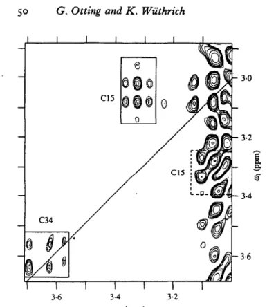
Fig. s- Contour plot of the H^2-H'ra cross-peak region for cysteinyl side chains in rat metallothionein-2 from the difference spectrum of a ['Hj'HJ-COSY experiment with 2X-filter (X = U3Cd; same experimental conditions as in Fig. 4). The position of the diagonal is indicated with a solid line. Resonances, originating from cysteines bound to two U3Cd ions are framed and identified with the sequence positions. The band of t, noise at w2 = 3-0-3-1 ppm stems from the CSH2 resonances of Lys residues. (From Worgotter et al. 1988.)
coupling constants 4J(1H,113Cd). Fig. 5 shows the difference spectrum from a
['rVHJ-COSY with zX-filter (X = U3Cd) recorded with rat [113Cd7
]-metallothionein. Eight of the total of 20 cysteinyl side chains in this protein are coordinated with two metal ions. The spectral region shown in Fig. 5 contains the
H^-H''3 cross-peaks of Cysis and the diagonal peak of one of the C^-protons of
Cys34. Their presence in the 2X-filtered difference spectrum identifies these residues as bridging cysteines with resolved spin-spin couplings from the
C^-protons to two 113Cd ions.
Overall, metallothionein is a good example of a problem where nX-filters are a powerful technique. Similar situations are presented quite generally by intermolecular interactions between hydrogen-bearing organic compounds and diamagnetic metal ions with a nuclear spin I = \. Although this appears to be a narrow area for potential applications, there is a large selection of bivalent metal-dependent enzymes and other proteins where the metal present in the native
compound can be substituted by, for example, 113Cd. DNA-binding Zn-finger
proteins provide a good illustration (South et al. 1989). X-filter techniques might also be used for identification of less specific metal binding sites in proteins and
nucleic acids, possibly with 113Cd as a 'NMR probe'. In all these potential applications the use of a nX-filter in the 2D ['H.'HJ-NMR experiments would enable a characterization of the metal-binding sites with a single experiment recorded with the complex containing the NMR-active metal ion.
2.2 nX-haljT-filters
In biological macromolecules the one-bond coupling constants 1Jf(1H,18N) and
1
.7(1H,1SC) are both large compared to all other homonuclear and heteronuclear
coupling constants, and their variation among different 15NH fragments, or
different 13CH fragments, respectively, is small (Bystrov, 1976). In work with
isotope-labelled molecules, the X-half-filters can therefore be tuned for separating the diagonal peaks of X-bound protons and the cross-peaks with X-bound protons from all other peaks (Tables 1, 3). On this basis the combined use of
isotope-labelling with 13C or 15N and 2D ['H/HJ-NMR with X-half-filters opens
attractive ways to support both the determination of 3D protein structures and studies of intermolecular interactions with proteins and nucleic acids. As any
given proton has only a single one-bond coupling constant with 13C or 15N, we
limit the discussion to X-half-filters with n = 1.
We select the DNA-binding domain of the P22 C2 repressor (Fig. 6; Fig. 1 shows a ^H^Hj-NOESY spectrum of this protein in a complex with an operator
DNA duplex) as an example to illustrate the use of [1H,1H]-NOESY with 15
N-half-filter in support of the determination of a three-dimensional protein structure. The spectra recorded with X-half-filters can be helpful on all three levels of data collection, i.e. for the resonance assignments, the identification of secondary structure elements, and the determination of the 3D structure (Wuthrich, 1986). The feasibility of a protein structure determination by NMR depends primarily
on one's ability to assign the XH-NMR spectrum (Wuthrich et al. 1982).
Assignment of a protein 'H-NMR spectrum includes the identification of the XH
spin systems of the individual amino-acid residues, using through-bond, scalar spin-spin couplings. The spin systems are characteristic for the different amino-acid types, but since a protein usually contains multiple copies of any given amino acid (Fig. 6) the sequence positions cannot be determined from this information
alone. Therefore, using through-space, dipolar couplings observed as 1H-1H
NOEs, two or several sequentially neighbouring amino-acid spin systems are identified {sequential assignments). The peptide segments thus determined are then matched against the primary structure, and as a result the sequence-specific assignments are obtained. Clearly, the reliability of the sequential assignment procedure is reduced when all the amino-acid spin systems cannot be identified
(Wuthrich, 1986). In the P22 C2 repressor individual identification of the XH spin
systems of the long side chains of Leu, Glu, Gin, Met, Arg, Lys and Pro was difficult because of spectral overlap. In this situation, which can generally be anticipated for larger proteins and sometimes even for small proteins with unfavourable spectral properties, sequence-specific assignments were greatly
52 G. Otting and K. Wiithrich TXLXG XLXI AL i i i i .'/ MNTQLMGERIRARRKKLKiRQAALGKMVGVSNVAISQWER I 10 20 30 40 SO 60 70 SETEPNGENLLAtSKALQCSPDyLLKGpi^SQTNVAY
iiAL'' ALX YLLX tSL
C8H3
/ OH
\ CSH3
F i g . 6. A m i n o - a c i d sequence of Salmonella p h a g e P22 C2 repressor 1-76. T h e 10 L e u residues were labelled with " N in t h e e x t e n t of > 85 % . T h e letters above and below t h e s e q u e n c e identify s e g m e n t s of n e i g h b o u r i n g a m i n o - a c i d spin systems in t h e p r i m a r y s t r u c t u r e , w h i c h were identified by sequential a s s i g n m e n t s with ' H - ' H N O E s and led to sequence-specific assignments of all 10 leucines (see text). X stands for a residue from t h e g r o u p G l u , G i n , M e t , A r g , L y s and P r o , for which t h e spin systems were n o t individually identified. At t h e b o t t o m , the s t r u c t u r e of 1 5N-labelled L e u is s h o w n .
the use of 2D ['H/Hj-NMR with X-half-filters. This is readily apparent from the following example: A tripeptide segment with three long amino acid side chains identified by sequential assignments in the non-labelled protein matches the six tripeptides 4-6, 13-15, 14-16, 15-17, 16-18, and 64-66 in the P22 C2 repressor
sequence (Fig. 6). With the unique identification of the leucine spin systems by 15N
labelling the ambiguities of the sequence-specific assignments were greatly reduced, since only the two tripeptides 13-15 and 14-16 then contained three neighbouring residues with long side chains that had not been further specified (see Fig. 6).
Selective labelling of all 10 leucyl residues in P22 C2 repressor was achieved by
growth of the E. coli overexpression system on a minimal medium containing 15
N-leucine, and the other amino acids in unlabelled form. Using a [1H,1H]-TOCSY
experiment with 15N(w2)-half-filter (Fig. 7) the *H resonance assignments for the
amide protons and the Ca protons of the 10 leucyl residues were obtained (Otting
et al. 1986). With these assignments, which were based entirely on covalent, scalar
relations between the different nuclei, the [1H,1H]-NOESY spectrum with
15N(w
2)-half-filter was analysed. Fig. 8 shows the spectral region with the
Fig. 7. Difference spectrum from a ['H.'HJ-TOCSY experiment with 16N(wa)-half-filter of the DNA-binding domain 1-76 of P22 C2 repressor with all 10 leucyl residues labelled with 15
N (protein concentration 8 mM in H2O, pH 4-8, T = 20 °C, MLEV-17 (Bax & Davis, 1985) was used during the mixing time of 60 ms, filter delay T = 5-5 ms, tlmax = 28 ms, 'H frequency 360 MHz). The multiplets are antiphase along <u2. Positive and negative levels are
- 40 o. - 50 - 9 0 70 &l (ppm)
conventional ['H.'HJ-TOCSY experiment this region contains the resonances of the amide protons and the aromatic protons. In the difference spectrum recorded with 15 N(w2)-half-filter all peaks have been suppressed except for the diagonal peaks originating from the 15 N-bound amide protons of Leu. The fine structure components of the diagonal peaks are connected by straight lines, (b) Spectral region (&>: = 3-1-5-2 ppm, w2 = 6-5-9-3 PPm)- 1° conventional ['H.'Hj-TOCSY this region contains the direct cross-peaks between the amide protons and the C-protons, and some relay peaks of Ser and Thr. In the difference spectrum recorded with 15N(w2)-half-filter all peaks have been suppressed except for the direct cross-peaks between the amide protons and the C-protons of the 15N-labelled leucines. In (a) and (6) the peaks of the Leu residues are identified with the sequence locations. (From Otting et al. 1986.)
54 G. Otting and K. Wtithrich 64 0 Q Q21 L50 K16 D68 - 4-0
I
o. - 50 r 90 i 80 I 7-0 Oh (ppm)Fig. 8. Spectral region (wt = 3-i—S"3 ppm, w2 = 6-6-O/3 ppm) of a NOESY spectrum with ls
N(w2)-half-filter recorded in a solution of P22 C2 repressor 1-76 in which all 10 leucyl residues were labelled with l5N (Fig. 6; protein concentration 8 mM in H2O, pH 4-8, 20 °C, mixing time = 100 ms, filter delay = 5-5 ms, tlmtx = 42 ms, *H frequency 360 MHz). The
difference spectrum is shown. The multiplets are antiphase along w2. Positive and negative levels are plotted without distinction. The fine structure components of the cross-peaks are connected by horizontal lines. This spectral region contains NOEs between Ca-protons (aij = 3'x~5'3 ppm) and 15N-labelled amide protons. Dotted lines identify the 10 intra-residual 15
NH-C*H cross-peaks of the labelled leucyl residues. The cross-peaks from sequential and medium-range NOEs are identified by solid lines and labelled with the one-letter symbol and the sequence position of the unlabelled residue involved in the cross-peak. (From Senn et at. 1987 a.)
CaH-1 5NH cross-peaks of the difference spectrum from a pH/HJ-NOESY
experiment with 15N(w2)-half-filter. As the filter effect of the 15N(w2)-half-filter is
only active along the a»2-frequency axis (Table 1), the difference spectrum contains
all the cross peaks which connect the amide proton resonances of the 10 labelled leucines with the resonances of all protons that are close enough in space to give rise to a NOE with an amide proton of Leu. Although each cross-peak is split
along u)i by the one-bond coupling constant 1Jf(1H,1N), the spectrum is very much
simplified when compared to the conventional NOESY spectrum recorded with the unlabelled protein (Fig. 16 gives an impression of the complexity of such
spectra). In this spectral region the 10 intraresidual amide proton-Ca-proton
cross-peaks of Leu (dotted lines) were readily identified, since they have the same
Next, sequential NOEs were identified in Fig. 8 and in other regions of the same spectrum, resulting in the identification of the peptide segments indicated in Fig. 6, and hence in the desired sequence-specific assignments for all 10 leucyl residues.
On the basis of the sequence-specific assignments the p H / H J - N O E S Y experiment with 15N(w2)-half-filter provided direct information on the secondary polypeptide structure near several of the 15N-labelled leucyl residues. For example, both Leu-Leu dipeptide segments (Fig. 6) were found to be located in a-helices, as evidenced by the strong sequential NOEs <fNN throughout the polypeptide segments 49-54 and 63-66 (not shown), and by the NOE connectivities daN (48, 51) and <faN (50, 53) observed in Fig. 8 (Wiithrich et al.
1984). Similarly, several longer-range NOE cross-peaks with the leucyl amide protons could be detected in the simplified 15N-half-filter spectra, which contributes further to the input for the calculation of the complete 3D structure (Wuthrich, 1986, 1989).
At present, and foreseeably also in the future, protein structure determination in solution will depend on the measurement of parameters that are accessible only with ' H - N M R experiments (Wuthrich et al. 1982; Wuthrich, 1986). Supporting procedures using isotope-labelling and heteronuclear NMR techniques should therefore be designed primarily for the purpose of enabling easier access to *H-NMR data. This is exactly the function of heteronuclear niters. Thereby they have the special advantage of providing simplified ' H - N M R spectra and other help in the spectral analysis without the need of ever having to record or assign the heteronuclear NMR spectrum.
The considerations on the use of heteronuclear half-filters for protein structure determination can readily be extended to complexes between two or several molecules. Consider, for example, a stable complex formed between a protein and a low-molecular-weight effector, or the interaction of a DNA fragment with a drug molecule. Assume further that the low-molecular-weight ligand molecule is fully labelled with 13C. In such a system the observation in the p H / H J - N O E S Y spectra with 13C-half-filter of the NOEs with the 13C-bound protons may allow to identify the sites on the macromolecule that are in contact with the ligand, and to probe the local conformation around the 13C-bound ligand protons. Foreseeably, studies of stable intermolecular complexes of isotope-labelled ligands with biological macromolecules could well turn out to be the most attractive use of heteronuclear half-filters (Fesik et al. 1988; Fesik, 1988).
2.3 Heteronuclear half-filters and heteronuclear three-dimensional NMR
The difference spectrum derived from a 2D pH^HJ-NMR spectrum with
X-half-filter represents the projection of a corresponding 3D X-correlated [1H,1H]-NMR
data set (Fesik et al. 1989; Marion et al. 1989; Messerle et al. 1989) along the heteronuclear frequency axis onto a 2D plane with two proton frequency axes
(Fig. 9). For example, in the three-dimensional versions of the 15N-half-filter
56 G. Otting and K. Wuthrich (a) (ox C'H)
0
00
o
0
0
o 0
0
o o
0
o
(X) <B3('H) (b)Fig. 9. Schematic presentation of a heteronuclear 3D NMR spectrum (a) and the corresponding X(w2)-half-filtered 2D ['H.'HJ-NMR experiment (6). (For example, these could be a 3D 15N-correlated ^H^HJ-NOESY spectrum, and a 2D ['H^HJ-NOESY spectrum with 15N(w2)-half-filter.) The spectrum (6) corresponds to a projection of the peaks in the different u)1-<i)3 planes of spectrum (a) along the heteronuclear shift axis onto a single
w^'HJ-WjCHJ-plane. (Note that the w3-axis of the 3D NMR spectrum corresponds to the 6J2-axis of the 2D NMR spectrum.)
chemical shifts of the leucyl residues in P22 C2 repressor. In the spectral region of the pH/HJ-TOCSY spectrum shown in Fig. 7, each of these planes would contain a single cross-peak. In the 3D spectrum corresponding to Fig. 8, each
plane would contain a linear array of cross-peaks at the o>3 chemical shifts of the
individual leucyl amide protons, and with variable co1 chemical shifts. This latter
example would correspond to the scheme in Fig. 9.
Clearly, in the examples of Figs 7 and 8 the additional improvement of the spectral resolution afforded by the 3D experiment is not needed. In a uniformly 15
N-labelled protein, however, the spectral resolution of a ^H^HJ-NOESY
experiment with 15N-half-filter is usually only little improved relative to
conventional [1H,1H]-NOESY, but further decisive improvements of the
resolution can be achieved with the use of 3D NMR spectroscopy (Fesik et al. 1989). We conclude that any decision on the experiments to be used must be
tailored to the available isotope-labelling of the compounds studied. In each individual situation, the choice of the strategy to be used will depend primarily on the problem to be solved, but also on the efficiency of the procedures to be used and the financial expenditure. These general considerations indicate that the combinations of different types of selective isotope-labelling with heteronuclear half-filters in 2D ['H/HJ-NMR spectra, or random isotope-labelling with heteronuclear 3D NMR, respectively, will be valid alternatives in future practice.
3. FUNDAMENTAL CONSIDERATIONS ON HETERONUCLEAR FILTERS AND THEIR USE IN 2D p H / H J - N M R
3.1 Hardware requirements
2D pH^HJ-NMR spectra with heteronuclear filters are obtained with experimental schemes consisting of pulses at the radiofrequencies of the protons and the heteronuclei, and of delays. These are combined with proper phase cycling of the pulses and the receiver to select the desired coherence transfer pathways. All these facilities are available in commercial multinuclear high-resolution NMR spectrometer systems. The sensitivity of proton observation can be optimized with the use of a reverse probehead, which has two separate radiofrequency coils. The inner coil is tuned and matched to the proton frequency, and is used for both the generation of *H radiofrequency pulses and for *H signal detection. The outer coil is selectively tuned and matched to the frequency of the heteronucleus of interest, and is used only to deliver radiofrequency pulses at the heteronuclear frequency. In a broadband probe one can choose from a wide range of heteronuclear frequencies, whereas a selective probe may be constructed for optimal performance with a single heteronucleus,
for example, 15N or 13C.
3.2 Product operator description of heteronuclear filtering
In the product operator descriptions (Sorensen et al. 1983) of nX-filters we denote the heteronuclear spins as X, protons coupled to X as H, and protons not coupled to X as I. For X half-filters, X denotes again the heteronuclear spins, H designates X-bound protons with large one-bond couplings to which the filter delay is tuned, and I stands for protons that are not directly bound to a X nucleus. For simplicity of the presentation the calculations were performed for protons which are not coupled to any other protons and therefore give rise to diagonal peaks in the 2D pH/HJ-NMR spectrum, and effects that might arise from spin relaxation and from strong spin-spin coupling are neglected.
The effect of heteronuclear filters is based on the fact that the sign of proton magnetization in antiphase relative to a spin X can be inverted with the application of a 7r-pulse at the radiofrequency of X:
(n)j(X)
58 G. Otting and K. Wiithrich
The combination of two experiments A and B with and without a 7r(X)-pulse then represents a straightforward means for separating the heteronuclear antiphase magnetization iiixKz from in-phase proton magnetization lx:
experiment A: lx + 2HxXz >lx — 2HXXz,
experiment B: lx + zHxXz *• lx + zRxXz.
Two linear combinations of A and B yield the desired subspectra: The difference of the two data sets obtained from the experiments A and B selects the term zli^X^ while the sum contains the proton magnetization lx.
The 7r(X)-pulse which is inserted to achieve the subspectral editing is called the editing pulse. In practice the presence of a 77-(X)-pulse in experiment A and its absence in experiment B are achieved by the combination of two (7r/2)(X)-pulses,
(»r/2),(X)-(w/2),(X). (3) The phase of the first pulse in (3) is kept constant, and the (77y2U(X)-pulse is actually the editing pulse. An effective (7r)I(X)-pulse is obtained with \jr — x, and an effective zero degree pulse with \/r = — x. This scheme ensures that the same pulse power is used in both experiments, and it minimizes potential artefacts that might arise from combining two data sets recorded under non-identical conditions (Freeman et al. 1981).
With the phase cycle of a ^-quantum filter (Piantini et al. 1982), the pair of (7r/2)(X)-pulses [equation (3)] can be used to select proton coherences which are antiphase with respect to n heterospins X (Worgotter et al. 1988). For example, to select the coherences with an even number of X operators in the product operator representation, e.g. ^H^^, the phase of the first (77-/2)(X)-pulse is kept constant, while the phase i/r of the second, editing (7r/2)(X)-pulse is cycled through
x
> y> ~x> a nd —y- The data sets recorded with the phases x and —x, and those recorded with y and —y, respectively, are stored separately. Their summation and subtraction result in the sum spectrum and the difference spectrum, respectively. The signals in the difference spectrum come from those coherences which have passed the 2X-filter.
The most straightforward application of the pair of pulses in equation (3) for subspectral editing is realized with the nX-filters. nX-half-filter techniques use the pulse combination of equation (3) together with a filter delay.
3.3 Extension of 2D ^H^JH^-NMR experiments with heteronuclear filters
Heteronuclear filters can be inserted into any 2D f/H^HJ-NMR pulse scheme. In the phase cycling program of the resulting experiment with heteronuclear filter, the phase cycle of the basic pH/rfJ-NMR experiment is retained and has to. be extended by independent phase cycling of the additional pulses introduced by the heteronuclear filter. The total number of steps in the resulting overall phase cycle corresponds to the product of the number of phase cycling steps in the basic 1 H-NMR experiment and those in the heteronuclear filter.
For practical purposes, if complete phase cycling is attempted the phase cycling schemes of 2D ['H/HJ-NMR experiments with heteronuclear niters may result in unacceptably long measuring times. This is best illustrated with a numerical example corresponding to a typical experiment with a biological macromolecule: 500 free induction decays must be acquired to record a phase-sensitive 2D NMR data set that covers a sweep width of 10 ppm at 500 MHz with a maximum
evolution time, t1 max, of 50 ms. For an experiment with a 64-step phase cycle and
a repetition rate of 1 4 s per scan, the total measuring time is at least 500 x 64 x 1-4 s « 12 h. Experiments with w-fold longer phase cycles require n times longer measuring times. All the individual steps in the complete, 'optimal' phase cycling schemes for 2D ['H.'HJ-NMR experiments do, however, not have equal weight, and some steps may be omitted without causing major deterioration of the quality of the resulting spectra. In the descriptions of individual experiments with heteronuclear filters (sections 4-6 below), a sequence of phase cycling priorities is indicated with each of the experimental schemes. These priority lists indicate the order in which the individual 2-step phase cycles should preferably be applied. Only minor artefacts are expected to result from omission of steps given at the end of the priority list, whereas the phase cycling steps at the top of the list are indispensible for obtaining spectra of satisfactory quality.
As an illustration of these somewhat abstract comments on phase cycling in 2D pH/HJ-NMR with heteronuclear filters, the following subsections 3.3.1-3.3.3 describe, respectively, phase-cycling schemes that have proven to produce high-quality spectra with the conventional pH^HI-COSY and pH^HJ-NOESY experiments, and a discussion of two alternative priority lists for phase cycling with the heteronuclear filters. Considerations similar to those described for COSY and NOESY apply for other 2D ['H/Hj-NMR experiments, which can all be supplemented with heteronuclear filters.
3.3.1 Phase-cycling in the ^H^H^-COSY experiment
The experimental scheme of phase-sensitive COSY is shown in Fig. 10a together with the coherence transfer pathway selected by the standard phase cycling for this experiment (Bodenhausen et al. 1984). The experiment consists of two (n/2)
pulses with phases (j>^ and 02, which are separated by the evolution period tl and
succeeded by the detection period t2 (Aue et al. 1976; Ernst et al. 1987; Wuthrich,
1986). For each fx value the experiment is repeated eight times with different phase
settings of the radio frequency pulses and the receiver. These eight phase settings are listed in Fig. 106. To further improve the signal-to-noise ratio, the entire phase cycle may be repeated n times with accumulation of a total of Sn scans.
The phase cycling of Fig. 106 contains the three basic 2-step cycles of Fig. io(c-e). In step (c) the simultaneous incrementation of the phases of the two pulses and the receiver from x to y ensures the elimination of undesired quadrature images. (A quadrature image originating from unbalanced receiver channels in dual channel detection would lead to an anti-diagonal in the 2D spectrum). The phase cycle of Fig. 10c corresponds only to the
60 G. Otting and K. Wiithrich (a) T 1 T£ (b) 0| = x y x y -x -y -x -y 4>i = x y ~x -y x y -x -y rec = x y x y -x -y -x -y (c) (d) (e) <t>x=x -x rec = x -x
Fig. 10. (a) Experimental scheme of phase-sensitive ['H.'HJ-COSY. The two vertical bars represent n/z radiofrequency pulses. (The experimental schemes discussed in this review contain exclusively pulses of duration 77/2 or n. These two pulse lengths are distinguished by the thickness of the bars. The phases of the pulses are indicated above the pulse
symbols). t, is the evolution period. The triangle represents the free induction decay during the detection period tr To obtain a 2D data set s(tvt2), the experiment is repeated with
incrementation of tv typically for about 512 tt values. For each f, value multiple scans are
accumulated to improve the signal-to-noise ratio. During this accumulation the phases of the pulses and the receiver (rec) are cycled. The minimal number of scans for each /, value is thus determined by the length of the phase cycle used. The coherence transfer pathway selected by the phase cycle (b) is indicated below the pulse sequence, where p is the order of coherence. (6) Complete 8-step phase cycle used to obtain the coherence transfer pathway in (a) and to suppress unwanted quadrature images, (c) Phase cycle of 2-step CYCLOPS, (d), (e) Basic two-step phase cycles for the selection of the coherence transfer pathway.
first half of the conventional CYCLOPS phase cycle (Hoult & Richards, 1975) and will in the following be referred to as 2-step CYCLOPS. In the complete CYCLOPS phase cycle, simultaneous incrementation of the phases of all pulses
and the receiver in steps of 900 or 1200 (Hoult & Richards, 1975; Bodenhausen
et al. 1984) results in the suppression of DC-offsets (DC = direct current) in the free induction decays, in addition to the quadrature image suppression. The use of 2-step CYCLOPS is a compromise resulting in a 2-fold reduction of the length of the overall phase cycle, which will be of practical interest for the experiments with heteronuclear filters. The absence of DC-offset suppression is acceptable, as is shown by the following considerations: The Fourier transform of a DC-offset is
a spike at the carrier frequency, which will give rise to a narrow band of tx noise.
This t1 noise band at the carrier frequency of the 2D spectrum is negligibly weak
recorded in aqueous solution the radiofrequency carrier is placed at the frequency of the solvent resonance, this artefact will hardly be noticeable. It is further worth noting that the elimination of quadrature images by 2-step CYCLOPS depends critically on the reproducibility of the experimental conditions during the time period between the two steps of the phase cycle. Therefore, it is advisable to apply step CYCLOPS before the cycling of the other phases. The two additional 2-step phase cycles of Fig. 10 (d, e) select for the coherence transfer pathway indicated below the pulse sequence in Fig. 10 a.
The complete COSY phase cycle of Fig. 10b was generated by independent cycling of each of the steps of Fig. io(c-e). Thus, the first two steps of the COSY phase cycle correspond to 2-step CYCLOPS (Fig. 10c). The next two steps are
obtained by repetition of the first two steps, but with alternation of the phase <j>2
according to Fig. \od. Finally, the first four steps are repeated with simultaneous phase alternation for the first pulse and the receiver (Fig. ioe). The sequence in which the basic 2-step phase cycle elements are used in Fig. 106 corresponds to
the phase cycle priority list: 2-step CYCLOPS, 08, (/>1.
3.3.2 Phase cycling in the ^H^H^-NOESY experiment
Fig. 11 a shows the experimental scheme of a NOESY experiment. Three (n/z)
pulses with phases <f>x, <j>2 and 03 are separated by the evolution period tx and the
mixing period Tm, and succeeded by the detection period t2. Fig. 11 b shows the
phase cycling suggested for use in experiments with heteronuclear filters. As with COSY, 2-step CYCLOPS is used to shorten the phase cycle. Another compromise accepted with this scheme is that in contrast to the routinely used, more extensive phase cycle for NOESY (Bodenhausen et al. 1984), the present version does not suppress two-quantum coherence (Macura et al. 1981).
The phase cycle in Fig. 116 has been generated along similar lines as the aforementioned COSY phase cycle of Fig. 10 b. The individual 2-step phase cycles are shown in Fig. 11 (c-f). 2-step CYCLOPS (Fig. 11 c) is again used for quadrature image suppression. The overall phase program of Fig. 11 b is obtained
by additional independent alternation of the phases <j>z (Fig. 11 d), 02 (Fig. n e )
and <f>x (Fig. 1 if) with simultaneous alternation of the receiver phase. The
coherence transfer pathway selected by this simple alternation of the phases <f>lt <f>2
and 0j eliminates single quantum coherence during the mixing period.
3.3.3 Two alternative priority lists for the phase cycling in zD ^H^H with heteronuclear filters
As was mentioned at the outset of section 3.3, phase-cycling priority lists will be indicated with each individual experiment described in sections 4-6. Here, we discuss two alternative phase-cycling schemes. The choice between the two schemes will depend primarily on the overall measuring time available. In principle it is desirable to cycle the phase of the editing (?r/2)(X)-pulse [see equation (3)] first. This ensures that the data sets recorded with different phases of the editing pulse are not significantly affected by spectrometer instabilities,
62 G. Otting and K. Wiithrich (a) r i <h <h (b) <I>I=JC y x y x y x y -x -y -x -y -x -y -x -y <k=x y x y -x -y -x -y x y x y -x -y -x -y fa = x y -x -y x y -x -y x y -x -y x y -x -y rec = x y -x -y -x -y x y -x -y x y x y -x -y ,=.r y (e) <t>{=x x (f) ipt=x -x 03
rec =x y rec = x -x rec = x -x rec = x —x
Fig. I I . (a) Experimental scheme of phase-sensitive ['H.'HJ-NOESY. Tm is the mixing time. The coherence transfer pathway selected by the phase cycle (b) is indicated below the pulse sequence, (b) Complete i6-step phase cycle used to obtain the coherence transfer pathway in (a) and to suppress unwanted quadrature images, (c) Phase cycle of 2-step CYCLOPS. (d)-(f) Basic two-step phase cycles for the selection of the coherence transfer pathway.
which might contribute to deterioration of the result of their later summation or subtraction. However, in turn one then has to consider that coherent artefacts from insufficient relaxation between successive scans can accumulate if each scan for the data set with effective 7r(X)-editing pulse is always immediately followed by the corresponding scan for the data set without effective editing pulse. It has been shown by Worgotter et al. (1988) that these artefacts can be suppressed by repetition of the phase cycle with permuted order of the phase of the editing (7r/2)(X)-pulse. In the first half of this extended phase cycle, each scan for data set A is followed by the corresponding scan for data set B [equation (2)], while in the second half the scans for data set A are sampled after the corresponding scans for data set B. In the following this procedure will be referred to as 'repetition of the phase cycle with permuted order of the phase xfr of the editing (?r/2)(X)-pulse'. For experiments with 2X-filters or 2X-half-filters a 4-fold permutation of the phase cycle has been recommended (Worgotter et al. 1988).
Inevitably the repetition of the phase cycle with permuted order of t/r doubles the overall length of the phase cycle. In situations where this is not acceptable, an alternative phase-cycling priority list is recommended, with the phase xjr of the editing pulse as the last step rather than the first step. This alternative data collection scheme imposes more stringent requirements on the stability of the
'H
Fig. 12. Experimental schemes for (a) ['H.'Hl-COSY with X-filter, and (6) ['H/H]-NOESY with X-filter. The letters 'H and X, respectively, identify the entries for pulses and delays at the radiofrequencies of the protons and the heteronucleus.
spectrometer, but it avoids the bias in the data from insufficient relaxation between successive scans. A further reduction of this bias can be achieved, if two to four dummy scans are inserted between the sets of scans recorded with different phases of ft. These two alternative phase cycling schemes are further discussed in section 7 below.
4. 2D ['H.'HJ-NMR EXPERIMENTS WITH TlX-FI LTERS
4.1 Product operator description of X-filters in ^H^ffi-COSY and pH,1
!!]-NOESY
In 2D ['H/HJ-NMR experiments with X-filter the pair of (77/2)(X)-pulses is inserted during the mixing period (Worgotter et al. 1986). Fig. 12 shows the resulting experimental schemes of pH/KJ-COSY and ^H/HJ-NOESY with X-filter. The phase cycles of the proton pulses are the same as for the corresponding unfiltered experiments (Figs io, 11). The phase-cycle priority list for COSY with
X-filter is: ft, 2-step CYCLOPS, <p2, <fiv and repetition with permuted order of ft.
This leads to a 32-step phase cycle. For NOESY with X-filter we have the phase
cycling priority list: ft, 2-step CYCLOPS, <f>3, (f>2, $lt and repetition with permuted
order of ft. This results in a 64-step phase cycle.
Following equations (2) and (3), the data sets recorded with phases x and — x of the editing (7r/2)^(X)-pulse are stored separately. The difference spectrum obtained by subtraction of these two data sets (Tables 1 and 2) contains only peaks from proton resonances that have evolved into antiphase magnetization with
respect to the heteronucleus X during the evolution period tlt and are therefore
sensitive to the editing pulse. During the detection period t2 they refocus into
64 G. Otting and K. Wiithrich
X-coupled proton to other protons by the mixing scheme used in the 2D
NMR experiment, the resulting coherence is still antiphase with respect to the
X-spin, and will become observable during t2 only if these other protons also have
non-vanishing scalar couplings to the heterospin. Therefore, the difference spectrum contains exclusively cross peaks between pairs of protons which are both coupled to the same heteronucleus X (Tables 1, 2). In the sum spectrum obtained by addition of the two data sets recorded with \jr = x and — x, respectively, the observed peaks result from those product operator terms which are insensitive to the phase of the editing pulse.
Starting from equilibrium magnetization of a proton coupled to a X spin, H2,
and a proton not coupled to X, Iz, the product operator calculation shows that the
following terms are created at the start of the detection period: ['H.'HJ-COSY with Xfilter
2 »• sin Qtt cos njnx t^ Hx + cos Qt 1 sin nJHX tt zHx Xz
z • sin Qtt \x.
['H.'HJ-NOESY with X-Hlter
2 >• cos Qtx cos 7r%jX tx Hv ± sin Qtt sin nJHX t12Hy X
y cos Qtx ly.
(4)
(5)
The trigonometric coefficients depending on Qtx and _5HX tt describe the
evolution of the magnetization under the chemical shift Hamiltonian and under
scalar coupling, respectively, during the evolution time *x (Sorensen et al. 1983).
As in all product operator calculations in this text, only the coherence transfer pathway selected by the phase cycling used (see Figs 10, 11) has been taken into account in equations (4) and (5). The upper and lower signs in equations (4) and (5) correspond, respectively, to the phase settings \/r = X and ifr = —X of the editing (7r/2)(X)-pulse. The difference between the two data sets recorded with
different phase xjr of the editing (77/2)(X)-pulse selects the terms 2RXXZ [equation
(4)] and 2Hj,X2 [equation (5)], respectively, whereas the sum contains all in-phase
magnetization. The X-coupled proton spins H occur in the two subspectra with different multiplet fine structures. Thus the X-filter is a technique for editing the multiplet fine structure of X-bound protons (rather than complete cross-peaks or complete diagonal peaks) into different subspectra. Nonetheless, since the proton spins I are absent from the difference spectrum, this subspectrum may be considerably simplified for suitably isotope-labelled compounds (Fig. 4). In contrast, the sum spectrum contains all the peaks seen in the conventional 2D PH/HJ-NMR experiment (Table 1).
The product operator terms of equations [4] and [5] give an accurate description of the phases and the multiplet fine structures observed in the X-filtered experiments. The trigonometric factors contain the phase and multiplet
information for the tx domain, and the operator terms contain the corresponding
information about the t2 domain. We thus see that the sum spectrum and the
difference spectrum have the same phase in both dimensions: In the COSY experiment the trigonometric factors are all odd and the transverse operator terms are all along the x-axis. In the NOESY experiment the trigonometric factors are all even and the transverse operator terms are all along the j>-axis. For all
multiplets the fine structure in the sum spectrum is in-phase with respect to the
X spin in both dimensions, because the functional dependence on JHxfi is cosine
modulated and the operator terms represent in-phase magnetization. In contrast,
since the functional dependence on ,7HX*I 'S s m e modulated and the product
operator terms are antiphase with respect to the heteronucleus X, the peaks in the difference spectrum are antiphase in both dimensions with respect to the
heteronuclear coupling JBX. (Note that these heteronuclear antiphase multiplets
cannot be decoupled with heteronuclear broadband decoupling.) The corresponding conventional 2D ['H/HJ-NMR spectrum without X-filter can be thought of as the sum of the two subspectra obtained with the X-filter. For example, in NOESY [equation (5)] the diagonal peaks of X-bound protons would
be represented by the superposition of the in-phase multiplet cos Qt1 cos 7r7Hxfi Hy
with the antiphase multiplet —sin Qtx sin 7r7Hx*i 2^ J/XZ. Only half of the multiplet
components are thus observed in the standard 2D ['H^Hj-NMR spectra of X-coupled protons, and the resulting multiplet pattern is of E. COSY-type (Griesinger et al. 1986) with respect to the heteronuclear coupling. (This phenomenon was initially observed and analyzed in ^H^HJ-COSY spectra of
[U3Cd7]-metallothionein (Neuhaus et al. 1984)).
4.2 Experimental examples of ^H^Hj-COSY and [lH,xH]-NOESY with
X-filters
A practical application of ['H^Hl-COSY with X-filter is presented in Fig. 4, and Fig. 5 illustrates the use of a 2X-filter. In these experiments with the protein
[U3Cd7]-metallothionein, the identification of 113Cd-coupled protons was based on
the presence of the corresponding signals in the simplified difference spectra. An experimental verification of the quantitative predictions on the multiplet fine structures in 2D ['H/HJ-NMR experiments with or without X-filter [equations (4) and (5)] was obtained with an experiment recorded with a sample of U 3Cd2 +-EDTA using the experimental scheme of Fig. 126. The cross-peak shown in Fig. 13 manifests a NOE between two methylene protons, H and H',
which are also coupled to each other via the scalar coupling constant JHH-. In
addition, both protons have non-vanishing heteronuclear coupling constants, JHX
and jfHX, with the 113Cd ion. The iD multiplet pattern of each proton is a doublet
of doublets (Fig. 13 a, top panel). Equation (5) can be expanded to describe the multiplet fine structure of the NOESY cross-peaks of Fig. 13 by multiplication of
each term on the right by the factor cos 7ryHWt1, which describes the scalar
coupling between the two methylene protons. The proton-proton coupling JHH,
causes a splitting of the cross-peak multiplet into four components centred on the
corners of a square spanned by the coupling constant ^H- (see the top panel of Fig.
13a). Fig. 13a shows the E. COSY-type cross-peak pattern observed in an unfiltered ['H^HJ-NOESY experiment, which corresponds to the data set recorded with ^ = — x (Fig. 12 b). Fig. 136 shows the result obtained with \jr = x. The difference and the sum of the data of Fig. 13(0, b), are shown in Fig. i3(c, d). The overall absolute intensity of the fine structure components in the cross-peak of Fig. 13 (c, d), is the same as in the corresponding unfiltered experiment of
f si n T-' r
o
8
#
o
•
88
•
o
I
09 o 9 § 3-2 8 3-2 3 3-2 8 3-2 3 3-2 8 3-2 3 -30 5 - 3-1 0 a, a" 3-2 8 3-2 3 w2 (ppm ) Fig . 13 . Fo r legen d se e opposit e page .Fig. 13 a. However, in the X-filtered spectra the intensity is distributed among twice the number of multiplet components, so that the intensity of each individual component is halved.
5. 2D ['H/HJ-NMR EXPERIMENTS WITH X-HALF-FILTERS
For reasons outlined at the outset of section 2.2 the following treatment is limited to X-half-filters, although higher order wX-half-filters with n > 1 can in principle also be devised.
5.1 X-half-filter elements
The basic X-half-filter element (Otting et al. 1986) is shown in Fig. 14a. It
consists of a filter delay of duration r = i/2jfux, where jfHX is the heteronuclear
coupling constant recognized by the filter, and of pulses on both the proton
channel and the heteronuclear channel. The 7r(1H)-pulse in the centre of the delay
refocuses the chemical shift evolution of the protons. The phase £ of the 7r(1
H)-pulse is alternated between x and — x to compensate for deviations from the 1800
flip angle and for off-resonance effects (Fig. 14^; Bodenhausen et al. 1977). The receiver phase is kept constant upon alternation of the phase £ (Fig. 14^). The phase \\r of the last, editing (7r/2)(X)-pulse is alternated between x and — x
independently of the phase f. Its combined effect with the preceding (n/2)x
(X)-pulse is that of an effective (7r)I(X)-pulse for \jr = x, or a zero degree pulse for
ijr = — x. The receiver phase is not changed upon alternation of the phase 1/r, but the data recorded with rjr = x and rjf — —x are stored in different memory locations, so that they can be additively or subtractively combined after the recording of the complete data set to yield the desired subspectra (Table 1).
To describe the X-half-filter in terms of product operators we make use of the fact that the one-bond coupling constants ^ ^ H . X ) between X and the directly bonded protons are nearly identical for all fragments H-X, and are much larger than any other homonuclear or heteronuclear coupling constant involving the Fig. 13. Comparison of the experimental multiplet patterns (bottom) of the NOESY cross-peak connecting the acetate methylene protons H and H' in U3Cd2+-EDTA
(ethylenediaminetetraacetate) with the pattern predicted for an HH'X spin system with 17HH'I = 1^>'5> 17HXI = X3'6> anc> UH'XI = I2'° ^z (t0P Pa r t °f t n e figure). In the predicted spectra full and open circles indicate opposite signs of the multiplet components, (a)
pH^H]-NOESY. The analysis in terms of spin-spin coupling constants is indicated in the predicted spectrum. (6) ['H.'HJ-NOESY with an effective ^(n3Cd)-pulse applied simultaneously with the lH observation pulse (Fig. 126). (c) Difference spectrum from [lH,lH]-NOESY recorded with a U3Cd filter. In the experiment the signs of the individual fine structure components coincide with those predicted. The product operator expression for this cross-peak is given at the top. It can be derived from the second product operator term for X-coupled protons in equation (5), which describes the diagonal peaks, by multiplication with a factor accounting for the homonuclear scalar coupling between the two NOE-related protons H and H', cos irjm.tv (d) Sum spectrum from pH.'HJ-NOESY recorded with a 113Cd filter.
The product operator description for this cross peak can be derived from the first product operator term for X-coupled protons in equation (5). (From Worgotter et al. 1986.)
68 G. Otting and K. Wiithrich (a)
n
n m
n
SLn m
(c)Fig. 14. X-half-filter elements. The filter delay r is optimally tuned with r = i/zJHX. The
(7r/2)(X)-pulse with phase \[r is the editing pulse, (a) Basic filter element. (6) X-half-filter element with spin-lock purge pulse (denoted SL). The spin-lock purge pulse has the same phase as the immediately preceding (ff/zX'HJ-pulse of the 2D ['H/HJ-NMR
experimental scheme into which the half-filter element is inserted. Its duration is typically 1-2 ms. (c) X-half-filter element with refocusing delay, (d) 2-step phase cycle of the ff(l H)-pulse.
protons H. Therefore, if the delay T is chosen to be i/21Jf(1H,X), the evolution under the scalar coupling Hamiltonian during this relatively short time period can be neglected for all coupling constants other than 1^f(1H,X). If the X-bound protons are designated H and all other protons are I, then we have that in-phase proton magnetization Kx + lx evolves as follows:
T/2-7Tc<lH)-77I(X)-T/2


![Fig. 4. (a) Conventional phase-sensitive ['H^HJ-COSY spectrum, and (6) difference spectrum from ['H.'HJ-COSY with U3 Cd-filter of rat [ U3 Cd 7 ]-metallothionein-2 (D 2 O, protein](https://thumb-eu.123doks.com/thumbv2/123doknet/14888962.648288/11.745.93.597.103.771/conventional-sensitive-spectrum-difference-spectrum-filter-metallothionein-protein.webp)