Abstract. The dystrophin glycoprotein complex (DGC) is
a multimeric protein assembly associated with either the
X-linked cytoskeletal protein dystrophin or its autosomal
homologue utrophin. In striated muscle cells, the DGC
links the extracellular matrix to the actin cytoskeleton and
mediates three major functions: structural stability of the
plasma membrane, ion homeostasis, and transmembrane
signaling. Mutations affecting the DGC underlie major
forms of congenital muscle dystrophies. The DGC is
prominent also in the central and peripheral nervous
sys-tem and in tissues with a secretory function or which form
barriers between functional compartments, such as the
blood-brain barrier, choroid plexus, or kidney. A
consid-erable molecular heterogeneity arises from cell-specific
expression of its constituent proteins, notably short
C-ter-minal isoforms of dystrophin. Experimentally, the
genera-tion of mice carrying targeted gene delegenera-tions affecting the
DGC has clarified the interdependence of DGC proteins
for assembly of the complex and revealed its importance
for brain development and regulation of the ‘milieu
intéri-eur’. Here, we focus on recent studies of the DGC in brain,
blood-brain barrier and choroid plexus, retina, and kidney
and discuss the role of dystrophin isoforms and utrophin
for assembly of the complex in these tissues.
Keywords. Blood-brain barrier, choroid plexus, dystrophin, Dp71, epithelial cell, endothelial cell, homeostasis,
kidney, retina, targeted gene deletion, transmembrane signaling, utrophin.
Introduction
The dystrophin glycoprotein complex (DGC) comprises
five classes of proteins (dystroglycans, syntrophins,
dys-trobrevins, sarcoglycans, and sarcospan) made of several
members or isoforms, and assembled with either
dystro-phin or its autosomal homologue utrodystro-phin (Fig. 1). The
DGC has been studied mainly in the context of muscle
dystrophies and cardiomyopathies [1–3]. It is critical
for integrity of muscle fibers by linking the actin
cyto-skeleton to the extracellular matrix (ECM) [4–8]. More
recently, its roles as a signaling complex and as a
scaf-fold for membrane proteins have gained preeminence.
Furthermore, the DGC has been recognized to be
mo-lecularly heterogeneous and present in numerous tissues,
notably in the central and peripheral nervous system, and
in tissues with secretory function or forming barriers
be-tween functional compartments, such as the blood-brain
barrier (BBB), choroid plexus (CP), or kidney. While the
functional role of ‘non-muscle’ DGC remains to be
clari-fied in most of these organs, there is compelling evidence
for its involvement in brain development, synapse
forma-tion and plasticity, as well as water and ion homeostasis.
The analysis of mice carrying spontaneous or targeted
mutations affecting specific DGC components has
clari-fied the interdependence of DGC proteins for assembly
of the complex. These studies have also shown that
de-spite functional redundancy, dystrophin isoforms and
Review
Role of dystrophin and utrophin for assembly and function
of the dystrophin glycoprotein complex in non-muscle tissue
T. Haenggi
+and J.-M. Fritschy*
Institute of Pharmacology and Toxicology, University of Zurich, Winterthurerstrasse 190, 8057 Zürich
(Switzerland), Fax +41 44 635 6874, e-mail: fritschy@pharma.unizh.ch
Received 4 October 2005; received after revision 14 March 2006; accepted 5 April 2006
Online First 15 May 2006
* Corresponding author.
+ Present address: Department of Clinical and Experimental Epi-lepsy, Institute of Neurology, University College London, Queen Square, London, WC1N 3BG, UK.
DOI 10.1007/s00018-005-5461-0 © Birkhäuser Verlag, Basel, 2006
utrophin are likely to fulfill distinct roles in non-muscle
tissue. Here, we will briefly summarize major features of
the DGC in skeletal muscle cells and present an overview
of recent developments about the DGC in the brain, BBB,
retina, and kidney. The major focus of this review is the
role of dystrophin isoforms and utrophin for proper
as-sembly and function of the DGC in these tissues.
The DGC in skeletal muscle
The major components of the DGC have been isolated
and characterized best in skeletal muscle cells and will
be presented briefly (Fig. 1) before discussing their role
and localization in ‘non-muscle’ tissue. The sarcoglycan
complex and sarcospan, which are transmembrane
pro-teins linked to the DGC (Fig. 1), will not be considered
in this review.
Dystrophin
The dystrophin gene, located on the X chromosome,
spans approximately 2.5 Mb and is composed of 79 exons
[9, 10]. Duchenne muscular dystrophy (DMD) is caused
by mutations causing a frame-shift and abortion of
pro-tein translation. Three independently regulated
promot-ers, in muscle, brain, and specifically Purkinje cells in the
cerebellum, control expression of full-length dystrophin
[11–17]. In skeletal muscle, dystrophin predominates at
the sarcolemma but is also found at the troughs of the
postsynaptic membrane along with voltage-gated sodium
channels [17, 18]. Several short dystrophin isoforms arise
from differential promoter usage. Transcripts from four
internal promoters encode proteins of 260, 140, 116, and
71 kDa (Dp260, Dp140, Dp116, and Dp71) [19]. Dp71
is subjected to alternative splicing of exons 71–74 and/
or 78, generating at least four Dp71 isoforms [20] with
widespread distribution in various non-muscle tissues
[21–32]. Full-length dystrophin binds actin near its N
terminus and dystroglycan, thereby providing a structural
link between the membrane and the cytoskeleton (Fig. 1).
Dp71 also carries an actin-binding site [33], suggesting
that this short C-terminal isoform fulfills similar
func-tions.
Utrophin
Utrophin was discovered as a cDNA isolated from fetal
muscle with high homology to the DMD gene [34]. It is
expressed in nearly all tissues, including skeletal muscle,
with particularly high levels in lung, kidney, nervous
sys-tem, and vascular endothelial and smooth muscle cells
[25, 31, 32, 35–38], but its function remains largely
un-known. Most studies have focused on adult skeletal
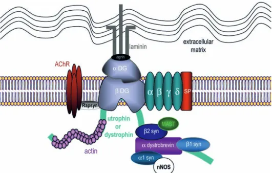
mus-Figure 1. Schematic organization and composition of the DGC at the neuromuscular junction. Dystrophin or utrophin bind to actin
fila-ments via their N terminus. At the C terminus, dystrophin or utrophin are associated with integral and peripheral membrane proteins that can be classified as the dystroglycan complex, the sarcoglycan-sarcospan complex, and the cytoplasmic complex. The cytoplasmic complex includes isoforms of syntrophin (α1-, β1-, β2-syn) and α-dystrobrevin. The sarcoglycan-sarcopsan complex comprises isoforms of sarco-glycan (α,β,γ,δ) and sarcospan (SP). The extracellular component of the dystroglycan complex, α-dystroglycan (α-DG), binds to agrin and laminin in the extracellular matrix and the transmembrane isoform β-dystroglycan (β-DG). In turn, β-dystroglycan binds to dystrophin or utrophin, thus completing the link between the actin-based cytoskeleton and the extracellular matrix. Rapsyn is involved in acetylcholine receptor (AChR) clustering. Signaling proteins such as the microtubule-associated serine/threonine kinase (MAST) or the neuronal nitric oxide synthase (nNOS) are recruited to the DGC via PDZ-binding domains.
cle, where two full-length isoforms of utrophin,
differ-ing in their initial few amino acids, have been identified
(A- and B-utrophin) [39, 40]. Utrophin is expressed in
several structures within skeletal muscle tissue
(includ-ing blood vessels and nerves) but the dominant utrophin
isoform in muscle fibers is A-utrophin. It is confined to
the neuromuscular junction (NMJ) and is closely
associ-ated with acetylcholine receptors (AChRs) at the crests of
the postsynaptic membrane [17, 18, 41–44]. In the
pres-ent review, no further distinction will be made between
the two full-length utrophin isoforms, which are simply
referred to as utrophin.
Dystroglycan
Dystroglycan was the first member of the DGC to be
cloned [45]. The dystroglycan gene, DAG1, comprises
two exons and encodes a single polypeptide that is
post-translationally cleaved to yield two glycoproteins [46, 47].
Dystroglycan, which anchors the DGC to the sarcolemma
and interacts with multiple components of the ECM, is
a central component of the complex. Its function
criti-cally depends on multiple glycosylation sites which are
regulated in a tissue-specific manner [48]. Full chemical
deglycosylation of dystroglycan results in loss of
ligand-binding activity [5], and abnormal glycosylation is
asso-ciated with several congenital muscular dystrophies and
abnormal central nervous system (CNS) development [2,
49–51].
α
-Dystroglycan (
α
-DG) is located extracellularly
where it functions as a laminin/agrin receptor involved in
basal membrane formation and synaptogenesis [52–56].
β
-Dystroglycan (
β
-DG) spans the membrane and binds
either dystrophin or utrophin at a WW domain near its C
terminus. Caveolin 3 binds to the same domain [57],
sug-gesting a potential competitive interaction modulating the
membrane anchoring of the DGC [58].
β
-DG also binds
rapsyn, a protein of the NMJ that is required for clustering
of AChRs [59, 60], as well as signaling molecules, such
as Grb2, a growth factor receptor-bound adapter protein
[61, 62]. The concept that
β
-DG acts as a scaffolding
pro-tein has recently been strengthened by the demonstration
that it binds to both MEK and ERK, thereby modulating
the ERK-MAP signaling cascade [63].
However, the significance of dystroglycan extends far
be-yond striated muscle, as it is the most broadly expressed
DGC component in both developing and adult tissues [2,
64–66], typically in cell types apposed to a basal lamina
[67]. Furthermore, dystroglycan plays an important role
in the peripheral nervous system, regulating Schwann cell
function and the organization of nodes of Ranvier [68,
69]. Finally, the binding of
α
-DG to neurexins,
neuron-specific cell surface proteins, suggests a novel role for
dystroglycan as an intercellular cell adhesion molecule
in neurons [70].
Syntrophin
The syntrophin (syn) family is composed of five
mem-bers,
α
1-,
β
1-,
β
2-,
γ
1-, and
γ
2-syn encoded by different
genes.
α
1,
β
1,
β
2, and
γ
2 are expressed in skeletal muscle
[71–73].
β
2-syn is restricted to the NMJ, whereas
α
1-,
β
1-, and
γ
2-syn are also localized along the sarcolemma
[73, 74]. Accordingly,
α
1- and
β
1-syn are associated with
dystrophin and
β
2-syn with utrophin [75]. Syntrophins
are also expressed in other tissues, such as brain [26, 76–
79], retina [80, 81], kidney [32, 82, 83], and liver [84–86].
The
γ
-syntrophins are most abundant in the brain with
γ
1-syn being neuron specific [73].
Syntrophins carry a PDZ-binding domain interacting
with a variety of signaling molecules and membrane
pro-teins (Fig. 1), including neuronal nitric oxide synthase
(nNOS) [87], aquaporin 4 (AQP4) [88], inwardly
rectify-ing K
+channels [89], muscle voltage-gated sodium
chan-nels [77, 90], and stress-activated protein kinase 3 [91].
In addition,
β
2-syn recruits the serine/threonine kinases
MAST and SAST to the DGC [92]. The multiple
protein-protein interactions mediated by syntrophins underscore
the role of the DGC as a scaffold regulating surface
ex-pression of channel proteins and subcellular localization
of signaling complexes.
Dystrobrevin
Dystrobrevin is a member of the dystrophin-related
pro-tein family with significant homology to the C terminus of
dystrophin [93, 94]. Two isoforms,
α
- and
β
-dystrobrevin
(
α
- and
β
-DB), are encoded by different genes [95–97].
The
α
-DB gene gives rise to at least five splice variants:
α
-DB1, -2, and -3 are present at the sarcolemma;
α
-DB1
is restricted to the NMJ whereas
α
-DB2 has a
distribu-tion similar to that of dystrophin [97–99]. Both isoforms
bind directly to dystrophin and utrophin through
recipro-cal coiled-coil regions in each protein [100, 101].
β
-DB is
absent from striated muscle but is expressed in many
non-muscle tissues where it associates with Dp71 and utrophin
[26, 29]. A potential signaling molecule containing two
MAGE homology domains has recently been identified
which selectively binds
α
-DB and is colocalized with the
DGC in brain, muscle, and peripheral nerves [102].
Mutant mice models for studying assembly and
function of the DGC
Although no human mutations have been found in genes
encoding dystroglycan, syntrophin or dystrobrevin [2,
50], mouse lines carrying targeted gene deletions for
these DGC proteins have been generated to study their
function. These mutants are presented here in the context
of the role and assembly of the DGC in striated muscle
and will be discussed in more detail further on.
Dystrophin-null mice
mdx mice [103] lack full-length dystrophin due to a
spon-taneous point mutation in exon 23 of the DMD gene [104].
These mice exhibit moderate signs of skeletal muscle
dystrophy but show little weakness and have a near
nor-mal lifespan. This mild phenotype is due in part to
com-pensatory up-regulation of utrophin [105, 106]. However,
in the absence of dystrophin,
α
-DB1 and -2, as well as
α
1-,
β
1-, and
β
2-syn disappear from the sarcolemma but
remain at the NMJ [106], where they probably associate
with utrophin [75]. The sarcolemmal loss of
α
1-syn also
affects nNOS and AQP4, which are not properly targeted
to the membrane in mdx mice [107, 108]. Human
stud-ies have reported similar findings, showing that
α
1-syn,
α
-DB1, and
α
-DB2 were reduced in muscle from DMD
patients [109, 110]. Despite the mild phenotype of mdx
mice, a typical diagnostic criterion for muscular
dystro-phies remains: creatine kinase plasma levels are elevated
in mdx mice [103].
In mdx mice, short C-terminal isoforms of dystrophin,
such as Dp71, are not affected. Mice lacking all
dys-trophin isoforms (mdx
3Cv) have been generated to
over-come this limitation [111, 112]. However, the presence
of dystrophin-expressing ‘‘revertant’’ muscle fibers has
been reported in both mdx and mdx
3Cvmice [16], probably
due to exon-skipping events occurring during all stages
of development. DMDmdx-
β
geo mice carry a transgene
inserted 3
′
to exon 63 of the dystrophin gene, affecting
translation of all dystrophin isoforms, including Dp71
[113]. These mice develop a dilated esophagus and also
cardiac hypertrophy. Both mdx
3Cvand DMDmdx-
β
geo
mice display essentially the same muscle pathology as
mdx mice but have additional defects reflecting the loss
of DGC in non-muscle tissue and resulting in a shorter
lifespan.
Finally, mutant mice carrying targeted deletions of the
DMD gene have been generated [114]. DMD-null males
are sterile and exhibit severe degeneration and
regenera-tion of myofibers in striated muscle, as seen in DMD
patients [114]. In contrast, mutants with a brain-specific
inactivation of full-length dystrophin exhibited no
his-tological abnormality in striated muscle nor in various
non-muscle tissues and were indistinguishable from their
wild-type littermates [114]. However, a detailed
behav-ioral evaluation of these mice will be necessary to
con-firm a lack of neurological phenotype.
Utrophin-null mice
Utrophin
0/0mice have no morphological defects, breed
normally, and have a normal lifespan [115]. Dystrophin
expression remains unchanged at the sarcolemma but is
up-regulated at the NMJ [116]. In contrast,
β
-DG and
dys-trobrevin are normally distributed in utrophin-deficient
skeletal muscle. Morphologically, the NMJ of utrophin
0/0mice is normal at birth but fewer postsynaptic folds
de-velop thereafter, along with a modest decrease in AChR
density [116, 117]. Therefore, utrophin is dispensable for
clustering of AChRs at the NMJ, although utrophin is lost
from the NMJ in the absence of AChRs [118, 119]. In
conclusion, utrophin appears to be dispensable for
assem-bly of a DGC but contributes to proper maturation of the
postsynaptic apparatus [60, 117].
Utrophin-dystrophin double-knockout mice
The functional redundancy between utrophin and
dystro-phin has been confirmed in utrodystro-phin
0/0/mdx
double-mu-tant mice, which show major symptoms of DMD,
includ-ing severe muscle weakness, pronounced growth
retarda-tion as well as reduced lifespan [120]. In addiretarda-tion,
β
-DG
is down-regulated, whereas dystrobrevin and
β
2-syn are
undetectable at the NMJ. Nonetheless, laminin-
β
2, agrin,
and rapsyn are unaffected at synapses of utrophin
0/0/mdx
mice, indicating that postsynaptic differentiation can
oc-cur not only in the absence of both utrophin and
dystro-phin but also when the DGC is largely disrupted [120].
However, a compensation by dystrobrevin or
dystrophin-related protein 2 [121] has not been excluded in these
mice.
Syntrophin- and dystrobrevin-null mutations
In
α
1-syn
0/0mice,
β
1- and
β
2-syn are up-regulated
whereas utrophin is lost from the NMJ, suggesting a
mandatory association [75, 122]. In addition, AChRs and
ACh esterase are significantly decreased, whereas nNOS
is absent from the postsynaptic membrane and the
sar-colemma [122]. Mice lacking
β
2-syn have no apparent
muscle phenotype,
except for elevated AChR number at
the NMJ [123]. That the absence of this protein, which
binds to several signaling molecules, does not cause a
more severe phenotype is surprising. Even in mice
lack-ing both of these
syntrophin isoforms there is no evidence
of muscle dystrophy although they run significantly less
on
voluntary exercise wheels than wild-type mice of either
parent
strain [123]. In the absence of damage to muscle
fibers, this deficit may be due to an unrelated
defect,
per-haps affecting metabolism or neuronal function. Analysis
of the NMJ of
α
1/
β
2-syn-null mice has revealed
struc-tural
defects similar in nature but more severe than those
observed
in the
α
1
-syn
0/0mice. These
alterations occurred
despite the presence of
normal levels of dystrophin,
dys-trobrevin, and sodium channels [123]. Altogether, these
observations point to extensive functional redundancy
between
α
1- and
β
2-syn.
Analysis of
α
-DB
0/0mice has revealed a dual role for
α
-DB in the pathogenesis of muscle dystrophy and in AChR
stabilization at the NMJ [124, 125]. These mice develop
a mild form of dystrophy without disruption of the DGC
at the sarcolemma, indicating that dystrophy might also
develop as a result of impaired DGC-dependent
signal-ing.
α
-DB may be part of a scaffolding or signaling
com-plex required to assemble components of the
postsynap-tic membrane during synapse formation [126]. Finally,
mice lacking
β
-DB do not suffer from dystrophy since
this isoform is not expressed in skeletal muscle.
Never-theless, the DGC is altered at the membrane of cortical
renal tubules and hepatic sinusoids [82], underscoring the
importance of
β
-DB in non-muscle tissues.
Mutations affecting dystroglycan
Dystroglycan is essential for embryonic development,
as reflected by disruption of the Reichert’s membrane
surrounding the embryo, resulting in early lethality of
null mutant mice [65]. In vitro, embryonic stem cells of
DAG1-null mice form embryoid bodies with a disrupted
basement membrane. However, when allowed to
differen-tiate further, these cells can give rise to skeletal muscle,
cardiac muscle, and neurons [127]. Furthermore,
chi-meric mice are rescued from early embryonic lethality
and exhibit normal striated muscle differentiation [127].
These mice have severely reduced levels of utrophin and
AChRs at the NMJ, confirming the critical role of
dystro-glycan in the formation of the NMJ [47].
Importantly, dystroglycan function is severely impaired
by glycosylation defects. Mutations in at least six genes
encoding glycosylation enzymes are associated with
congenital muscular dystrophies or myopathies [51,
128–132] commonly termed dystroglycanopathies [50].
The mutations are associated with hypoglycosylation
of
α
-DG and concomitant loss of binding to laminin,
agrin, neurexin, or perlecan [133–137]. For instance, the
Large gene, which encodes a putative bifunctional
gly-cosyltransferase, is mutated in the myodystrophy (myd)
mouse [133] and in congenital muscular dystrophy type
1D (MDC1D) [138]. myd mice exhibit deficits in
neu-ronal migration (lissencephaly), runting, an abnormal
gait, cardiomyopathy, and have a shortened lifespan.
Surprisingly, dystrophin and other DGC members are
still present at the sarcolemma of muscle cells devoid of
dystroglycan [139, 140], suggesting alternative
mecha-nisms for membrane anchoring of the DGC [50]. The
significance of dystroglycanopathies in the brain will
be discussed below.
Altogether, the results summarized in this section show
that muscle dystrophies are associated with
loss-of-func-tion mutaloss-of-func-tions independently affecting multiple members
of the DGC. The phenotype of most animal models is
not identical to that of patients with congenital muscle
dystrophies, probably due to the complex, tissue-specific
roles assumed by the DGC. These results also show that
the function and assembly mechanisms of the DGC in
skeletal muscle cells cannot be generalized to
non-mus-cle tissues because most functions depend on partner
pro-teins with a cell-specific distribution and regulation.
The DGC in non-muscle tissues
In striated muscle cells, three major functional domains
can be distinguished in the DGC: (i) the actin-binding
domain of utrophin or dystrophin which links the
com-plex to the cytoskeleton; (ii) the transmembrane protein
β
-DG and its peripheral isoform
α
-DG which anchor the
DGC to the cell membrane and provide contact with the
ECM; (iii) the binding sites to signaling proteins located
on
β
-DG, syntrophin, and dystrobrevin. In non-muscle
tissues, the DGC typically contains fewer components
as-sociated with a short C-terminal isoform of dystrophin,
such as Dp71 or with utrophin, although the failure to
detect some DGC members may be due to technical
limi-tations. Nevertheless, the DGC is usually concentrated at
membranes facing a basal lamina, suggesting an
impor-tant signaling function, and its components carry binding
sites for membrane channels or transporters. Below, we
review recent developments in the molecular
composi-tion, localizacomposi-tion, and functional role of the DGC in four
major organs and tissues: brain, BBB and CP, retina, and
kidney, in which the DGC has been characterized
exten-sively in mutant mice.
Brain
Studies of the DGC in brain have addressed mainly its
role in mediating brain abnormalities and mental
retarda-tion affecting numerous patients with congenital muscle
dystrophies, as well as its role in synapse formation and
plasticity [141–146]. Members of the DGC are present
in specific neurons, astrocytes, and radial glia [19, 23,
26, 30, 35, 37, 64, 145, 147–155], usually associated
with either dystrophin isoforms or with utrophin. Thus,
full-length dystrophin is neuron specific and is present
in the hippocampus, cerebral cortex, and cerebellum,
as-sociated with dystroglycan, as well as dystrobrevin and
syntrophins [26, 76, 151, 156, 157]. Dp140 is present
se-lectively in the CNS and kidney and is particularly highly
expressed prenatally; it is distributed primarily in
astro-cytic processes, outlining blood vessels and in the
menin-ges [158]. Dp71 is present in dentate gyrus granule cells
and in the olfactory bulb [23, 159] and is predominantly
found in astrocytes (see below). Utrophin has a
distribu-tion in neurons largely complementary to dystrophin,
be-ing highly abundant in brainstem [37]. Moreover,
utro-phin is highly expressed in brain microvessel endothelial
cells (see below).
Dystroglycan has a widespread distribution in the brain,
including regions lacking dystrophin and utrophin [64,
155]. Besides its distribution in neurons with other DGC
members, dystroglycan is highly abundant in astrocytes,
such as the Bergman glia [151, 160] and glial limitans [49,
135]. In neurons, the DGC is localized postsynaptically at
inhibitory synapses, colocalizing with GABA
Areceptors
[153, 157, 158]. However, other studies have shown that
dystrophin,
β
-DB, and syntrophins are enriched in
prepa-rations of postsynaptic densities (PSDs) [147, 161, 162],
suggesting that both inhibitory and excitatory synapses
might contain a DGC. The functional significance of the
DGC in PSDs is further underscored by the binding of
nNOS to
α
1-syn [78, 87]. In turn, nNOS binds to PSD-93
and PSD-95, which are involved in N-methyl D-aspartate
(NMDA) receptor clustering [87]. Therefore, the DGC
might recruit nNOS in excitatory synapses, where it is
activated by NMDA-receptor-mediated Ca
2+influx. This
hypothesis is further supported by the demonstration that
nNOS mRNA expression depends on dystrophin in
cul-tured human neurons [163].
To understand whether alterations in the DGC in neurons
underlie cognitive impairments in DMD patients, mdx
mice have been extensively studied for defects of
neuro-nal function. At the morphological level, the absence of
full-length dystrophin is accompanied by impaired
syn-aptic clustering of GABA
Areceptors [153], suggesting a
role for dystrophin in regulating GABAergic
transmis-sion in a subset of inhibitory synapses. Thus, this deficit
has been associated with a reduction in inhibitory
trans-mission in Purkinje cells [164] and with altered short-
and long-term synaptic plasticity in CA1 pyramidal cells
[165, 166]. While some studies have observed no
im-pairment in spatial learning or long-term potentiation in
the absence of dystrophin [167, 168], a reassessment of
mdx mice has revealed altered long-term retention, but
not acquisition in both spatial and non-spatial learning
tasks [166]. mdx
3Cvmice show enhanced anxiety-related
behaviors and reduced locomotion but are otherwise no
more impaired than mdx mice in learning and memory
tasks [169], which is in line with the predominant
ex-pression of full-length dystrophin in hippocampal
pyra-midal cells.
Pathological studies of brains of DMD patients with
cog-nitive impairments have shown reductions in brain weight,
preferential loss of neuronal populations that normally
express dystrophin, and small cortical ischemic infarcts
[141], suggesting that the absence of dystrophin increases
neuronal susceptibility against hypoxia-induced injury.
In line with this hypothesis, CA1 pyramidal neurons in
a hippocampal slice preparation of mdx mice have been
found to be more vulnerable to hypoxia [170] and could
be protected by pretreatment with diphenylhydantoin, an
anticonvulsant that blocks both sodium-dependent action
potentials and low-threshold transient calcium channels.
This increased neuronal vulnerability might contribute to
the development of cognitive deficits in DMD patients
[170].
The role of utrophin in brain is not known. Interestingly,
unlike in muscle cells, utrophin is not aggregated at
post-synaptic sites but is localized along the membrane of
neuronal somata and proximal dendrites [37],
suggest-ing that its function is unrelated to synaptic transmission.
A potential neuroprotective role of utrophin has been
uncovered in a mouse model of temporal lobe epilepsy
induced upon unilateral injection of kainic acid into the
dorsal hippocampus of adult mice [171], and in which
ex-tensive dispersion and hypertrophy of granule cells occur
in the dentate gyrus. These changes are accompanied by
a prominent overexpression of utrophin in granule cells
[159]. Utrophin
0/0mice exhibit an increased sensitivity
to kainate-induced excitotoxicity, as shown by increased
mortality and faster progression of the lesion [172] and a
significant reduction in the number of hypertrophic
gran-ule cells, suggesting that utrophin contributes to protect
these neurons against pathological insults, in particular
stimuli leading to cellular hypertrophy [172].
By far the major functional contribution of the DGC in
brain is assumed by dystroglycan, as revealed by the
pro-found brain malformations occurring in muscle-eye-brain
disease and in Fukuyama congenital muscular dystrophy
[173, 174], two congenital muscle dystrophies associated
with
α
-DG loss of function due to mutations in
glycosyl-ation enzymes [135]. Similar brain malformglycosyl-ations could
be reproduced experimentally in myd mice [135] and in
mutant mice with a brain-specific deletion of DAG1
un-der the control of an astrocyte-specific promoter [49],
demonstrating that dystroglycan requires glycosylation
for proper function and is essential for regulating cell
mi-gration and differentiation during development, as well
as synaptic plasticity in adult brain. In the brain of myd
mice, other members of the DGC, including dystrophin,
were not targeted appropriately to postsynaptic sites and
to glial endfeet [135], underscoring the role of
dystrogly-can-mediated function for this process, unlike in striated
muscle cells. Despite this deficit, dystroglycan-deficient
neurons in culture form synapses containing GABA
Are-ceptors and gephyrin clusters opposite GABAergic
termi-nals [158], indicating that the DGC is not required for the
development of these synapses.
Altogether, these findings reveal that the DGC expressed
in cells of the astrocytic lineage plays an essential role
during brain development, whereas the neuronal DGC,
which is localized selectively in specific subsets of
syn-apses in adult brain, most likely modulates synaptic
func-tion and plasticity, and might be neuroprotective against
ischemic damage and other stimuli leading to
hyper-trophy. This conclusion is in line with reports that
full-length dystrophin expression becomes detectable during
the third postnatal week in rodents [37], after neuronal
migration and synaptogenesis are largely completed. The
reason why the DGC is restricted to specific neuronal
populations in the brain is open to speculation.
BBB and CP
Endothelial cells of brain microvessels form the BBB,
thereby contributing to the protection of the brain against
variations in the chemical composition of the blood
[175]. Astrocytes contribute to the formation of the BBB
during development by inducing tight junctions between
endothelial cells [176]. Brain vessels, including
capillar-ies, are a prominent site of expression of DGC proteins,
expressed in both endothelial cells and astrocytic endfeet
[26, 37, 79, 177–179]. In addition, DGC proteins are also
present in the CP [31], suggesting a potential role in
wa-ter homeostasis and regulation of transport mechanisms
across the BBB, as well as cerebrospinal fluid
produc-tion.
Utrophin is abundantly expressed along with
β
2-syn in
brain endothelial cells [31, 37] but not in astrocytic
end-feet (Fig. 2) [31]. In these studies, no other DGC
mem-ber protein, including Dp71 and dystroglycan, has been
detected in endothelial cells of brain capillaries, unlike
previous reports describing the presence of
dystrogly-can in brain blood vessels [96, 155]. The discrepancy
between these results might reflect a possible
heteroge-neity between capillaries and arterioles. In any case, the
molecular composition of the DGC in endothelial cells
appears to be simpler than in striated muscle or in
neu-rons.
No
β
2-syn immunoreactivity is detectable in brain blood
vessels from utrophin
0/0mice, demonstrating a direct
as-sociation of these proteins in endothelial cells [31], as in
striated muscle. However, the loss of utrophin and
β
2-syn
did not affect the localization of the MRP2-type of ABC
transporter or the glucose transporter 1 (GLUT1), which
are present in the luminal membrane of endothelial cells
in the brain [31]. So far, we do not know whether specific
signaling proteins or transporters are associated with the
DGC in endothelial cells. No alteration in staining for
utrophin and
β
2-syn could be observed in brain blood
ves-sels of mdx
3Cvmice [31], in agreement with the absence
of detectable Dp71 in endothelial cells of wild-type mice
(Fig. 2). However, the absence of Dp71 from astrocytic
endfeet (see below) during development has been
sug-gested to affect the development of the BBB, leading to
an altered expression of the tight junction marker zonula
occludens 1 (ZO-1) in old mdx mice [180]. These authors
therefore speculate that altered cross-talk between glial
endfeet and endothelial cells in the absence of dystrophin
might contribute to the neurological dysfunctions
associ-ated with DMD [181].
Astrocytic endfeet surrounding brain blood vessels
ex-hibit prominent Dp71 expression (Fig. 2) along with
β
-DG, syntrophin isoforms, and
α
-DB1 [26, 31, 182–184].
High-resolution immunoelectron microscopy studies have
demonstrated that AQP4 and the inwardly rectifying K
+channel, Kir4.1, are localized selectively in the astrocytic
membrane that is in direct contact with the basal lamina
facing the blood vessel [179, 185–187]. Both proteins
bind to the PDZ domain of
α
1-syn [185], suggesting that
the DGC anchors these proteins at the membrane. The
importance of this association has been demonstrated
in
α
-syn
0/0mice, in which the membrane localization of
AQP4 and Kir4.1 is disrupted, causing a delay in
clear-ance of extracellular K
+after neuronal activation and an
increase in seizure susceptibility [178, 185, 188, 189].
In mdx
3Cvmice, no DGC proteins can be detected at
peri-vascular endfeet of astrocytes [31], suggesting a complete
disruption of the DGC affecting also the localization of
AQP4 [190]. However, earlier studies reported that
syn-trophin, dystrobrevin, and dystroglycan were not altered
in these mice [26, 112]. While these discrepancies might
reflect incomplete penetrance of the mutation, expression
of DGC proteins, as detected by Western blotting, may
remain unaltered in mutant mice even when their
local-ization is disrupted.
In CP epithelium, a DGC distinct from those found in
brain microvessels has been described [31, 37, 191]. It
is formed by utrophin along with
β
1- and
β
2-syn and
β
-DB [31]. Unexpectedly,
β
-DG was not detected in CP
epithelial cells, whereas
α
-DG was targeted apically [31]
despite the presence of a basal lamina between the
epi-thelial cells and the underlying endothelium. Therefore,
it remains unclear how the DGC is anchored at the
epithe-lial cell membrane and whether it is linked to the ECM. In
CP of utrophin
0/0mice,
β
1
- and
β
2-syn were undetectable
whereas
β
-DB was mislocalized to an intracellular
com-partment, suggesting that these proteins are differentially
dependent on utrophin for proper membrane targeting
[31]. These alterations had no apparent consequences
for the morphology of epithelial cells, although no
com-pensation by full-length dystrophin or Dp71, which are
normally not detectable in the CP, could be observed in
utrophin
0/0mice [31].
Taken together, these observations suggest the presence
of at least three distinct DGCs in the BBB and CP. The
analysis of mutant mice shows a clear dependence of
DGC proteins for the presence of either Dp71 or
utro-phin for proper assembly of the complex, and provides
no evidence for compensatory up-regulation of another
member of the DGC. While the function of the DGC at
the BBB and in the CP remains to be uncovered, its role as
a scaffold for membrane anchoring of AQP4 and Kir4.1
channels is well established in astrocytic endfeet.
Retina
In analogy to the brain, multiple molecularly distinct
DGCs are found in the retina, notably in photoreceptors,
neurons, Muller glial cells (MGCs), and blood vessels
[192–194]. The predominant dystrophin isoforms are
full-length dystrophin, Dp260, Dp140, and Dp71; utrophin is
also present, mainly at the same sites as Dp71 [195–200].
In the outer plexiform layer (OPL), where photoreceptors
form synapses with horizontal and bipolar cells, the DGC
contains dystrophin, dystroglycan,
α
1-syn, and
α
-DB
[80, 199, 201]. The organization of the DGC differs from
that of other neurons, being localized presynaptically in
photoreceptor terminals [197, 198, 202–207]. Possible
species differences exist, with both a pre- and a
postsyn-aptic DGC being present in the OPL of rabbit and porcine
retina [81, 207]. A DGC of similar composition is found
also at the IPL (synapses between bipolar/amacrine cells
and ganglion cells), although
α
-DG was not detectable
[199]. In contrast, an atypical DGC is found at the outer
segment (OS) of photoreceptors, containing
β
-DG and
α
1-syn but lacking either dystrophin or utrophin [199].
A major DGC is present in the inner retina where MGC
endfeet join to form the inner limiting membrane (ILM),
separating the retina from the vitreous body. Across this
barrier, K
+ions are released into the vitreal space through
Kir4.1 channels, which are concentrated at the MGC
endfeet [208], contributing to K
+buffering [209]. In
ad-dition, AQP4 is localized at the perivascular membrane
of MGC endfeet [210] to control retinal water transport
[211]. Several members of the DGC have been detected
in MGCs (
β
-DG,
α
1-,
β
1-syn,
α
-DB) and their endfeet
(Dp71, utrophin, dystroglycan) [28, 199–201, 208, 212].
The complex associates with Kir4.1 and AQP4, similar
to the DGC found in perivascular astrocytic endfeet in
brain, and most likely fulfills a similar functional role
[213].
The segregation of various DGCs to different retinal
lay-ers has allowed detailed analyses of the role of dystrophin
isoforms for their assembly and subcellular localization.
For example, in retina of mdx
3Cvmice,
β
-DG protein levels
are reduced in the ILM and OPL [199], whereas
α
1-syn
and
α
-DB are not affected [199]. Furthermore, clustering
of Kir4.1 is disrupted in MGC endfeet of mdx
3Cv[208];
unexpectedly, it is not affected in
α
1-syn
0/0mice [212],
pointing to a differential mechanism compared with brain
blood vessels. Partial disruption of the DGC has also been
reported in the retina of Dp71-null mice, which exhibit
reduced levels of
β
-DG at the ILM, whereas
α
1-syn is
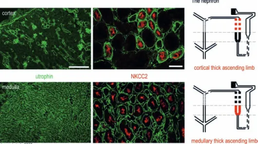
Figure 3. Differential expression and cellular distribution of utrophin in mouse kidney. A schematic drawing of the nephron is depicted
on the right panel. The cortical and medullary thick ascending limbs are colored in red. Low-resolution immunofluorescence images (left panel) depict utrophin (green) distribution in the cortex and medulla. In the cortex, utrophin staining is heterogeneous, labeling glomeruli and few cortical segments. Using a renal segment-specific marker, the Na+/K+/Cl– cotransporter 2 (NKCC2) (red, right panel), which is
polarized along the apical membrane of cortical and medullary thick ascending limbs, we could identify a utrophin-positive segments. In the medulla, utrophin exhibits a homogenous distribution and double staining with NKCC2 revealed medullary thick ascending limbs as utrophin-positive segment (lower middle panel). More renal markers have been investigated [32]. Scale bars, 200 μm (utrophin), 30 μm (NKCC2).
Figure 2. Segregated distribution of utrophin (green) and Dp71
(red) at the BBB and in astrocytic endfeet (GFAP-staining, blue). A schematic representation of a blood vessel surrounded by endfeet is depicted on the left. Utrophin immunoreactivity is localized in en-dothelial cells, whereas Dp71 overlaps with the GFAP signal in glial endfeet forming a ring around the blood vessel. Scale bar, 5 μm.
not affected [200]. Unlike in mdx
3Cvmice,
β
-DG
expres-sion is normal at the OPL, possibly due to association
with Dp260 [200]. Therefore, Dp71 is important for the
assembly of the DGC selectively in MGC endfeet. This
conclusion is supported by the fact that
β
-DG is disrupted
at the OPL of mice lacking full-length dystrophin and
Dp260 [214, 215].
The retina also provides a functional read-out applicable
for both mutant mice and DMD patients. Indeed,
electro-retinogram (ERG) anomalies are among the
best-charac-terized non-muscular manifestations of DMD. Analysis
of the dark-adapted ERG has revealed a reduction in the
amplitude of the b-wave response in 80% of DMD
pa-tients [216–219]. A prolonged implicit time of the b-wave
has been observed in ERG of mice lacking full-length
dystrophin and Dp260 [215], whereas Dp71-null mice
showed no significant change [200]. By comparing ERG
alterations in patients with distinct mutations, Pillers et
al. [220] suggested that in addition to Dp260, other
C-terminal isoforms contribute to the generation of the
b-wave. This hypothesis was confirmed by the
demonstra-tion that mdx
3Cvhave b-waves with reduced amplitude and
increased implicit time [221].
As expected from studies in the brain, myd mice exhibit
major morphological alterations in the retina, affecting
MGCs as well as neurons. A similar phenotype was seen,
in addition, in a novel mutant mouse line, Large
vlsmice
[134, 222]. These mice carry a mutation in a new allele
of Large, named veils (vls), and share phenotypic
char-acteristics with the myd mutation [222]. These findings
confirm that defective dystroglycan glycosylation
con-tributes to retinal abnormalities.
Kidney
Epithelial cells in the nephron express numerous DGC
proteins, forming several distinct DGCs. As the nephron
is organized in distinct segments to sequentially
reab-sorb ions and solutes from the glomerular ultrafiltrate,
it represents an attractive organ to study the localization
and specific distribution of DGC proteins [29, 32, 66,
223–225]. Dystroglycan is expressed early by epithelial
cells in the developing kidney, whereas in adult tissue
only low levels are detectable [66, 67], suggesting that
dystroglycan is more important for morphogenesis of
renal epithelial cells than during the adult stage.
Simi-larly to dystroglycan, Dp140 is only expressed during
kidney development [226]. Low levels of Dp71 are
de-tectable in adult kidney [32, 82, 224, 226–230], although
a specific splice variant has been reported to be
abun-dant, in particular in the cortex [29]. In contrast,
utro-phin is prominent in all segments of the nephron except
proximal tubules (Fig. 3) [32]. On the subcellular level,
utrophin is specifically localized along the basal, but not
lateral, membrane of tubular epithelial cells,
demonstrat-ing that it is restricted to sites of contact with the basal
lamina (Fig. 3). In contrast, the DGC is polarized along
the basolateral membrane of cultured kidney epithelial
cells [231], suggesting altered targeting in vitro in the
absence of the basal lamina. Using specific markers to
identify distinct segments of the nephron, utrophin has
been shown to be associated with different members
of the DGC in a segment-specific manner (Fig. 3). In
particular,
α
1- and
β
1-syn have a restricted distribution,
whereas
β
2-syn and
β
-DB are ubiquitous [29, 32]. These
findings indicate that Dp71 isoforms and utrophin are
the major partners of the DGC in the nephron and that
the functional specialization of the tubule is reflected in
the segment-specific distribution of certain DGC
pro-teins.
Possible alterations in DGC assembly and targeting have
been investigated in mdx
3Cvmice to test the role of Dp71.
β
2-syn staining was altered in cortical renal tubules,
Bowman’s capsule and glomeruli, whereas the
localiza-tion of
β
-DB,
α
-DB-1, utrophin,
α
1-syn and
β
1-syn was
not affected [29]. These findings suggest differential
de-pendence of
β
2-syn and other DGC proteins on Dp71
for complex formation. In a complementary study using
utrophin
0/0mice, we have demonstrated that
β
2-syn
local-ization is not impaired in cortical segments in the absence
of utrophin, whereas it is lost in all segments
express-ing high utrophin levels in wild-type mice [32]. Again,
other DGC proteins were either not affected in mutant
mice (
β
1- and
α
1-syn) or were upregulated (Dp71,
β
-DG,
and dystrobrevin) (Fig. 4), indicating that compensatory
mechanisms are activated to preserve most of the DGC in
either mdx
3Cvor utrophin
0/0mice [32].
To directly demonstrate this compensatory
up-regula-tion, utrophin-deficient mice were cross-bred with mdx
3Cvmice to generate utrophin
0/0/mdx
3Cvdouble mutants. These
mice have a reduced lifespan [120], and only few reach
the adult stage. Nevertheless, analysis of the nephron has
revealed a complete disruption of the DGC, highlighting
the functional redundancy between utrophin and
dystro-phin in cells coexpressing both proteins [32].
The complex, segment-specific molecular organization
of the DGC in the nephron suggests multiple functional
roles related to ion transport mechanisms. In analogy to
skeletal muscle cells, where the DGC provides membrane
stability during muscle contractions [4], renal epithelial
cells might also have a DGC to resist the high osmotic
pressure of the hypertonic interstitial fluid surrounding
medullary tubules [232]. The high abundance of utrophin
in these segments of the nephron, unlike in the renal
cor-tex, supports this idea. Although Dp71 is upregulated in
utrophin
0/0mice, the compensation is only partial because
β
2-syn is lost from the DGC. The reduced life expectancy
of double-mutant mice where no compensation is possible
in the kidney might be due to renal dysfunction. However,
this hypothesis remains to be tested.
Finally, for proper function of renal epithelial cells, ion
channels, exchangers, and transporters must be targeted
to either the apical or basolateral membrane [233]. The
DGC may be involved in anchoring renal transporters
and channels to the basal membrane. So far, however, no
protein has been identified for which the DGC serves as
a scaffolding protein, similarly to AQP4 and Kir4.1 in
as-trocytes.
Conclusions
The characterization of the DGC in non-muscle tissues
has revealed an unexpected heterogeneity in molecular
composition, in particular with respect to the presence
of
α
- or
β
-DG. The mechanism of membrane anchoring
and/or communication with the ECM is therefore not
es-tablished for some prominent DGCs, such as those seen
in microvascular endothelial cells and the CP. However,
negative results are not necessarily conclusive, and the
failure to detect dystroglycan in some tissues might be
due to technical reasons. Furthermore, some
biochemi-cal methods are of limited use in heterogeneous tissues
with a cell-specific expression of DGC proteins. It is
important to note that in neurons and astrocytes, as well
as kidney tubular epithelial cells, the localization of the
DGC precisely matches the presence of a basal lamina,
suggesting that communication with the ECM via
α
-DG
is essential for specifying the subcellular localization of
the DGC. This feature might, for example, explain the
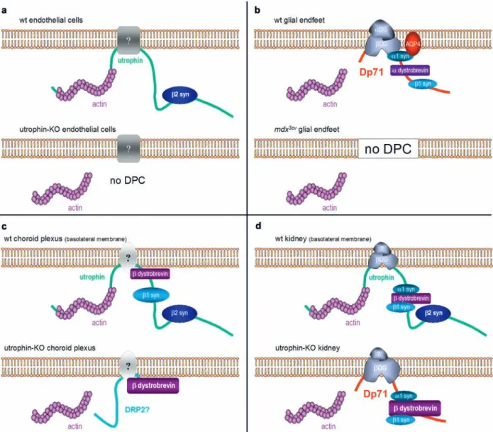
Figure 4. Molecular composition of the DGC in four tissues of wild-type and either utrophin0/0 or mdx3Cv mice. Note the mandatory
as-sociation of β2-syn with utrophin in kidney, CP, and endothelial cells. In utrophin0/0 kidney, the DPC is partially rescued by compensatory
upregulation of Dp71, but it is unclear whether it binds the actin cytoskeleton. In blood vessels (endothelial and glial endfeet), no com-pensation occurs and the DPC is disrupted, along with AQP4. An intermediate situation occurs in the CP, where β-DB is partially retained, possibly due to upregulation of dystrophin-related protein 2 (DRP2).
remarkably specific targeting of the DGC in astrocytic
endfeet. In the CP, the DGC is present basolaterally in
epithelial cells, arguing against the presence of
dystro-glycan in this tissue.
The analysis of mutant mice has revealed that in all cell
types coexpressing a dystrophin isoform with utrophin,
compensatory up-regulation takes place in the absence of
the homologue protein (Fig. 4), obscuring the
interpreta-tion of the analysis of single mutant mice. In contrast,
these compensatory mechanisms do not take place in
tis-sues expressing only dystrophin (glial endfeet) or only
utrophin (CP epithelial cells, vascular endothelial cells).
The analysis of double-mutant mice lacking dystrophin
and utrophin is hampered by the severe phenotype,
lim-ited breeding capacity, and reduced life expectancy of
these animals.
A major convergent finding emerging from the analysis
of the DGC in non-muscle tissue is that syntrophins
re-quire the presence of either dystrophin or utrophin for
assembly of the DGC and membrane localization. Owing
to the presence of several ‘simple’ DGCs containing a
re-duced number of proteins, a mandatory association of
β
2
-syn with utrophin, as well as
α
-DB,
α
1
-syn, and
β
1-syn
with dystrophin, has been demonstrated in three distinct
tissues (Fig. 4). The role of
β
-DB is less clear since it does
not disappear from the CP of utrophin
0/0mice and
associ-ates with Dp71 in the absence of utrophin in the kidney.
Therefore, although dystroglycan is a key member of the
DGC, interacting with signaling proteins, dystrophin and
utrophin appear to be essential for the formation of the
complex, with very few exceptions so far [234].
Several membrane-associated proteins have been
iden-tified, notably AQP4 and Kir4.1, which depend on the
DGC for proper targeting and localization. However, no
generalization is possible since major transporters, such
as members of the ABC transporter or the glucose
trans-porter families, which colocalize with the DGC in several
tissues, are not affected in mutant mice. It is therefore
dif-ficult to predict functional deficits that might arise from
an altered expression of DGC proteins. However, the
ex-istence of functional redundancy between dystrophin
iso-forms, dystrobrevin, and utrophin might represent a strong
stimulus for exploiting further compensatory mechanisms
to alleviate the symptoms of muscle dystrophy.
Acknowledgements. Our work is supported by the Swiss National Science Foundation. We are grateful to our colleagues M. C. Schaub, R. Züllig, B. Bornhauser, and I. Knüsel for their contribu-tion at various phases of the project and for advice and support. We thank C. Sidler, F. Parpan and T. Grampp for excellent technical assistance.
1 Blake, D. J., Weir A., Newey, S. E. and Davies, K. E. (2002) Function and genetics of dystrophin and dystrophin-related proteins in muscle. Physiol. Rev. 82, 291–329.
2 Durbeej M. and Campbell, K. P. (2002) Muscular dystrophies involving the dystrophin-glycoprotein complex: an overview
of current mouse models. Curr. Opin. Genet. Dev. 12, 349– 361.
3 Cohen N. and Muntoni F. (2004) Multiple pathogenetic mechanisms in X linked dilated cardiomyopathy. Heart 90, 835–841.
4 Petrof, B. J., Shrager, J. B., Stedman, H. H., Kelly, A. M. and Sweeney, H. L. (1993) Dystrophin protects the sarcolemma from stresses developed during muscle contraction. Proc. Natl. Acad. Sci. USA 90, 3710–3714.
5 Ervasti, J. M. and Campbell, K. P. (1993) A role for the dys-trophin-glycoprotein complex as a transmembrane linker be-tween laminin and actin. J. Cell Biol. 122, 809–823.
6 Mora M., Morandi L., Piccinelli A., Gussoni E., Gebbia M., Blasevich F., Dworzak F. and Cornelio F. (1993) Dystrophin abnormalities in Duchenne and Becker dystrophy carriers: correlation with cytoskeletal proteins and myosins. J. Neurol. 240, 455–461.
7 Campbell, K. P. (1995) Three muscular dystrophies: loss of cytoskeleton-extracellular matrix linkage. Cell 80, 675–679. 8 Lapidos, K. A., Kakkar R. and McNally, E. M. (2004) The
dystrophin glycoprotein complex: signaling strength and in-tegrity for the sarcolemma. Circ. Res. 94, 1023–1031. 9 Koenig M., Hoffman, E. P., Bertelson, C. J., Monaco, A. P.,
Feener C. and Kunkel, L. M. (1987) Complete cloning of the Duchenne muscular dystrophy (DMD) cDNA and prelimi-nary genomic organization of the DMD gene in normal and affected individuals. Cell 50, 509–517.
10 Love, D. R., Bloomfield, J. F., Kenwrick, S. J., Yates, J. R. and Davies, K. E. (1990) Physical mapping distal to the DMD lo-cus. Genomics 8, 106–112.
11 Nudel U., Zuk D., Einat P., Zeelon E., Levy Z., Neuman S. and Yaffe D. (1989) Duchenne muscular dystrophy gene product is not identical in muscle and brain. Nature 337, 76–78. 12 Chelly J., Hamard G., Koulakoff A., Kaplan, J. C., Kahn A.
and Berwald-Netter Y. (1990) Dystrophin gene transcribed from different promoters in neuronal and glial cells. Nature 344, 64–65.
13 Klamut, H. J., Gangopadhyay, S. B., Worton, R. G. and Ray, P. N. (1990) Molecular and functional analysis of the muscle-specific promoter region of the Duchenne muscular dystrophy gene. Mol. Cell Biol. 10, 193–205.
14 Boyce, F. M., Beggs, A. H., Feener C. and Kunkel, L. M. (1991) Dystrophin is transcribed in brain from a distant up-stream promoter. Proc. Natl. Acad. Sci. USA 88, 1276–1280. 15 Gorecki D., Geng Y., Thomas K., Hunt, S. P., Barnard, E. A.
and Barnard, P. J. (1991) Expression of the dystrophin gene in mouse and rat brain. Neuroreport 2, 773–776.
16 Makover A., Zuk D., Breakstone J., Yaffe D. and Nudel U. (1991) Brain-type and muscle-type promoters of the dystro-phin gene differ greatly in structure. Neuromuscul. Disord. 1, 39–45.
17 Berthier C. and Blaineau S. (1997) Supramolecular organi-zation of the subsarcolemmal cytoskeleton of adult skeletal muscle fibers: a review. Biol. Cell 89, 413–434.
18 Sealock R., Butler, M. H., Kramarcy, N. R., Gao, K. X., Mur-nane, A. A., Douville K. and Froehner, S. C. (1991) Localiza-tion of dystrophin relative to acetylcholine receptor domains in electric tissue and adult and cultured skeletal muscle. J. Cell Biol. 113, 1133–1144.
19 Gorecki, D. C., Monaco, A. P., Derry, J. M., Walker, A. P., Barnard, E. A. and Barnard, P. J. (1992) Expression of four alternative dystrophin transcripts in brain regions regulated by different promoters. Hum. Mol. Genet. 1, 505–510.
20 Gonzalez E., Montanez C., Ray, P. N., Howard, P. L., Gar-cia-Sierra F., Mornet D. and Cisneros B. (2000) Alternative splicing regulates the nuclear or cytoplasmic localization of dystrophin Dp71. FEBS Lett. 482, 209–214.
21 Jung D., Filliol D., Metz-Boutigue, M. H. and Rendon A. (1993) Characterization and subcellular localization of the
dystrophin-protein 71 (Dp71) from brain. Neuromuscul. Dis-ord. 3, 515–518.
22 Tamura T., Yoshioka K., Jinno Y., Niikawa N. and Miike T. (1993) Dystrophin isoforms expressed in the mouse retina. J. Neurol. Sci. 115, 214–218.
23 Gorecki, D. C. and Barnard, E. A. (1995) Specific expression of G-dystrophin (Dp71) in the brain. Neuroreport 6, 893–896. 24 Ueda H., Tsukahara S., Kobayashi T. and Ohno S. (1995) Im-munocytochemical study of dystrophin-related protein in the rat retina. Ophthal. Res. 27, 219–226.
25 Imamura M. and Ozawa E. (1998) Differential expression of dystrophin isoforms and utrophin during dibutyryl-cAMP-induced morphological differentiation of rat brain astrocytes. Proc. Natl. Acad. Sci. USA 95, 6139–6144.
26 Blake, D. J., Hawkes R., Benson, M. A. and Beesley, P. W. (1999) Different dystrophin-like complexes are expressed in neurons and glia. J. Cell Biol. 147, 645–658.
27 Austin, R. C., Morris, G. E., Howard, P. L., Klamut, H. J. and Ray, P. N. (2000) Expression and synthesis of alternatively spliced variants of Dp71 in adult human brain. Neuromuscul. Disord. 10, 187–193.
28 Claudepierre T., Mornet D., Pannicke T., Forster V., Dalloz C., Bolanos F., Sahel J., Reichenbach A. and Rendon A. (2000) Expression of Dp71 in Müller glial cells: a comparison with utrophin- and dystrophin-associated proteins. Invest. Ophthal-mol. Vis. Sci. 41, 294–304.
29 Loh, N. Y., Newey, S. E., Davies, K. E. and Blake, D. J. (2000) Assembly of multiple dystrobrevin-containing complexes in the kidney. J. Cell Sci. 113, 2715–2724.
30 Aleman V., Osorio B., Chavez O., Rendon A., Mornet D. and Martinez D. (2001) Subcellular localization of Dp71 dystro-phin isoforms in cultured hippocampal neurons and forebrain astrocytes. Histochem. Cell. Biol. 115, 243–254.
31 Haenggi T., Soontornmalai A., Schaub, M. C. and Fritschy, J. M. (2004) The role of utrophin and Dp71 for assembly of different dystrophin-associated protein complexes (DPCs) in the choroid plexus and microvasculature of the brain. Neuro-science 129, 403–413.
32 Haenggi T., Schaub, M. C. and Fritschy, J. M. (2005) Molecu-lar heterogeneity of the dystrophin-associated protein com-plex in the mouse kidney nephron: differential alterations in the absence of utrophin and dystrophin. Cell Tissue Res. 319, 299–313.
33 Howard, P. L., Klamut, H. J. and Ray, P. N. (1998) Identifica-tion of a novel actin binding site within the Dp71 dystrophin isoform. FEBS Lett. 441, 337–341.
34 Love, D. R., Hill, D. F., Dickson G., Spurr, N. K., Byth, B. C., Marsden, R. F., Walsh, F. S., Edwards, Y. H. and Davies, K. E. (1989) An autosomal transcript in skeletal muscle with ho-mology to dystrophin. Nature 339, 55–58.
35 Blake, D. J., Schofield, J. N., Zuellig, R. A., Gorecki, D. C., Phelps, S. R., Barnard, E. A., Edwards, Y. H. and Davies, K. E. (1995) G-utrophin, the autosomal homologue of dystro-phin Dp116, is expressed in sensory ganglia and brain. Proc. Natl. Acad. Sci. USA 92, 3697–3701.
36 Lumeng, C. N., Phelps, S. F., Rafael, J. A., Cox, G. A., Hutchinson, T. L., Begy, C. R., Adkins E., Wiltshire R. and Chamberlain, J. S. (1999) Characterization of dystrophin and utrophin diversity in the mouse. Hum. Mol. Genet. 8, 593– 599.
37 Knuesel I., Bornhauser, B. C., Zuellig, R. A., Heller F., Schaub, M. C. and Fritschy, J. M. (2000) Differential expres-sion of utrophin and dystrophin in CNS neurons: an in situ hy-bridization and immunohistochemical study. J. Comp. Neurol. 422, 594–611.
38 Sogos V., Curto M., Reali C. and Gremo F. (2002) Develop-mentally regulated expression and localization of dystrophin and utrophin in the human fetal brain. Mech. Ageing Dev. 123, 455–462.
39 Burton, E. A., Tinsley, J. M., Holzfeind, P. J., Rodrigues, N. R. and Davies, K. E. (1999) A second promoter provides an al-ternative target for therapeutic up-regulation of utrophin in Duchenne muscular dystrophy. Proc. Natl. Acad. Sci. USA 96, 14025–14030.
40 Weir, A. P., Burton, E. A., Harrod G. and Davies, K. E. (2002) A- and B-utrophin have different expression patterns and are differentially up-regulated in mdx muscle. J. Biol. Chem. 277, 45285–45290.
41 Byers, T. J., Kunkel, L. M. and Watkins, S. C. (1991) The sub-cellular distribution of dystrophin in mouse skeletal, cardiac, and smooth muscle. J. Cell Biol. 115, 411–421.
42 Khurana, T. S., Watkins, S. C., Chafey P., Chelly J., Tome, F. M., Fardeau M., Kaplan, J. C. and Kunkel, L. M. (1991) Immunolo-calization and developmental expression of dystrophin related protein in skeletal muscle. Neuromuscul. Disord. 1, 185–194. 43 Ohlendieck K., Ervasti, J. M., Matsumura K., Kahl, S. D.,
Leveille, C. J. and Campbell, K. P. (1991) Dystrophin-related protein is localized to neuromuscular junctions of adult skel-etal muscle. Neuron 7, 499–508.
44 Bewick, G. S., Nicholson, L. V., Young C., O’Donnell E. and Slater, C. R. (1992) Different distributions of dystrophin and related proteins at nerve-muscle junctions. Neuroreport 3, 857–860.
45 Ibraghimov-Beskrovnaya O., Ervasti, J. M., Leveille, C. J., Slaughter, C. A., Sernett, S. W. and Campbell, K. P. (1992) Primary structure of dystrophin-associated glycoproteins linking dystrophin to the extracellular matrix. Nature 355, 696–702.
46 Chamberlain J. (1999) The dynamics of dystroglycan. Nat. Genet. 23, 256–258.
47 Winder, S. J. (2001) The complexities of dystroglycan. Trends Biochem. Sci. 26, 118–124.
48 Durbeej M., Henry, M. D. and Campbell, K. P. (1998) Dystro-glycan in development and disease. Curr. Opin. Cell Biol. 10, 594–601.
49 Moore, S. A., Saito F., Chen J., Michele, D. E., Henry, M. D., Messing A., Cohn, R. D., Ross-Barta, S. E., Westra S., Wil-liamson, R. A., Hoshi T. and Campbell, K. P. (2002) Deletion of brain dystroglycan recapitulates aspects of congenital mus-cular dystrophy. Nature 418, 422–425.
50 Michele, D. E. and Campbell, K. P. (2003) Dystrophin-glyco-protein complex: post-translational processing and dystrogly-can function. J. Biol. Chem. 278, 15457–15460.
51 Haliloglu G. and Topaloglu H. (2004) Glycosylation defects in muscular dystrophies. Curr Opin Neurol. 17, 521–527. 52 Jacobson C., Montanaro F., Lindenbaum M., Carbonetto S.
and Ferns M. (1998) α-Dystroglycan functions in acetylcho-line receptor aggregation but is not a coreceptor for agrin-MuSK signaling. J. Neurosci. 18, 6340–6348.
53 Montanaro F., Gee, S. H., Jacobson C., Lindenbaum, M. H., Froehner, S. C. and Carbonetto S. (1998) Laminin and α -dys-troglycan mediate acetylcholine receptor aggregation via a MuSK-independent pathway. J. Neurosci. 18, 1250–1260. 54 Montanaro F., Lindenbaum M. and Carbonetto S. (1999) α
-Dystroglycan is a laminin receptor involved in extracellular matrix assembly on myotubes and muscle cell viability. J. Cell Biol. 145, 1325–1340.
55 Henry, M. D., Satz, J. S., Brakebusch C., Costell M., Gustafs-son E., Fassler R. and Campbell, K. P. (2001) Distinct roles for dystroglycan, beta1 integrin and perlecan in cell surface laminin organization. J. Cell Sci. 114, 1137–1144.
56 Jacobson C., Cote, P. D., Rossi, S. G., Rotundo, R. L. and Carbonetto S. (2001) The dystroglycan complex is necessary for stabilization of acetylcholine receptor clusters at neuro-muscular junctions and formation of the synaptic basement membrane. J. Cell Biol. 152, 435–450.
57 Song, K. S., Scherer, P. E., Tang Z., Okamoto T., Li S., Chafel M., Chu C., Kohtz, D. S. and Lisanti, M. P. (1996) Expression