Journal of
SURGERY
< //. © 1997 by the Societe Internationale de ChirurgieIncreased Surface Expression of CD18 and CD11b in Leukocytes after
Tourniquet Ischemia during Elective Hand Surgery
Paul M. Sutter, M.D.,2°3 Giulio C. Spagnoli, M.D.,z Axel Marx, M.D.,2°3 Lorenz Gurke, M.D.,' Hans Troeger, M.D.,' Renato Fricker, M.D.,' Felix Harder, M.D.,' Michael Heberer, M.D.2
'Department of Surgery, Kantonsspital Basel, Spitalstrasse 21, 4031 Basel, Switzerland
2Department of Surgical Research, Kantonsspital Basel, 4031 Basel, Switzerland
3Department of Surgery, Ospedale Civico Lugano, Via Tesserete 46, 6900 Lugano, Switzerland
Abstract. The surface expression of /32-integrins was investigated in leukocytes from patients undergoing ischemia induced by tourniquet application for elective hand surgery. Blood samples were obtained before initiation, at the end of ischemia, and after 15 minutes of reperfusion from ischemic and contralateral arms of five patients. Comparable expression of CD18, CD11a, CD11b, and CD11c could be detected by immunofluorescence in leukocytes from samples drawn from either arm before tourniquet application. In contrast, a significant increase in the expression of CD18 was detectable in monocytes, granulocytes, and lymphocytes from the ischemic arm compared with that in the nonisch-emic contralateral control, at the end of the ischemia time (80 ± 16 minutes). A significantly increased expression of CDl1b, but not CD11a or CD11c, determinants was also observed in granulocytes and mono-cytes. Concomitantly, a significant reduction in the percentages of gran-ulocytes in samples from ischemic areas was detectable. After 15 minutes of reperfusion, differences in the expression of these adhesion molecules were no longer significant. The expression of the genes encoding inter-leukins IL-la, IL-1, and IL-6 and tumor necrosis factor alpha (TNFa) proinflammatory cytokines was also studied by reverse polymerase chain reaction (rPCR) in peripheral blood mononuclear cells (PBMCs) ob-tained from the same samples in three patients. IL-1/3 or IL-6 gene expression was never observed. Expression of IL-la and TNFa genes, as detected in two patients, was not related with induction of ischemia. However, in these patients expression of one or both these genes was observed in samples derived from the ischemic but not the control arm after 15 minutes of reperfusion. These data document that overexpression of adhesion molecules and sequestration of leukocytes take place follow-ing short ischemia times, as routinely applied clinically for minor surgical procedures.
lschemia/reperfusion injury represents a potential complication of vascular injury, trauma, and transplantation surgery. Activation of leukocytes, endothelium, or both leading to increased cell adhe-sion and extravasation can result in damage of the vessel walls and surrounding tissues, particularly during the reperfusion phase. Furthermore, cytokines and other mediators can be produced, possibly responsible for distant effects targeting the liver and the pulmonary capillary area [1, 2].
Experimental models have addressed in detail the molecular bases of ischemia reperfusion damage. Leukocyte infiltration, as
Correspondence to: P.M. Sutter, M.D., Surgical Research Laboratory, Z.L.F., 20 Hebelstrasse, 4031 Basel, Switzerland
detectable in postischemic tissues, appears to largely mediate injury induction. Indeed, leukocyte depletion has been shown to decrease the severity of reperfusion damage, and monoclonal antibodies (mAbs) specific for CD18 protect tissues from post-ischemic injury [3-8].
Clinically, tourniquet-induced ischemia is routinely applied during limb surgery to ensure a relatively bloodless operative field. If prolonged in time, it may lead to skeletal muscle damage and impaired postoperative function [9-11]. However, clinical data concerning the activation of adhesion molecules in leuko-cytes during ischemia and reperfusion are sparse to date.
In this work we took advantage of tourniquet application in elective hand surgery to investigate the surface expression of 132-integrins in different leukocyte populations under ischemic and postischemic conditions. Concomitantly, the induction of proin-flammatory cytokines' gene expression was assayed by the reverse polymerase chain reaction (rPCR).
We report here that a significant increase in the expression of Mac-1 (CD1Ib/CD18) (32- integrin is detectable in granulocytes and monocytes from arms undergoing relatively short (60-101 minutes) ischemia times. Under these conditions, proinflamma-tory cytokines do not appear to play a role the induction of these events.
Patients and Methods
Experimental Design
Five patients undergoing elective hand surgery (Table 1) in the absence of systemic diseases were studied. All patients except one were anesthetized with regional brachial block anesthesia. No Esmarch bandage exsanguination was applied. Heparinized ve-nous blood samples were obtained from both arms before initia-tion of surgery but after inducinitia-tion of anesthesia in the limb undergoing surgery. Subsequently, a tourniquet was applied on the upper arm at a pressure of 300 mmHg. Duration of ischemia ranged between 60 and 101 minutes (80 ± 16 minutes). At the end of surgery and immediately before releasing the tourniquet, a
Patient
Age
(years) Sex Surgery Anesthesia
Duration of ischemia (min)
1 22 M Internal fixation of scaphoid fracture Regional 90
2 19 M Flexor tenolysis (little finger) Regional 60
3 36 F Ulnar head resection Regional 81
4 43 M Scapholunate ligament reconstruction Regional 101
5 49 F Excision of a hemangioma at the back of the hand General 70
Table 2. Pairs of primers used in this study
Gene Sequence Size"
/3-Actin Sense: TGACGGGGTCACCCACACTGTGCCCATCTA 661
Antisense: CTAGAAGCATTGCGGTGGACGATGGAGGG
IL-1a Sense: ATGGCCAAAGTTCCAGACATGTTTG 808
Antisense: GGTTTTCCAGTATCTGAAAGTCAGT
IL-1/3 Sense: CTTCATCTTTTGAAGAAGAACCTATCTTCTT 331
Antisense: AATI'I"ITGGGATCTACACTCTCCAGCTGTA
IL-6 Sense: ATGAACTCCTTCTCCACAAGCGC 628
Antisense: GAAGAGCCCTCAGGCTGGACTG
TNFa Sense: ATGAGCACTGAAAGCATGATCCGG 694
Antisense: GCAATGATCCCAAAGTAGACCTGCCC "Expected size of the amplified fragment expressed as number of basepairs
second blood sample was obtained from both arms. A third sample was obtained from both limbs 15 minutes after the termination of ischemia. In some experiments another blood specimen was obtained 1 minute after release of the tourniquet.
Phenotypic Studies
Phenotypic studies were performed on whole blood to avoid separation procedures likely to affect the expression of adhesion molecules [12]. Briefly, 50 µl of heparinized venous blood were incubated in the presence of fluorochrome-labeled mAbs recog-nizing CD14, CD45, CD11b (Becton-Dickinson, San Jose, CA, USA). The mAbs directed against CD11a, CD11c, CD18, and CD54 (ICAM-1) were obtained from Serotec (Oxford, UK). Erythrocytes were lysed (FACS lysis buffer, Becton-Dickinson) after 15 minutes of incubation at room temperature. Following two washes, cells were resuspended in phosphate-buffered saline (PBS) 0.2% paraformaldehyde, and specific fluorescence was quantitatively evaluated on a FACScan fluorocytometer equipped with Lysis lI software (Becton-Dickinson). The instrument's set-tings were adjusted by taking advantage of CaliBRITE beads and AutoComp software (both from Becton-Dickinson). Student's
t-test for paired samples was used in the statistical analysis of data
throughout this study.
PCR Studies
Peripheral blood mononuclear cells were obtained from hepa-rinized venous blood samples by centrifugation on density gradi-ents (Lymphoprep; Nycomed, Oslo, Norway). Total cellular RNA was extracted and reverse-transcribed as described previously [13]. The cDNA thus obtained was assayed in 35-cycle PCR
reactions in the presence of pairs of primers specific for -actin housekeeping gene as the positive control and interleukins IL-la, IL-1/3, and IL-6 and tumor necrosis factor alpha (TNFa) genes (Table 2). PCR products were then electrophoresed in the presence of ethidium bromide on agarose gels that were subse-quently photographed under ultraviolet (UV) transillumination. The specificity of the amplified genes was validated by restriction mapping [13].
Results
Effects of Ischemia on Leukocyte Populations
Absolute counts of leukocytes did not significantly differ in blood samples drawn from the ischemic or the control arm at any of the time points under investigation (data not shown). Percentages of granulocytes, monocytes, and lymphocytes in blood samples were evaluated by taking advantage of physical cytometric parameters [forward scatter (FSC) and side scatter (SSC)] and phenotypic characteristics. Monocytes are CD14/CD45 + cells, whereas lym-phocytes are CD14/CD45 bright cells and granulocytes appear as CD14/CD45 dim cells. No differences in the relative percentages of granulocytes, monocytes, and lymphocytes were observed in samples drawn from either limb before ischemia. In contrast, at the end of ischemia, as reported in Figure 1, a significant decrease of the granulocytes' percentages could be observed in samples obtained from the ischemic arm (57.7 ± 9.7%) compared with values simultaneously obtained from the nonischemic limb (71.1 ± 9.5%). In parallel, increased lymphocytes' percentages were detected in the same specimens (38.2 ± 9.1% versus 24.2 ± 8.6%). No significant modifications of the monocytes' percentages were detectable under these conditions. After 15 minutes of
Fig. 1. Differential percentages of leukocytes. Percentages of granulocytes, monocytes, and lymphocytes in blood samples from ischemic and nonischemic areas. Heparinized blood was obtained at the indicated times from ischemic limbs (shaded columns) and control limbs (open columns). The percentages of granulocytes (G), monocytes (M), and lymphocytes (L) among the total leukocytes were evaluated by staining with fluorochrome-labeled anti-CD14 and anti-CD45 mAbs. Data from the five patients under investigation are reported as mean values ± standard deviations. Asterisks indicate significant differences (p < 0.05). For other details see Materials and Methods.
reperfusion, comparable percentages of granulocytes, monocytes, or lymphocytes were observed in samples obtained from the two limbs (Fig. 1).
CD18 Expression in Leukocytes from Ischemic Areas
Surface expression of CD18 was quantitatively evaluated in granulocytes, monocytes, and lymphocytes from ischemic and nonischemic arms before application of the tourniquet, at the end of ischemia, and after 15 minutes of reperfusion. As shown in Figure 2, CD18 expression in leukocyte populations was compa-rable in samples obtained from the two arms before ischemia (206.8 ± 72.2 versus 174.0 ± 42.8 for granulocytes, 387.4 ± 156.8 versus 349.2 ± 95.7 for monocytes, 239.4 ± 42.5 versus 243.8 ± 23.1 for lymphocytes). A significant increase of CD18 expression concerning all leukocyte populations was detected in samples derived from the ischemic arm at the end of ischemia compared with those obtained from the contralateral limb (321.2 ± 50.2 versus 188.8 ± 37.5 for granulocytes, 653 ± 54.4 versus 426.0 ± 121.6 for monocytes, 299.0 ± 44.5 versus 228.2 ± 36.0 for lymphocytes). After 15 minutes of reperfusion, regarding granu-locytes and monocytes, apparent differences in the expression of CD18 in samples obtained from the ischemic arm compared with the untreated arm (331.4 ± 72.5 versus 234.8 ± 66.9 for granu-locytes, 567.4 ± 157.4 versus 443.2 ± 120.8 for monocytes) were no longer significant. Regarding lymphocytes, virtually identical values were observed in cells from the two arms (222.2 ± 48.7 versus 220.0 ± 56.9).
Fig. 2. Surface expression of CD18 in leukocytes from ischemic and nonischemic areas. Expression of CD18 was evaluated quantitatively, as the mean fluorescence intensity, in granulocytes (G), monocytes (M), and lymphocytes (L) from blood samples obtained at the indicated times from ischemic arms (shaded columns) and nonischemic arms (open columns). Data from the five patients, expressed in arbitrary units, are reported as mean values ± standard deviations. Asterisks indicate significant differ-ences (p < 0.05). For other details see Materials and Methods.
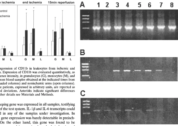
a-chains was also investigated. As shown in Figure 3, CD11b expression before ischemia was comparable in leukocyte popula-tions in samples from either arm. In contrast, CD11b expression was significantly increased in granulocytes (499.8 ± 341.7 versus 213.0 ± 149.8) and monocytes (532.8 ± 358.6 versus 237.3 205.9) from the ischemic arm compared with cells derived from the contralateral limb at the end of ischemia. At close resem-blance to CD18, apparent differences detectable after 15 minutes of reperfusion (474.4 ± 281.7 versus 284.6 ± 214.4 for granulo-cytes and 531.8 ± 358.1 versus 345 ± 291.8 for monogranulo-cytes) were no longer significant. On the other hand, no modifications of CD11b expression were observed on lymphocyte surfaces at any of the times considered. Remarkably, no significant differences in the expression of CD11a and CD11c were detectable in leukocyte populations at any time (data not shown).
ICAM-1 /CD54 Expression in Leukocytes from Ischemic or Nonischemic Areas
Because ICAM-1/CD54 represents a major ligand molecule for CD11a and CD11b integrins, its expression was analyzed in leukocytes from ischemic and nonischemic limbs. At a striking difference with CD18, no significant variation in the surface expression of ICAM-1/CD54 could be detected in granulocytes, monocytes, or lymphocytes following ischemia compared to the expression in cells from the nonischemic limb (data not shown).
Cytokine Gene Expression in Mononuclear Cells from Ischemic or Nonischemic Areas
Effects of Ischemia on Expression of a-Chains of /32- Integrins
on Leukocyte Surfaces Expression of IL-la, IL-1J3, IL-6, and TNFa genes was
investi-gated by 35-cycle rPCR in RNA from peripheral blood mononu-Because ischemia appeared to increase surface expression of clear cells (PBMCs) obtained simultaneously with the samples CD18, the common 13-chain of (32-integrins, the expression of used for phenotypic studies. Three patients were studied. The
Fig. 3. Surface expression of CD11b in leukocytes from ischemic and nonischemic areas. Expression of CD11b was evaluated quantitatively, as the mean fluorescence intensity, in granulocytes (G), monocytes (M), and lymphocytes (L) from blood samples obtained at the indicated times from ischemic arms (shaded columns) and nonischemic arms (open columns). Data from the five patients, expressed in arbitrary units, are reported as means ± standard deviations. Asterisks indicate significant differences (p < 0.05). For other details see Materials and Methods.
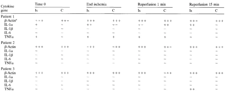
(3-actin housekeeping gene was expressed in all samples, testifying to the integrity of the test system. IL-113 and IL-6 transcripts could not be detected in any of the samples under investigation. In patient 1, IL-la gene expression was barely detectable in preisch-emia samples. On the other hand, this gene was found to be relatively highly expressed in samples from both ischemic and control arms at the end of ischemia and after 1 minute of reperfusion as well as in PBMCs from the ischemic limb only after 15 minutes of reperfusion. In contrast, no expression could be observed in the 15-minute reperfusion sample from the nonisch-emic control arm (Fig. 4, Table 3). Regarding TNFa gene, in this patient weak expression was observed in all samples, with the exception of PBMCs obtained after 15 minutes of reperfusion from the control arm. Interestingly, TNFa expression was also detectable in PBMCs from patient 3 obtained from the ischemic arm after 15 minutes of reperfusion (Table 3).
Discussion
The mechanisms underlying ischemia/reperfusion-associated
in-jury have been thoroughly investigated in a number of
experimen-tal models [14, 15]. Emerging evidence concordantly attributes a key role to leukocytes in the generation of postischemic damage. Overexpression of adhesion molecules and production of proin-flammatory cytokines would lead to leukocyte infiltration of postischemic tissues, a key step in the induction of reperfusion injury [16]. Furthermore, leukocyte—endothelial cell adhesion is thought to create a microenvironment favoring the release of toxic products, including reactive oxidant metabolites [17]. In this context, evidence that administration of CD18-specific mAbs can prevent generation of mucosal damage following intestinal isch-emia [7-9] suggests a major relevance of these molecules in the induction of ischemia/reperfusion-related injury.Despite myriad experimental data, however, little information is available on these events as detectable in clinical situations. Significant neutropenia and reduction of the percentages of granulocytes expressing CD11b/CD18 and CD11c/CD18 was
ob-Fig. 4. Cytokine gene expression in peripheral blood mononuclear cells (PBMCs) from ischemic and nonischemic areas. Total cellular RNA was extracted from Ficoll-purified PBMCs and reverse-transcribed. The cDNA thus obtained was amplified by 35-cycle PCR in the presence of primers specific for /3-actin (A), IL-la (B), and TNF-a (C). PCR products were electrophoresed in the presence of ethidium bromide and photo-graphed under UV transillumination. Samples were obtained before initiation of ischemia (lanes 1 and 2), at the end of ischemia (lanes 3 and 4), after 1 minute of reperfusion (lanes 5 and 6), and after 15 minutes of reperfusion (lanes 7 and 8). Lanes 1, 3, 5, and 7 contain samples from the ischemic arm, whereas lanes 2, 4, 6, and 8 contain samples from the contralateral arm. For other details see Materials and Methods.
served in patients affected by peripheral arterial occlusive disease following physical exercise [18].
In this work we analyzed the expression of p2 integrins in a group of patients undergoing elective hand surgery, implying application of a tourniquet and an ischemia time ranging between 60 and 101 minutes. Differently from most experimental models, however, we specifically addressed the expression of adhesion molecules at the end of ischemia and after a short (15-minute) reperfusion.
In keeping with observations in animal models, we found that relatively short ischemia times, as required for technical reasons during minor surgical procedures, induce overexpression of CD18 on the surface of granulocytes, monocytes, and lymphocytes. In parallel, a concomitant, significant increase in the expression of CD11b, but not CD11a or CD11c, is detectable on granulocytes and monocytes. Local sequestration of granulocytes was also observed, as witnessed by their decreased percentages in effluent blood. Considering the relatively rapid kinetics of these phenom-ena, translocation of adhesion molecules from intracellular
gran-Table 3. Expression of proinflammatory cytokine genes in PBMCs from ischemic and nonischemic areas.
Time 0 End ischemia Reperfusion 1 min Reperfusion 15 min
Cytokine
gene Is C Is C Is C Is C
Patient 1 (3-Actin" +++ +++ +++ +++ +++ +++ +++ +++ IL-la + + ++ ++ ++ ++ ++ IL-1(3 — — — — IL-6 — — — — — — — — TNFa + + + + + + + — Patient 2 (3-Actin +++ +++ +++ +++ +++ +++ +++ +++ IL-la — — — — — — — — IL-lp — — — — — — _ — IL-6 — — — — — — — — TNFa — — — — — Patient 3 (3-Actin +++ +++ +++ +++ +++ +++ +++ +++ IL-la — — — — — — — IL-1(3 — — — — — — — — IL-6 — — — — — — — — TNFa — — — — — ++ —
Total cellular RNA was extracted from PBMCs obtained from blood samples drawn at the indicated times from ischemic (Is) or contralateral (C) arms and reverse-transcribed.
"cDNA was assayed during 35-cycle PCR in the presence of the appropriate pairs of primers (see Table 2). Amplification products were run on agarose gels in the presence of ethidium bromide and photographed under UV transillumination.
—:
no detectable PCR product; +—+ + +: intensity (in arbitrary units) of the fluorescence emitted by amplified genes under investigation following electrophoresis in the presence of ethidium bromide (see also Fig. 4).ules to the cell surfaces, as observed in experimental models [19], is highly likely.
Granulocytes and monocytes obtained from the ischemic arm after 15 minutes of reperfusion still display an apparently higher expression of CD18 and CD11b than did their counterparts sampled simultaneously from the nonischemic limb. These differ-ences, however, are no longer significant. This could be related to the "dilution" of the blood from the ischemic area into the systemic circulation.
It has been suggested that cytokine production by leukocytes trapped in ischemic areas might activate both leukocytes and endothelial cells, resulting in the overexpression of adhesion molecules and their ligands [20]. White blood cell sequestration would ensue. Our data on cytokine gene expression in PBMCs do not support this hypothesis. In fact, no IL-1f3 or IL-6 transcripts could be amplified. As for IL-la and TNFa genes, in two of three patients no expression was detectable in mononuclear cells from the ischemic arm at the end of ischemia. Furthermore, expression of these genes in patient 1 did not appear to be directly related to the ischemia inasmuch as it was also observed in samples from the contralateral, nonischemic arm. Clearly, we cannot exclude that other relevant cytokines, possibly produced by other cell types, might be involved.
It is worth noting, however, that in two of three patients we observed a discrepancy in cytokine gene expression in PBMCs from blood sampled from the two arms after 15 minutes of reperfusion. Based on these data and taking into account the high sensitivity of rPCR, it is tempting to speculate that activated cells, expressing cytokine genes, could be sequestered in the ischemic areas following adhesion to activated endothelial cells and slowly released in the systemic blood flow.
Taken together, our data document that after routine minor surgical procedures, usually devoid of significant postsurgery
complications, molecular events typically involved in the genera-tion of postischemic damage are observed.
Resume
L'expression de surface des integrins (32 des leucocytes a ete
analysec chez les patients operes de la main ayant une ischemie induite par un garrot applique an niveau du bras. On a preleve du sang aux deux bras, chez cinq patients, avant et 15 minutes apres l'ischemie provoquee. Par immunofluorescence on a mis en evidence 1'expression des integrins CD18, CD11a, CD11b et CD11c. L'expression avant l'ischemie etait comparable dans les deux bras. Par contre, on a retrouve
a
la fin de l'ischemie provoquee (80 ± 16 minutes) une augmentation significative de 1'expression de CD18 dans les monocytes, les polynucleaires et les lymphocytes du bras sous garrot, compare au bras libre controle. On a egalement retrouve une augmentation de 1'expression de CD11b, mais pas de CD11a ou de CD11c, dans les polynucleaires et les monocytes. Dans le meme temps, on a detecte une reduction significative du pourcentage des polynucleaires dans les zones ischemiques. Apres 15 minutes de reperfusion, les differ-ences dans 1'expression de ces molecules d'adhesion n'etaient plus significatives. L'expression des genes qui codent les cytokines proinflammatoires IL-1-a, IL-1/3, IL-6 and TNF-a a ete egale-ment etudiee par une reaction en chaine de polymerase inversee dans les cellules mononucleees du sang peripherique (PBMC) obtenuesa
partir de ces memes echantillons chez trois patients. L'expression des genes IL-113 ou IL-6 n'a jamais ete observee. L'expression des genes IL-la et TNF-a, detectec chez deux patients, n'etait pas en rapport avec l'ischemie. Apres 15 minutes de reperfusion, l'expression d'un ou des deux genes pouvaient etre detectee dans les echantillons provenant du bras ischemie mais pas dans le bras controle. Ces donnees confirment que la surex-pression des molecules d'adhesion et la sequestration desleuco-cytes ont lieu apres des temps d'ischemie relativement courts, tels que ceux qui sont entraines par les procedes chirurgicaux mineurs de tous les jours.
Resumen
Se investigo la expresion de superficie de las integrinas-b2 en leucocitos de pacientes sometidos a isquemia por aplicacion de un torniquete para cirugia electiva de la mano. En 5 pacientes se tomaron muestras de sangre antes de la iniciacion y al final de la isquemia y luego de 15 minutos de reperfusion de la isquemia yen el brazo contralateral. Se detecto expresion comparable de CD18, CD11a, CD11b y CD11c mediante immunofluorescencia en leu-cocitos de las muestras tomadas de cualquiera de los dos brazos antes de la aplicacion del torniquete. En contraste, se detecto un incremento significativo de la expresion de CD18 en monocitos, granulocitos y linfocitos del brazo isquemico, en comparacion con el control contralateral no isquemico, al final del periodo de isquemia (80 B1 16 minutos). Tambien se observo una expresion significativamente aumentada de CD11b, pero no de determi-nantes de CD11a o CD11c, en granulocitos y monocitos. En forma concomitante, se detecto una reduccion significativa en el porcen-taje de granulocitos en muestras de territorios isquemicos. Luego de 15 minutos de reperfusion, la diferencia en la expresion de estas moleculas de adhesion dejo de ser significativa. La expresion de los genes, que encodan la citocinas proinflamatorias IL-1EO, IL
-la,
IL-6 y FNT-E0 tambien fue estudiada por medio de la tecnica de rPCR en celulas sanguineas mononucleares perifericas obtenidas a partir de las mismas muestras, en tres pacientes. No se observo expresion del gen IL-la
o IL-6. La expresion de los genes de IL-lEO y FNT-E0, segnn fue detectada en dos pacientes, no se relaciono con la induccion de isquemia. Sin embargo, en estos pacientes la expresion de uno o de los dos genes pudo ser observada en muestras derivadas del brazo isquemico pero no en las del brazo control, despuaes de 15 minutos de reperfusion. Estos datos documentan que la sobre-expresion de moleculas de adhesion y el secuestro de leucocitos tienen lugar luego de cortostiempos de isquemia, tales como se aplican en el curso de
procedimientos quirurgicos menores.
Acknowledgments
This work was partially supported by a grant from the Swiss National Science Foundation (grant 3200-043639.95/1 to L.G.). The expert technical assistance of Ms. Bianca Giesen is gratefully acknowledged.
References
1. Welbourn, C.R.B., Goldman, G., Paterson, I.S., Valeri, C.R., Shepro, D., Hectman, H.B.: Pathophysiology of ischemia-reperfusion injury: central role of the neutrophil. Br. J. Surg. 78:651, 1991
3. Barroso-Aranda, J., Schmid-Schonbein, G.W., Ifach, B.W., Engler, R.L.: Granulocytes and no-reflow in irreversible hemorragic shock. Circ. Res. 63:437, 1988
4. Korthuis, R.J., Grisham, M.B., Granger, D.N.: Leukocyte depletion attenuates vascular injury in postischemic skeletal muscle. Am. J. Physiol. 254:H823, 1988
5. Carden, D.L., Smith, J.K., Korthuis, R.J.: Neutrophil mediated mi-crovascular dysfunction in postischemic canine skeletal muscle. Circ. Res. 66:1436, 1990
6. Vedder, N.B., Winn, R.K., Rice, C.L., Chi, E.Y., Arfors, K.E., Harlan, J.M.: A monoclonal antibody to adherence promoting leukocyte glycoprotein, CD18; reduces organ injury and improves survival from hemorrhagic shock and resuscitation in rabbits. J. Clin. Invest. 81:939, 1988
7. Ma, X.L., Tsao, P.S., Lefer, A.M.: Antibody to CD18 exerts endothe-lial and cardiac protective effects in myocardial ischemia and reper-fusion. J. Clin. Invest. 88:1237, 1991
8. Simpson, P.J., Todd, R.F., Fantone, J.C., Mickelson, J.K., Griffin, J.D., Lucchesi, B.R.: Reduction of experimental canine myocardial reper-fusion injury by a monoclonal antibody (anti-Mo 1, anti CD11b) that inhibits leucocyte adhesion. J. Clin. Invest. 81:624, 1988
9. Petrasek, P.F., Liauw, S., Romaschin, A.D., Walker, P.M.: Salvage of postischemic skeletal muscle by monoclonal antibody blockage of neutrophil adhesion molecule CD18. J. Surg. Res. 56:5, 1994 10. Silver, R., De La Garza, J., Rang, M., Koreska, J.: Limb swelling after
release of a tourniquet. Clin. Orthop. 206:86, 1986
11. Salam, A.A., Eyres, K.S., Cleary, J., EI-Sayed, H.H.: The use of a tourniquet when plating tibial fractures. J. Bone Joint Surg. [Br.] 73:86, 1991
12. Macey, M.C., McCarty, D.A., Vordermeyer, S., Newland, A.C., Brown, K.A.: Effects of cell purification methods on CD11b and
L-selectin expression as well as the adherence and activation of leucocytes. J. Immunol. Methods 181:211, 1995
13. Luscher, U., Filgueira, L., Juretic, A., Zuber, M., Luscher, N.J., Heberer, M., Spagnioli, G.C.: The pattern of cytokine gene expression in freshly excised human metastatic melanoma suggests a state of reversible anergy of tumor-infiltrating lymphocytes. Int. J. Cancer 57:612, 1994
14. Hernandez, L.A., Grisham, M.B., Twohig, B., Arfors, K.E., Harlan, J.M., Granger, D.N.: Role of neutrophils in ischemia reperfusion induced microvascular injury. Am. J. Physiol. 253:H699, 1987 15. Farb, A., Kolodgie, F.D., Jenkins, M., Virmani, R.: Myocardial infarct
extension during reperfusion after coronary artery occlusion: patho-logic evidence. J. Am. Coll. Cardiol. 21:2145, 1993
16. Vollmar, B., Menger, M.D., Glasz, J., Leiderer, R., Messmer, K.: Impact of leucocyte—endothelial cell interaction in hepatic ischemia reperfusion injury. Am. J. Physiol. 267:G786, 1994
17. Smith, J.K., Grisham, M.B., Granger, D.N., Korthuis, R.J.: Free radical defense mechanisms and neutrophil infiltration in postisch-emic skeletal muscle. Am. J. Physiol. 256:H789, 1989
18. Ciuffetti, G., Lombardini, R., Paltriccia, R., Santambrogio, L., Man-narino, E.: Human leucocyte—endothelial interactions in peripheral arterial occlusive disease. Eur. J. Clin. Invest. 24:65, 1994
19. Zimmermann, G.A., Prescott, S.M., McIntyre, T.M.: Endothelial cell interactions with granulocytes: tethering and signaling molecules. Immunol. Today 13:93, 1992
20. Ascer, E., Gennaro, M., Cupo, S., Mohan, C.: Do cytokines play a role in skeletal muscle ischemia and reperfusion? J. Cardiovasc. Surg. 33:588, 1992