Prolactin Mediated Intracellular Signaling in Mammary
Epithelial Cells
Nancy E. Hynes,1-3 Nathalie Cella,1 and Markus Wartmann2
Prolactin binds to a member of the cytokine receptor superfamily. The cytoplasmic domain of the prolactin receptor (PrlR)4 displays no enzymatic activity yet prolactin treatment leads to the induction of protein tyrosine phosphorylation. PrlR is associated with JAK2, a protein tyrosine kinase whose activity is stimulated following receptor dimerization. JAK2 subsequently phosphorylates PrlR and other cellular proteins which are recruited to the activated receptor complex. Among the JAK2 substrates is the transcription factor Stat5 whose phosphorylation mediates the transcriptional activation of p-casein gene expression. In this review we discuss the prolactin induced signaling pathways which mediate differentiation of the mammary gland. KEY WORDS: p-casein gene transcription; JAK; Stat; MAP kinase; She; SHP-2.
INTRODUCTION
Differentiation of the mammary gland requires the coordinated action of growth factors and hormones which promote morphological development and milk protein production in the lactating gland (1). Much of our current understanding of the intracellular signaling pathways activated by prolactin stems from experi-ments with mammary epithelial cells, cells that
differ-entiate in response to this hormone. In addition, the Nb2 lymphoma cells, which are dependent upon pro-lactin for proliferation have provided another valuable system for examining prolactin action. The prolactin receptor (PrlR) is a member of the cytokine receptor superfamily (2). These receptors have no intrinsic kinase activity. However, they are associated with members of the Janus kinase (JAK) family of protein tyrosine kinases. Following ligand-induced receptor aggregation, JAK is activated and phosphorylates cel-lular proteins on tyrosine residues. Transcription fac-tors of the signal transducer and activator of transcription (Stat) family are among the most important of the JAK substrates. Stats are latent cyto-plasmic transcription factors which, when phosphory-lated on tyrosine, activate transcription of their target genes.
The promoter region of the gene encoding the milk protein p-casein has binding sites for numerous transcription factors which confer both positive and negative regulation on its expression (3-8). One of these factors, indispensable for hormonal induction of P-casein gene transcription, binds to a conserved, IFN-•v activated (GAS)-like sequence present in the pro-moter of casein genes from different species (3). This
1 Friedrich Miescher Institute, P.O. Box 2543, CH-4002 Basel,
Switzerland.
2 Ciba-Geigy Ltd., CH-4002 Basel, Switzerland.
3 To whom correspondence should be addressed at: Friedrich
Miescher Institute, Maulbeerstrasse 66, CH-4058 Basel, Switzer-land, e-mail: hynes@fmi.ch
4 Abbreviations: Mammary gland factor (MGF); signal transducer
and activator of transcription (Stat); IFN--y activated sequence (GAS); prolactin receptor (PrlR); receptor tyrosine kinase (RTK); Janus kinase (JAK); Src homology 2 (SH2); growth hormone (GH); erythropoietin receptor (EpoR); phosphotyrosine-binding domain (PTB); electrophoretic mobility shift assay (EMSA); gran-ulocyte-macrophage colony stimulating factor (GM-CSF); mito-gen-activated protein (MAP); extracellular-signal-regulated kinase (ERK); Son of Sevenless (SOS); interferon (IFN); protein tyrosine phosphatase (FTP); extracellular matrix (ECM); protein kinase C (PKC); myelin basic protein (MBP).
19
factor is the mammary gland factor (MGF), a member of the Stat family, also known as Stat5 (9; reviewed in 10).
The discovery of the JAK-STAT pathway by sci-entists working on interferon (IFN) responsive tran-scription factors (reviewed in 11) has provided fresh insight into prolactin-mediated mammary differentia-tion. Intracellular signaling molecules downstream from the PrlR associated JAK are the main topic of this review. For an excellent review on other signaling pathways activated by prolactin the reader is referred to Reference 12.
PROLACTIN INDUCED SIGNALING IN MAMMARY EPITHELIAL CELLS
The molecular events triggering the process of mammary differentiation have been examined by employing an in vitro cell culture system. HC11 mam-mary epithelial cells synthesize (i-casein in response to the lactogenic hormones glucocorticoids, insulin and prolactin (13-15), all three of which are required for (3-casein gene expression (16). This observation suggests that multiple signals converge on the (3-casein gene and contribute to its transcriptional activation. HC11 cells have been used to explore the pathways by which this occurs. Where possible, the results obtained with mammary cells will be discussed in the context of the mitogenic effects of prolactin on Nb2 cells.
The Prolactin Receptor
PrlR has multiple forms, identical in the extracel-lular ligand binding domain but differing in their cyto-plasmic portion (17,18). The short and long form of the receptor have cytoplasmic domains of 57 and 357 amino acids, respectively. HC11 cells, like the mouse mammary cells from which they were isolated, express the long form of the PrlR (19). Nb2 cells express an
intermediate form of PrlR in which 198 amino acids of the central cytoplasmic portion of the long form are genetically deleted (20).
The initial intriguing results on prolactin mediated signaling came from an examination of the ability of the various PrlRs to induce 3-casein gene transcription. Ectopic expression of the three PrlRs revealed that the short form of the receptor does not mediate [3-casein
gene transcription, whereas the intermediate and long forms of PrlR are able to activate transcription of this gene (21,22). Similarly, when the three forms of PrlR were tested for induction IRF-1, a prolactin-induced early response gene in Nb2 cells, only the long and
intermediate forms were able to activate its transcrip-tion (23) and were mitogenically competent in cells requiring prolactin for growth (24).
JAK2
Despite the fact that cytokine receptors have no intrinsic tyrosine kinase activity, ligand triggered dimerization of PrlR leads to the rapid appearance of phosphotyrosine-containing proteins (25). Cytokine receptors associate with and activate the JAK family of protein tyrosine kinases. There are four JAKs: JAK1, JAK2, JAK3 and Tyk2. These proteins have two char-acteristics which distinguish them from the Src-family of cytoplasmic tyrosine kinases: JAKs possess a kinase and a kinase-like domain and they do not have a phos-photyrosine-binding, Src homology 2 (SH2) domain (reviewed in 26). The cytoplasmic domain of PrlR is non-covalently associated with JAK2 (27,28). Interest-ingly, JAK2 appears to be associated with both the PrlR (29) and Erythropoietin receptor (EpoR) (30) even in the absence of ligand binding. In contrast, JAK2 bind-ing to the growth hormone (GH) receptor is dependent on GH binding (31).
The region of the PrlR required for JAK2 binding has been examined and the difference in the signaling potential of the long, intermediate and short forms of PrlR can be explained by their differential ability to activate JAK2. In the membrane-proximal cytoplasmic domain of cytokine receptors there are two regions, boxl and box2, which share limited similarity among the receptors. The long and intermediate forms of PrlR have both conserved domains, whereas the short form contains only the proline-rich boxl. Experimental evi-dence obtained from transfecting natural forms of PrlR as well as mutant receptors into various cells shows that the cytoplasmic, juxtamembrane region of PrlR binds JAK2. Furthermore, PrlRs which are lacking boxl and box2 neither mediate JAK2 binding nor its activation in response to prolactin (24,32). Hormone induced activation of JAK2 is also impaired in cells expressing EpoR mutants lacking boxl (30). The abil-ity of a particular form of PrlR to bind JAK2 provides an explanation for the different signaling potential of the receptors.
Stat5
When prolactin binds the intermediate and long forms of PrlR there is an increase in the phosphotyro-sine content of JAK2 reflecting its elevated kinase activity. Activated J AK2 in turn phosphorylates the PrlR on Tyr residues (27,33). The phosphorylated residues of the receptor have the potential to serve as docking sites for SH2 and phosphotyrosine-binding domain (PTB)-containing intracellular signaling molecules.
Stat proteins, which are latent cytoplasmic tran-scription factors, are an important class of PrlR binding proteins. JAKs are the upstream activators of Stats. To date, seven Stats encoded by different genes have been identified: Statl, 2, 3, 4, 5a, 5b, and 6. It is now recognized that many cytokines and peptide growth factors activate the JAK-Stat pathway. JAKs phospho-rylate Stats on a single Tyr residue leading to their activation, a process involving Stat dimerization, nuclear translocation and DNA binding. Tyr phosphor-ylated Stats specifically bind DNA at GAS-like sites leading to transcriptional activation of target genes (Fig. 1) (reviewed in 11).
The phosphorylation of Stat5(MGF) on Tyr694 is essential for the prolactin response since replacement of Tyr694 by Phe prevents Stat5 Tyr phosphorylation, DNA binding and transcriptional activation of the (3-casein gene (34). Prolactin induced activation of StatS has been reported in mouse, rat and rabbit mammary cells (35-37). Two Stat5 proteins, Stat5a and Stat5b, the products of two closely related genes, are expressed in mouse mammary epithelial cells (38) and prolactin mediates the Tyr phosphorylation of both StatSa and Stat5b in HC11 cells (Fig. 2). Stat5 Tyr phosphoryla-tion is maximal after 5-10 min. of prolactin treatment then drops and remains constant over at least 2 days of lactogen treatment (Fig. 2 and N. Cella, unpublished). Stat5 appears to be the main activator of p-casein transcription in HC11 cells since it is the only Stat detected in an electrophoretic mobility shift assay (EMSA) using a specific GAS-like oligonucleotide from the p-casein gene promoter (N. Cella unpublished results). Prolactin induced activation of Stall, Stat3, and Stat5 has been observed in Nb2 lymphoma cells
and in T47D human breast tumor cells (39-41). How-ever, we have not observed prolactin-induced Stat1 aclivation in HC11 cells (N. Cella unpublished results). Whether or not Stall or Slal3 are aclivated by prolactin in mammary cells in vivo is not known.
Phosphorylated Tyr residues serve as docking sites for cytoplasmic proteins which, when recruited
to an activated receptor tyrosine kinase (RTK), mediate the stimulalion of numerous intracellular signaling pathways. Compared to RTKs where a wealth of knowledge exists concerning the phosphotyrosine resi-dues recognized by different signaling molecules (42), there is only limited knowledge about the recognition specificity of proteins which bind the PrlR. Attempts have been made to identify the phosphorylated Tyr residue(s) of the PrlR which binds Stat5. There are five conserved Tyr residues in the cytoplasmic domain of the long form of the PrlR. Using the numbering for the rat PrlR, these are: Tyr237, 402, 479, 515, and 580 (41). A PrlR in which Tyr580 is replaced by Phe has a reduced ability to trancriptionally activate the p-casein gene following its ecotopic expression in transfected cells (43). This suggesls that Stat5 binding to Tyr580 of the PrlR is important for its JAK2-medi-ated phosphorylation. It is noteworthy that the interme-diate form of the PrlR, present in Nb2 cells has the
equivalent of Tyr 580 and is capable of transmitting a mitogenic as well as a lactogenic signal.
Stat5 couples to many members of the cytokine superfamily of receptors, including the EpoR, the IL-2 receptor and the granulocyte-macrophage colony stimulating factor (GM-CSF) receptor (11). This allows a comparison of Tyr580 with the other phos-phorylated Tyr residues which potentially bind Stat5. The sequence surrounding Tyr580 of the PrlR -LDY580LDPT- exhibits homology with the sequence
surrounding Tyr343 of the EpoR -DTY343
LVLD-(44,45) and TyrSlO of the IL-2 receptor p-chain -DAY510LSLQ- (46), two other sites which have been
shown to be involved in Stat5 binding. However, the sequence surrounding Tyr392 of the IL-2 receptor P-chain -DAY392CTFP- which has also been implicated
in Stat5 activation shows less similarity to the sequence surrounding Tyr580. In addition, a PrlR lacking Tyr580 and containing only 94 amino acids in the cytoplasmic domain, was able to mediate Tyr phosphorylation of Stat5 (41) and transmit a growth stimulus when intro-duced into 32D cells (24). Finally, mutants of the GM-CSF receptor have been tested for their ability to medi-ate phosphorylation of Stat5 and transcriptional activa-tion of the p-casein promoter. Interestingly, a GM-CSF receptor mutant devoid of Tyr residues but able to activate JAK2 is capable of mediating Stat5 phos-phorylation and stimulating P-casein transcription (47).
Although the experiments with mutated receptors present an artificial situation, some important conclu-sions can be drawn from them. Activation of a JAK
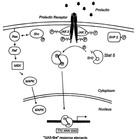
Fig. 1. Prolactin induced signaling. Binding of prolactin to the extracellular domain of the receptor induces dimerization and activation of the Tyr kinase JAK2 associated with the cytoplasmic domain of PrlR. Some of the substrates for JAK2 are indicated. These include: PrlR, StatS, She, and SHP-2. Tyr phosphorylation of StatS, a latent cytoplasmic transcription factor, promotes its dimerization, nuclear translocation and transcriptional activation of target genes following binding to GAS-like recognition sites. Tyr phosphory-lated She connects PrlR to the MAP kinase pathway via binding to the Grb-2/SOS complex (indicated by arrows). This complex in turn, activates Ras and the downstream signaling molecules Raf, MEK, and MAPK. SHP-2 has a positive effect on (3-casein gene transcription via its specific protein tyrosine phosphatase activity. The figure is a composite of information on prolactin signaling in mammary cells and in Nb2 lymphoma
cells. For details and references see the text.
appears to be absolutely essential for signal transmis-sion and in the normal situation a Stat very likely binds a particular phosphotyrosine site on a cytokine receptor. However, this interaction is not necessary for Stat phosphorylation and it is possible that JAK may in some cases function both as a docking protein and as a Tyr kinase for a Stat. Tyr phosphorylation of the receptor may not be essential for all aspects of signal-ing and, at least in an artificial system, appears to be expendable for transcriptional activation of (3-casein (47). It is interesting to speculate that some of the conserved Tyr residues present in the cytoplasmic
domain of the PrlR may couple to proteins involved in "fine-tuning" the prolactin signal. The Nb2 cell line
which expresses the intermediate form of the PrlR is very sensitive to prolactin yet its PrlR is not heavily phosphorylated on Tyr residues (25). The intermediate PrlR may lack conserved Tyr residues potentially important for down-regulation of the prolactin signal. The MAP Kinase Pathway
Mitogen-activated protein (MAP) kinase cas-cades are conserved intracellular signaling pathways
Fig. 2. Prolactin mediated Tyr phosphorylation of Stat5 and She, and activation of p42 ERK2 MAP kinase. Stat5a, She and the p42 ERK2 isoform of MAP kinase were immunoprecipitated from control, prolactin (P) and lactogenic hormone (DIP) treated HC11 cells. The Stat5a (upper panel) and She (middle panel) samples were electrophoresed, blotted and probed with a phosphotyrosine specific mAb. Tyr phosphorylation on Stat5 is maximum after 10 min of treatment and drops by 60 min. There is a basal level of Tyr phosphorylation on She under the conditions used in the experiment (c) and only the p52 and p46 She isoforms display an increase in phosphotyrosine following both treatments. An in vitro kinase assay was performed with p42 ERK2 MAP kinase using myelin basic protein (MBP) as a substrate (lower panel). MAP kinase activity is more strongly stimulated by DIP than P due to the combined effect of insulin and prolactin in the lactogenic hormone mix.
by which cells respond to a variety of external stimuli including hormones, cytokines and growth factors (48). In mammalian cells the 44 kDa and 42 kDa extracellular-signal-regulated kinases (ERKs) are members of the archetype MAP kinase cascade. Gener-ally, ERK1 and ERK2 are activated by the sequential stimulation of Ras, a small GTP binding protein, Raf-1, a 74 kDa Ser/Thr kinase and MEK, a dual specificity
kinase which activates ERK1 and ERK2 by phosphor-ylation on a both a Thr residue and a Tyr residue. ERK1 and ERK2 are broad-specificity kinases which recognize Ser/Thr in context of Pro residues. There is great interest in MAP kinase cascades since, among the substrates of ERKs, are transcription factors whose activity is altered by phosphorylation. Thus, MAP kinases transmit a signal from outside the cell into the nucleus thereby influencing its phenotype (Fig. 1).
The MAP kinase pathway is coupled to activated RTKs via the She adaptor protein. She binds to phos-photyrosine residues in many RTKs via its PTB bind-ing domain which recognizes -NXXYP- (49). She in
turn is phosphorylated on Tyr and serves as a docking site for the Grb-2/Son of Sevenless (SOS) complex. The relocalization of SOS, the Ras-GTP exchange fac-tor, to the inner surface of the plasma membrane allows it to exchange GDP on Ras for GTP, thus activating Ras and the downstream components of the MAP kinase cascade (Fig. 1). She is phosphorylated on Tyr follow-ing addition of prolactin to HC11 cells (Fig. 2) and Nb2 lymphoma cells (50). None of the conserved Tyr
residues in the PrlR are in a consensus PTB binding domain. This suggests that She may bind activated, phosphorylated JAK2 and directly serve as a substrate. In fact, the JAK2 sequence has a Tyr residue in a potential PTB recognition site (51). Although it is not yet known whether this Tyr is autophosphorylated by JAK2, it has been observed that She coimmunoprecipi-tates with JAK2 (50). In addition to She phosphoryla-tion, treatment of HC 11 cells with lactogenic hormones leads to the rapid activation of Raf-1, MEK1 and ERK2 (52 and Fig. 2). Nb2 cells treated with prolactin display
an elevation in Ras-GTP levels and Raf-1 kinase activ-ity (50,53). There is also a rapid appearance of ERK1 in the nucleus of these cells (54). See the chapter by Das and Vonderhaar for a more detailed discussion of the role of MAP kinase in prolactin induced mitogenesis.
Whilst experimental evidence attests to activation of the MAP kinase cascade by prolactin, the role of ERKs in prolactin induced mammary differentiation is not yet clear. Potential MAP kinase substrates are Stat5 itself as well as other transcription factors which play a role in lactogenic hormone induced transcription of milk protein genes. Although it is clear that Tyr-phosphorylation is necessary for Stat DNA binding, whether or not this phosphorylation is sufficient to promote transcriptional activation is now being investi-gated in various systems including mammary cells. Due to the diversity of signals which lead to Stat
activation, as well as the numerous genes whose tran-scription is stimulated in response to a particular Stat, it is unlikely that there will be a simple answer.
The COOH termini of Stat la and Stat3 have a consensus MAP kinase site -PXS/TP- which is phos-phorylated in response to interferon (IFN). A mutant Stat la in which the Ser in the consensus MAP kinase site was changed to Ala binds DNA as well as wild type. However, transcriptional activation of an IFN-y responsive gene was reduced in cells expressing the mutant (55). Currently only indirect evidence suggests that it is MAP kinase which phosphorylates this site (discussed in 56). However, these experiments clearly show that Ser phosphorylation can contribute to the transcriptional activation potential of Stats.
Stat5 is phosphorylated on Tyr and Ser residues in lactogen-treated HC11 cells (52), in IL-2 stimulated T cells (57) and in liver cells from GH treated rats (58). Since the MAP kinase signaling pathway is acti-vated by prolactin, the role of this pathway in the differentiation of HC11 cells was examined using the MAP kinase kinase (MEK) specific inhibitor PD98059 (59). Pretreatment of HC11 cells with PD98059 led to repression of lactogenic-hormone induced ERK2 MAP kinase activity but had no effect on the Ser phosphorylation of Stat5, on its DNA binding activity or on lactogenic hormone induced transcriptional acti-vation of the (i-casein promoter luciferase construct (52). These data demonstrate that MAP kinase activa-tion is not involved in Ser phosphorylaactiva-tion of Stat5 or in the transcriptional induction of the (3-casein gene mediated by lactogenic hormones.
Protein Tyrosine Phosphatase SHP-2
JAK2 mediates the initiation of the prolactin sig-nal. Since Tyr phosphorylation is transient, it is likely that protein tyrosine phosphatases (PTPs) are involved in some aspect of signal termination. However, little is known about the mechanism of signal termination and it should be noted that PTPs have both positive and negative effects on the propagation of signals initi-ated by RTKs (60). Among the multiple PTPs (60) there is a cytosolic subclass which appears to play a role in the transduction of cytokine induced signals. This subclass has two members, SHP-1 (previously called: SH-PTP1 or PTP1C) and SHP-2 (previously called: SH-PTP2 or PTP1D), Both have SH2 domains at their N-termini. SHP-1 has a restricted expression
pattern and is mainly found in hematopoietic cells, while SHP-2 is more widely expressed (61).
SHP-2 appears to play a role in prolactin signal-ing. In Nb-2 cells SHP-2, like JAK2, is associated with the PrlR even in the absence of hormone. Mutant PrlRs with no Tyr residues to serve as docking sites are also associated with SHP-2 (62). Further, in cells expressing an inactive mutant of SHP-2, prolactin induced tran-scription of ^-casein is lower than in cells expressing wild type SHP-2, suggesting that SHP-2 has a positive effect upon the prolactin induced signal (62). These results contrast dramatically with the role of SHP-1 in cytokine signaling. SHP-1 binds to a specific phospho-tyrosine residue in the COOH terminus of the EpoR and is not associated with the receptor in non-stimu-lated cells (63). SHP-1 binding to the EpoR is corre-lated with a decrease in JAK2 phosphorylation and cells expressing a mutant EpoR unable to bind SHP-1 are hypersensitive to Epo. These data suggest a negative role for SHP-1 in regulation of the Epo signal and show that despite the similarity in structure, 1 and SHP-2 have distinct functions in cytokine signaling. It is inter-esting that Nb2 cells which express the intermediate form of PrlR are hypersensitive to prolactin, suggesting that another PTP which binds to the region of the PrlR missing in the intermediate form, might be responsible for "down-regulation" of the prolactin signal.
EXTRACELLULAR MATRIX AND
PROLACTIN
The induction of milk protein gene expression in the mammary gland requires multiple signals including peptide and steroid hormones as well as signals ema-nating from the extracellular matrix (ECM) (64). In primary cultures of mammary gland cells prolactin-dependent Stat5 DNA binding and transcriptional acti-vation of milk protein genes is observed only in cells plated on a laminin-containing ECM (35). This implies that there is a hierarchy of signaling in mammary cells and that the ECM disposes the cells to respond to lactogenic hormones. The HC11 cells have retained this characteristic of primary cells. Sparse cultures of HC11 cells do not synthesize p-casein in response to lactogenic hormones (14). The cells must be grown to confluency in medium containing specific growth factors before becoming competent to respond to pro-lactin (15,16). Growing cultures of HC11 cells deposit an ECM which influences their ability to produce (3-casein in response to lactogenic hormones (65).
Inter-estingly, Stat5 is present in sparse cultures of HC11 cultures. However, prolactin treatment of these cells does not activate Stat5 DNA binding activity (7; N. Cella, unpublished results). This could in part be due to the lack of an appropriate matrix, reflecting the results seen with the primary mammary cells. The fact that Stat5 cannot be induced to bind its cognate GAS-like sequence in the 3-casein gene promoter under these conditions explains the lack of ^-casein produc-tion in sparse cultures of lactogen-treated HC11 cells.
DISCUSSION AND FUTURE QUESTIONS The description of the cytokine inducible JAK-Stat pathway has provided an answer to some of the long standing questions concerning prolactin-mediated signaling. For example, the mechanism underlying the different signaling potentials of the various forms of PrlR can now be explained by their ability to bind and activate JAK2. As is usual for any novel intracellular signaling pathway, there are now new problems to be solved. The results described earlier suggest that there might be two classes of PTPs involved in propagating the prolactin signal, one important for positive regula-tion and one involved in down-regularegula-tion of the signal. It will be interesting to examine endogenous SHP-2 regulation in mammary epithelial cells as well as to determine whether other PTPs might bind PrlR directly. In addition, further analyses of primary mam-mary cells may reveal an interesting connection between ECM and the prolactin inducible JAK2-Stat5 pathway. The role of Ser phosphorylation in Stat5 tran-scriptional activation also remains to be clarified. The Ser residue as well as the kinase which phosphorylates the site are still unknown. Stat5 proteins display con-sensus phosphorylation sites for protein kinase C (PKC), MAP kinase, casein kinase 2 (CK2) and cyclin dependent kinases (see 36 for a listing of some of these sites). The experiments with the MEK specific inhibitor PD98059 show that MAP kinase is not responsible for the Ser phosphorylation of Stat5. How-ever, there is experimental evidence implicating PKC (68,69) and CK2 (70) in the control of Stat5 activity.
In vitro treatment of nuclear extracts from lactating
mammary glands with PKC (69) or with CK2 (70) leads to enhanced Stat5 DNA binding. These experi-ments suggest that Ser phosphorylation together with Tyr phosphorylation may promote optimal DNA bind-ing activity of Stat5.
In addition to addressing new questions, some of the older experiments in the literature can now be reexamined in light of our current knowledge of the JAK2-Stat5 pathway. In the past, mammary cells trans-formed via ectopic expression of oncogenes have been used to probe the normal intracellular signaling path-ways involved in differentiation. The Ha-ras, v-ra/and
neu oncogenes all transform HC11 cells. However,
only the first two interfere with lactogen-induced 0-casein expression (66,67). Transformed cells undergo many alterations and p-casein gene expression is con-trolled by glucocorticoids, prolactin as well as other transcription factors (3-8). Thus, it is conceivable that the oncogenes interfere with more than one factor important for p-casein gene expression. However, Stat5 DNA binding activity is impaired in Ha-ras and v-ra/transformed cells (67) suggesting that these two oncogenes do interfere at some step in the prolactin-induced JAK-Stat pathway. In the future it would be interesting to use oncogenes expressed under the con-trol of an inducible promoter to examine early events in the transformation process.
ACKNOWLEDGMENTS
We thank Dr. Bernd Groner for many helpful discussions. We acknowledge Dr. Roger Beerli, Dr. Carmen Hagios, and Dr. Michael Fritsche for help with the figures and Dr. John Daly for comments on the manuscript. REFERENCES 1, 2. 3. 4, 5. 6
Y. J. Topper and C. S. Freeman (1980). Multiple hormone interactions in the developmental biology of the mammary gland. Physiolog. Rev. 60:1049-1106.
J. F. Bazan (1990). Structural design and molecular evolution of a cytokine receptor superfamily. Proc. Natl. Acad. Sci.
U.S.A. 87:6934-6938.
M. Schmitt-Ney, W. Doppler, R. K. Ball, and B. Groner (1991). B-casein gene promoter activity is regulated by the hormone mediated relief of transcriptional repression and a mammary gland-specific nuclear factor. Mol. Cell. Biol. 11:3745-3755. C. S. Lee and T. Oka (1992). A pregnancy-specific mammary nuclear factor involved in the repression of the mouse B-casein gene transcription by progesterone. J. Biol. Chem. 267: 5797-5801.
S. Altiok and B. Groner (1993). Interaction of two sequence-specific single-stranded DNA-binding proteins with an essen-tial region of the B-casein gene promoter is regulated by lacto-genic hormones. Mol. Cell. Biol. 13:7303-7310.
V. S. Meier and B. Groner (1994). The nuclear factor YY1 participates in repression of the B-casein gene promoter in mammary epithelial cells and is counteracted by the mammary
7. 8. 9. 10. 11. 12. 13. 14. 15. 16. 17. 18. 19. 20. 21. 22. 23.
gland factor during lactogenic hormone induction. Mol. Cell.
Biol. 14:128-137.
T. Welte, K. Garimorth, S. Philipp, and W. Doppler (1994). Prolactin-dependent activation of a tyrosine phosphorylated DMA binding factor in mouse mammary epithelial cells. Mol.
Endocrinol. 8:1091-1102.
B. Raught, W. S.-L. Liao, and J. M. Rosen (1995). Developmen-tally and hormonally regulated CCAAT/enhancer-binding pro-tein isoforms influence S-casein gene expression. Mol.
Endocrinol. 9:1223-1232.
H. Wakao, F. Gouilleux, and B. Groner (1994). Mammary gland factor (MGF) is a novel member of the cytokine regulated transcription factor gene family and confers the prolactin response. EMBO J. 13:2182-2191.
B. Groner and F. Gouilleux (1995). Prolactin-mediated gene activation in mammary epithelial cells. Curr. Opin. Genetics
Develop. 5:587-594.
C. Schindlerand J. E. Darnell (1995). Transcriptional responses to polypeptide ligands: the JAK-STAT pathway. Ann. Rev.
Bio-chem. 64:621-651.
W. Doppler (1994). Regulation of gene expression by prolactin.
Rev. Physiol. Biochem. Pharmacol. 124:93-130.
R. K. Ball, R. R. Friis, C. A. Schoenenberger, W. Doppler and B. Groner (1988). Prolactin regulation of B-casein expression and of a cytosolic 120 kDa protein in a cloned mouse mammary epithelial cell line. EMBO J. 7:2089-2095.
W. Doppler, B. Groner, and R. K. Ball (1989). Prolactin and glucocorticoid hormones synergistically induce expression of transfected rat B-casein gene promoter constructs in a mam-mary epithelial cell line. Proc. Natl. Acad. Sci. U.S.A. 86:104-108.
D. Tavema, B. Groner, and N. E. Hynes (1991). Epidermal growth factor receptor, platelet-derived growth factor receptor and c-erbB2 receptor activation all promote growth but have distinctive effects upon mouse mammary epithelial cell differ-entiation. Cell Growth Different. 2:145-154.
G. R. Merlo, D. Graus-Porta, N. Cella, B. M. Marte, D. Taverna, and N. E. Hynes (1996). Growth, differentiation and survival of HC11 mammary epithelial cells: diverse effects of receptor tyrosine kinase-activating peptide growth factors. Eur. J. Cell.
Biol. 70:97-105.
J. M. Boutin, C. Jolicoeur, H. Okamura, J. Gagnon, M. Edery, M. Shirota, D. Banville, I. Dusanter-Fourt, J. Dijiane, and P. A. Kelly (1988). Cloning and expression of the rat prolactin receptor, a member of the growth hormone/prolactin receptor gene family. Cell 53:69-77.
J. A. Davis and D. I. H. Linzer (1989). Expression of multiple forms of the prolactin receptor in mouse liver. Mol.
Endocri-nol. 3:674-680.
K. Buck, M. Vanek, B. Groner, and R. K. Ball (1992). Multiple forms of prolactin receptor messenger ribonucletc acid are specifically expressed and regulated in murine tissues and the mammary cell line HC11. Endocrinology 130:1108-1114. S. Ali, I. Pellegrini, and P. A. Kelly (1991). A prolactin-depen-dent immune cell line (Nb2) expresses a mutant form of
prolac-tin receptor. J. Biol. Chem. 266:20110-20117.
L. Lesueur, M. Edery, S. Ali, J. Paly, P. A. Kelly, and J. Djiane (1991). Comparison of long and short forms of the prolactin receptor on prolactin-induced milk protein gene transcription.
Proc. Natl. Acad. Sci. U.S.A. 88:824-828.
M. Edery, C. Levi-Meyrueis, J. Paly, P. A. Kelly, and J. Djiane (1994). A limited cytoplasmic region of the prolactin receptor critical for signal transduction. Mol. Cell. Endocrinol. 102:39-44.
K. D. O'Neal and L-Y. Yu-Lee (1994). Differential signal trans-duction of the short, Nb2 and long prolactin receptors. J. Biol.
Chem. 269:26076-26082. 24. 25. 26. 27. 28. 29. 30, 31. 32. 33. 34. 35. 36. 37. 38. 39.
L. DaSilva, O. M. Z. Howard, H. Rui, R. A. Kirken, and W. L. Farrar (1994). Growth signaling and JAK2 association mediated by membrane-proximal cytoplasmic regions of pro-lactin receptors. J. Biol. Chem. 269:18267-18270.
H. Rui, J. Y. Djeu, G. A. Evans, P. A. Kelly, and W. L. Farrar (1992). Prolactin receptor triggering: Evidence for rapid tyro-sine kinase activation. J. Biol. Chem. 267:24076-24081. J. N. Ihle, B. A. Witthuhn, F. W. Quelle, K. Yamamoto, W. E. Thierfelder, B. Kreider, and O. Silvennoinen (1994). Signaling by the cytokine receptor superfamily: JAKs and STATs.
TIBS 19:222-227.
I. Dusanter-Fourt, O. Muller, A. Ziemiecki, P. Mayeux, B. Drucker, J. Djiane, A. Wilks, A. G. Harpur, S. Fisher, and S. Gisselbrecht (1994). Identification of JAK protein tyrosine kinases as signaling molecules for prolactin. Functional analy-sis of prolactin receptor and prolactin-erythropoietin receptor chimera expressed in lymphoid cells. EMBO J. 13:2583-2591. H. Rui, R. A. Kirken, and W. L. Farrar (1994). Activation of receptor-associated tyrosine kinase JAK2 by prolactin. J. Biol.
Chem. 269:5364-5368.
G. S. Campbell, L. S. Argetsinger, J. N. Ihle, P. A. Kelly, J. A. Rillema, and C. Carter-Su (1994). Activation of JAK2 tyrosine kinase by prolactin receptors in Nb2 cells and mouse mammary
gland explants. Proc. Natl. Acad. Sci. U.S.A. 91:5232-5236. B. A. Witthuhn, F. W. Quelle, O. Silvennoinen, T. Yi, B. Tang, O. Miura, and J. N. Ihle (1993). JAK2 associates with the erythropoietin receptor and is tyrosine phosphorylated and acti-vated following stimulation with erythropoietin. Cell 74:227-236.
L. S. Argetsinger, G. S. Campell, X. Yang, B. A. Witthuhn, O. Silvennoinen, J. N. Ihle, C. Carter-Su (1993). Identification of JAK2 as a growth hormone receptor-associated tyrosine kinase. Cell 74:237-244.
J.-J. Lebrun, S. Ali, A. Ullrich, and P. A. Kelly (1995). Proline-rich sequence-mediated Jak2 association to the prolactin recep-tor is required but not sufficient for signal transduction. J. Biol.
Chem. 270:10664-10670.
M. ). Waters, N. Daniel, C. Bignon, and J. Djiane (1995). The rabbit mammary gland prolactin receptor is tyrosine-phosphor-ylated in response to prolactin in vivo and in vitro. J. Biol.
Chem. 270:5136-5143.
F. Gouilleux, H. Wakao, M. Mundt, and B. Groner (1994). Prolactin induces phosphorylation of Tyr694 of Stat5 (MGF), a prerequisite for DNA binding and induction of transcription.
EMBO J. 13:4361-4369.
C. H. Streuli, G. M. Edwards, M. Delcommenne, C. B. A. Whitelaw, T. G. Burden, C. Schindler, and C. J. Watson (1995). Stat5 as a target for regulation by extracellular matrix. J. Biol.
Chem. 270:21639-21644.
A. V. Kazansky, B. Raught, S. M. Lindsey, Y.-F. Wang, and J. M. Rosen (1995). Regulation of mammary gland factor/Stat5a during mammary gland development. Mol. Endocrinol. 9:1598-1609.
N. Tourkine, C. Schindler, M. Larose, and L.-M. Houdebine (1995). Activation of STAT factors by prolactin, interferon--y, growth hormones, and a tyrosine phosphatase inhibitor in rabbit primary mammary epithelial cells. J. Biol. Chem. 270:
20952-20961.
X. Liu, G. W. Robinson, F. Gouilleux, B. Groner, and L. Hennighausen (1995). Cloning and expression of Stat5 and an additional homologue (Stat5b) involved in prolactin signal transduction in mouse mammary tissue. Proc. Natl. Acad. Sci.
U.S.A. 92:8831-8835.
M. David, E. F. Petricoin III, K.-I. Igarashi, G. M. Feldman, D. S. Finbloom, and A. C. Lamer (1994). Prolactin activates the interferon-regulated p91 transcription factor and the Jak2 kinase by tyrosine phosphorylation. Proc. Natl. Acad. Sci.
40 41 42 43 44, 45, 46, 47. 48. 49. 50. 51. 52. 53. 54.
C. Pallard, F. Gouilleux, M. Charon, B. Groner, S. Gisselbrecht, and I. Dusanter-Fourt (1995). Interleukin-3, erythropoietin, and prolactin activate a Stat5-like factor in lymphoid cells. J. Biol.
Chem. 270:15942-15945.
L. DaSilva, H. Rui, R. A. Erwin, O. M. Z. Howard, R. A. Kirken, M. G. Malabarba, R. H. Hackett, A. C. Lamer, and W. L. Farrar (1996). Prolactin recruits STAT1, STAT3, and STATS independent of conserved receptor tyrosines TYR402, TYR479, TYR515, and TYR580. Mol. Cell. Endocrinol. 117:131-140.
Z. Songyang, S. E. Shoelson, J. McGlade, P. Olivier, T. Pawson, X. R. Bustelo, M. Barbacid, H. Sabe, H. Hanafusa, T. Yi, R. Ren, D. Baltimore, S. Ratnofsky, R. A. Feldman, and L. C. Cantley (1994). Specific motifs recognized by the SH2 domains of CSK, 3BP2, FES/FPS, GRB2, HPC, SHC, SYK, and VAV.
Mol. Cell. Biol. 14:2777-2785.
J.-J. Lebrun, S. Ali, V. Goffin, A. Ullrich, and P. A. Kelly (1995). A single phosphotyrosine residue of the prolactin recep-tor is responsible for activation of gene transcription. Proc.
Natl. Acad. Sci. U.S.A. 92:4031-4035.
F. W. Quelle, D. Wang, T. Nosaka, W. E. Thierfelder, D. Stravo-podis, Y. Weinstein, and J. N. Ihle (1996). Erythropoietin induces activation of Stat5 through association with specific tyrosines on the receptor that are not required for a mitogenic response. Mol. Cell. Biol. 16:1622-1631.
S. Gobert, S. Chretien, F. Gouilleux, O. Muller, C. Pallard, I. Dusanter-Fourt, B. Groner, C. Lacombe, S. Gisselbrecht, and P. Mayeux (1996). Identification of tyrosine residues within the intracellular domain of the erythropoietin receptor crucial for STAT5 activation. EMBO J. 15:2434-2441.
J. X. Lin, T. S. Migone, M. Tsang, M. Friedmann, J. A. Weath-erbee, L. Zhou, A. Yamauchi, E. T. Bloom, J. Mietz, S. John,
et al. (1995). The role of shared receptor motifs and common
Stat proteins in the generation of cytokine pleiotropy and redun-dancy by 1L-2, IL-4, IL-7, 1L-13, and IL-15. Immunity 2:331-339.
A. L.-F. Mui, H. Wakao, A.-M. O'Farrell, N. Harada, and A. Miyajima (1995). Interleukin-2, granulocyte-macrophage colony stimulating factor and interleukin-5 transduce signals through two STAT5 homologs. EMBO J. 14:1166-1175. S. L. Pelech (1996). Signaling pathways: kinase connections on the cellular intranet. Current Biol. 6:551-554.
W. M. Kavanaugh and L. T. Williams (1994). An alternative to SH2 domains for binding tyrosine-phosphorylated proteins.
Science 266:1862-1865.
R. A. Erwin, R. A. Kirken, M. G. Malabarba, W. L. Farrar, and H. Rui (1995). Prolactin activates Ras via signaling proteins SHC, growth factor receptor bound 2, and son of sevenless.
Endocrinology 136:3512-3518.
O. Silvennoinen, B. A. Witthuhn, F. W. Quelle, J. L. Cleveland, T. Yi, and J. N. Ihle (1993). Structure of the murine JAK2 protein-tyrosine kinase and its role in interleukin 3 signal trans-duction. Proc. Natl. Acad. Sci. U.S.A. 90:8429-8433. M. Wartmann, N. Cella, P. Hofer, B. Groner, X. Liu, L. Hen-nighausen, and N. E. Hynes (1996). Lactogenic hormone acti-vation of Stat5 and transcription of the 3-casein gene in mammary epithelial cells is independent of p42 ERK2 MAP kinase activity. J. Biol. Chem. (in Press).
C. V. Clevenger, T. Torigoe, and J. C. Reed (1994). Prolactin induces rapid phosphorylation and activation of prolactin recep-tor-associated RAF-1 kinase in a T-cell line. J. Biol. Chem. 269:5559-5565.
Y.-P. Rao, D. J. Buckley, and A. R. Buckley (1995). Rapid activation of mitogen-activated protein kinase and p21ras by
55. 56. 57. 58. 59. 60. 61. 62. 63. 64. 65. 66. 67. 68. 69. 70.
prolactin and interleukin 2. Cell Growth Different. 6:1235-1244.
Z. Wen, Z. Zhong, and J. E. Darnell, Jr. (1995). Maximal activation of transcription by Statl and Stat3 requires both tyrosine and serine phosphorylation. Cell 82:241-250. J. N. Ihle (1996). STATs and MAPs: obligate or opportunistic partners in signaling. BioEssays 18:95-98.
C. Beadling, J. Ng, J. W. Babbage, and D. A. Cantrell (1996). Interleukin-2 activation of STAT5 requires the convergent action of tyrosine kinases and a serine/threonine kinase pathway distinct from the Rafl/ERK2 MAP kinase pathway. EMBO
J. 15:1902-1913.
P. A. Ram, S.-H. Park, H. K. Choi, and D. J. Waxman (1996). Growth hormone activation of Stat1, Stat5, and StatS in rat liver. J. Biol. Chem. 271:5929-5940.
D. R. Alessi, A. Cuenda, P. Cohen, D. T. Dudley, and A. R. Saltiel (1995). PD98059 is a specific inhibitor of the activation of mitogen-activated protein kinase kinase in vitro and in vivo. J. Biol. Chem. 270:27489-27494.
H. Sun and N. K. Tonks (1994). The coordinated action of protein tyrosine phosphatases and kinases in cell signaling.
Trends Biochem. Sci. 19:480-485.
T. Yi, J. L. Cleveland, and J. N. Ihle (1992). Protein tyrosine phosphatase containing SH2 domains: characterization, prefer-ential expression in hematopoietic cells, and localization to human chromosome 12p 12-13. Mol. Cell. Biol. 12:836-846. S. Ali, Z. Chen, J.-J. Lebrun, W. Vogel, A. Kharitonenkov, P. A. Kelly, and A. Ullrich (1996). PTP1D is a positive regulator of the prolactin signal leading to b-casein promoter activation.
EMBOJ. 15:135-142.
U. Klingmuller, U. Lorenz, L. C. Cantley, B. G. Neel, and H. F. Lodish (1995). Specific recruitment of SH-PTP1 to the erythropoietin receptor causes inactivation of JAK2 and termi-nation of proliferative signals. Cell 80:729-738.
C. D. Roskelley, A. Srebrow, and M. J. Bissell (1995). A hierarchy of ECM-mediated signaling regulates tissue-specific gene expression. Curr. Opin. Cell Biol. 7:736-747.
R. Chammas, D. Taverna, N. Cella, C. Santos, and N. E. Hynes (1994). Laminin and tenascin assembly and expression regulate HC11 mouse mammary cell differentiation. J. Cell Sci. 107:1031-1040,
N. E. Hynes, D. Taverna, I.-M. Harwerth, F. Ciardiello, D. S. Salomon, T. Yamamoto, and B. Groner (1990). Epidermal growth factor receptor, but not c-erbB2 activation prevents lactogenic hormone induction of the B-casein gene in mouse mammary epithelial cells. Mol. Cell. Biol. 10:4027-4034. B. Happ, N. E. Hynes, and B. Groner (1993). Ha-ros and
v-raf oncogenes, but not int-2 and c-myc, interfere with the
lactogenic hormone dependent activation of the mammary gland specific transcription factor. Cell Growth Different. 4:9-15.
B. M. Marte, T. Meyer, S. Stabel, G. J. R. Standke, S. Jaken, D. Fabbro, and N. E. Hynes (1994). Protein kinase C and mammary cell differentiation: involvement of Protein Kinase C a in the induction of ^-casein expression. Cell Growth
Different. 5:239-247.
A. Marti, B. Jehn, E. Costello, N. Keon, G. Ke, F. Martin, and R. Jaggi (1994). Protein kinase A and AP-1 (c-Fos/JunD) are induced during apoptosis of mouse mammary epithelial cells.
Oncogene 9:1213-1223.
M. Schmitt-Ney, B. Happ, R. K. Ball, and B. Groner (1992). Developmental and environmental regulation of a mammary gland specific nuclear factor essential for transcription of the gene encoding B-casein. Proc. Natl. Acad. Sci. U.S.A. 89:3130-3134.