1 Division for Hand-, Plastic- and Reconstructive Surgery, Department of Surgery, University Hospital Zurich, Switzerland,
2 Division of Trauma, Department of Surgery, University Hospital Zurich, Switzerland.
Received: October 16, 2005; accepted: January 12, 2006
Iatrogenic Neurovascular Entrapment Injuries
Caused by Reduction and Intramedullary
Fixation of Fractures of the Lower Limb
Volker Wedler
1, Ludwig Labler
2, Christian Köhler
1, Merlin Guggenheim
1, Walter Künzi
1,
Otmar Trentz
2Abstract
Background: Damage to the main neurovascular trunk
in the lower limb due to trauma has also been de-scribed in the literature. Little attention, however, has been directed toward the quantitative assessment of iatrogenic injury to peripheral neurovascular structures caused by the treatment of open fractures in the lower limb. Obtaining an angiography of the lower limb early in the course of treatment of such injuries is necessary.
Methods: We performed a retrospective analysis of
253 patients with open fractures. We divided into them into two groups, treated either with an intramedullary (A) or an extramedullary (B) approach. We furthermore noted whether a free tissue transfer was necessary in either group. Particular attention was directed to iatro-genic neurovascular injuries caused by reduction and intramedullary fixation.
Results: In group A, we found two cases of entrapment
of the anterior tibial artery and its concomitant veins, and the deep peroneal nerve. In group B, we recorded four cases of iatrogenic injuries to the common pero-neal nerve. Free tissue transfer was performed in 102 cases, 4 of which were emergency free flaps.
Conclusion: We wish to stress that ascertaining the
anatomy and patency of the vascular anatomy of the injured limb early during the primary assessment of the patient, either by palpation, portable Doppler flow-meter, duplex sonography, or angiography before, and preferably also after, reduction and intramedullary nail-ing of the fracture is in our view mandatory, before any treatment of the fracture is envisioned.
Key Words
Fracture care · Intramedullary nailing · Lower limb injuries · Microsurgery · Nerves
Eur J Trau ma 2006;32:381–6 DOI 10.1007/s00068-006-6127-9
Introduction
The principles of treatment of open fractures are well established: limitation of soft tissue damage, adequate debridement, prevention (or at least monitoring) of compartment swelling, identification of injury to neurovascular structures, fracture alignment, and sta-bilization, early initiation of weight bearing and joint movements. It goes without saying that the optimal treatment strategy should be decided upon primarily, depending on the classification of the injury [1]. In this respect, in addition to imaging examinations such as conventional radiographs, the physical assessment of the lower limb is of prime importance, with particular focus on neurological aspects, such as sensory and motor nerve function, and peripheral circulation. The results obtained by such an examination greatly influence the decision making process. Iatrogenic lesions can be brought about by any of the therapeutic actions neces-sary for the treatment of open fractures in the lower limb, such as reduction of the fracture, intramedullary advan-cement of the nail across the fracture line and locked intramedullary nailing of the fracture. Most closed frac-tures of the shaft of either femur or tibia can be treated successfully by internal fixation with medullary or
inter-locking nails. Associated extensive soft tissue damage mandates coverage with pedicled or microvascular free tissue transfer in an interdisciplinary approach. In case of open multifragment fractures, the indication of intra-medullary nailing versus other open or closed methods should be discussed with careful attention to the risk of a possible entrapment of the main vessels and nerves. Clo sed intramedullary nailing is preferable for trans-verse fractures that can be reduced and “hooked on” under the image intensifier. If interlocking nails are used, the indications can be extended to more unstable fractures. Protagonists of the intramedullary nailing methods claim that it significantly shortens the period of inactivity and diminishes the likelihood of angulation deformity and joint stiffness when compared with clo-sed techniques [2]. Iatrogenic injury to the major vessels is observed mainly in fractures around the knee. The artery and its accompanying veins may be cut, torn, compressed, or contused either by the initial trauma or subsequently by jagged bone fragments or malred-uction [3]. In case of a fracture of the tibia or in com-bination with a fractured fibula, the leg is in an exter-nal rotated position and the reduction of the fracture is performed by distraction and an internal rotation maneuver with verification of the correct alignment under the image intensifier. If intramedullary nai-ling is used for treatment of such fractures, iatrogenic injury to entrapped neurovascular structures can occur either through multiple fragments, which may be dis-located substantially in the course of the repositio-ning process (Figures 1a and 1b) or by advancing the nail through the medullary cavity across the fracture line (Figure 2). We therefore consider it to be of prime importance to ascertain both anatomy and patency of the vascular anatomy of the injured limb early during the primary assessment of the patient, either by palpation, portable Doppler flowmeter, duplex sonography, or angiography before and preferably also after reduction and intramedullary nailing of the fracture is perform-ed. In order to ascertain the patency of the posterior tibial artery, however, we have found relying on digital palpation alone to be unreliable, as the palpation of an arterial pulse either behind the medial ankle or on the dorsum of the foot can be the result of retrograde perfu-sion through the arterial arcus plantaris and therefore be deceiving. These examinations also provide the pla-stic surgeon with invaluable information concerning the level on which to perform microvascular anastomoses for free tissue transfer or the division of a pedicled flap.
Patients and Methods
Between January 1997 and March 2004, the Division of Traumatology treated 253 patients with open fractures (Gustillo I–IIIc), 182 male and 71 female. These patients were divided into two groups, group A (n = 53) treated with intramedullary nailing and group B with extrame-dullary techniques (n = 200). Of the 53 patients in group A, 39 were male and 14 female. The mean age was 43 years (19–89 years). In group B there were 182 males and 71 females with a mean age of 43 years, (ranged 4–93 years) like in group A. In group A and B together, 102 microvascular free tissue transfers were performed in cooperation with the Division for Hand-, Plastic- and Reconstructive Surgery to cover the associated soft tis-sue defects in an interdisciplinary approach, with the
Figures 1a and 1b. Draft of a tibial fracture and the interpositioning of the anterior tibial vessels (veins = blue, artery = red) and deep pero-neal nerve ( yellow) during the retention of the fracture.
trauma surgery team performing the reduction and fixa-tion of the fracture as well as the debridement of the open wound. In four of those cases, the free tissue trans-fer was performed as an emergency free flap. More often than not, in anticipation of the coverage of the defect by the plastic surgery team, the debrided wound was covered with a V.A.C.-dressing (V.A.C. KCI® Medical
Schweiz GmbH, Geroldswil, Switzerland). The plastic surgery team strived to cover the tissue defect between the third and eighth day posttraumatically, depending, however upon the go-ahead by the trauma surgery team, which was always in charge of patient management. In this paper, we focus entirely on the iatrogenic injuries caused during the treatment of patients in group A by the entrapment of the main neurovascular bundle.
Results
A retrospective review of these patients focused on iatrogenic neurovascular injuries caused by reduction and intramedullary fixation was undertaken to docu-ment the risk of injury with the intramedullary nailing technique compared with other techniques applied for treatment of open fractures of the leg. All fractures in group A were treated with copious irrigation and debri-dement initially and underwent intramedullary nailing at an average of 8 days postinjury (range 0–18 days). In group A, we found two cases of entrapment of the anterior tibial artery and its concomitant veins and the deep peroneal nerve, the latter resulting in asensibility of the second dorsal intermetatarsal space. In group B, we recorded four cases of iatrogenic injuries to the com-mon peroneal nerve with a subsequent motoric deficit and no injury to a main vessel. We would like to present a brief summary of the two patients in group A with en-trapment of the anterior tibial artery and its concomitant veins and, in one case, the deep peroneal nerve as well.
Case 1
A 71-year-old male patient sustained a rollover injury to his right foot caused by the wheel of a car during a traffic accident. The patient was evaluated according to the guidelines of advanced trauma live support (ATLS) by our trauma surgery team [4]. No addi-tional injuries were detected. The patient was alert and stable, but complained about pain in the lower right leg and foot. Physical examination of the extremity in question showed a decollement of the forefoot with a 2 × 3 cm2 wound over the first metacarpal bone.
Clini-cally, the development of a compartment syndrome of the forefoot was evident. In the absence of open wounds, contusions were still manifest at the fracture site on the distal right tibia. Neurological evaluation showed red-uced sensibility over the dorsum of the forefoot, but normal plantar sensibility. No clinical signs of vascular injury were present. Conventional radiographs demons-trated proximal fractures of the metacarpalia II–V, furthermore a fracture of the proximal phalanx of the great toe and a spiral fracture of the tibia between the middle and distal third of the tibia (Figure 3). Immedi-ate surgical release of the compartments on the right forefoot was followed-up with an aggressive debride-ment with copious irrigation of the soft tissues of the forefoot. Smears for bacterial culture were taken and antibiotic therapy with second-generation cephalospo-rines was initiated. The fractures of the metacarpalia
Figure 2. Advancement of the nail through the medullary cavity across the fracture line and damaging of the neu-rovascular bundle.
and the proximal phalanx of the great toe were sta-bilized intramedullary with Kirschner wires. The soft tissue incisions were temporarily covered with Epigard®
(Biovision GmbH, Ilmenau, Germany). The tibia fracture was reduced without a traction table and, after reaming; an intramedullary nail was inserted under fluoroscopic control. Distal interlocking screw holes were drilled in a freehand technique and screws of proper size were placed (Figure 4). The proximal interlocking was accomplished by means of a proximal locking guide (Figures 4a to 4c). The patient presented a wound defect of about 12 × 10 cm pretibial and the plastic surgeon performed a free latissi-mus dorsi transfer at day five postoperatively. Intraope-rative he found the neurovascular bundle with the ante-rior tibial artery, the concomitant veins and the deep peroneal nerve strangulated into the fracture (Figures 5a and 5b). A retrospective review also of this case, howe-ver, proved that the A. dorsalis pedis must have been perfused retrogradely through the arcus plantaris. The arterial and venous microanastomoses were performed 2 cm proximal to the entrance where the neurovascular bundle went into the medullary cavity. Wound healing of the soft tissue coverage was without any complica-tion and the patient was discharged to the rehabilitacomplica-tion center 2.5 weeks after the injury.
Case 2
A 42-years old healthy construction worker had his right lower leg caught under a falling steel beam, sustaining a Gustillo II type fracture of medial third of the tibia.
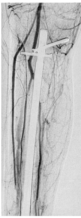
During the primary survey, no sensory deficits were recorded. Both the A. dorsalis pedis and the posterior tibial artery were palpable. Neither a Doppler Sonogram, nor a Doppler flow meter examination was performed. The primary debridement was carried out by the trauma surgery team on the same day and covered with a V.A.C.-dressing. On the third day, the fracture was reduced and fixated with an intramedullary nail. On the following day an angiography was performed and an interruption of the anterior tibial artery was diagnosed (Figure 6); there was every reason to believe that the falling steel beam inju-red the vessel. At the fifth day the tissue defect remai-ning measured 8 × 9 cm on the anterolateral tibia, with bone being exposed. The plastic surgery team covered the defect with a free latissimus dorsi muscle flap on day 7. The initial concept before the angiogram was perfor-med had been to perform end-to-end-anastomoses to the anterior tibial artery and a concomitant vein; the division of this neurovascular bundle, however, uncovered an ent-rapment of these structures within the fracture. As the entrapment went no more profound than the width of the cortical tibial bone, the nail within the medullary cavity had not further damaged these structures. As the design of the flap necessary to cover the defect made it unavoi-dable to place the vascular pedicle very proximally on the lower leg, an arterio- and venotomy at the level up the upper tibial third was performed and the anastomoses were placed end-to-end, as initially envisioned. The pro-found peroneal nerve was pro-found not to entrapped. A retrospective review also of this case, however, proved
Figure 3. X-ray of the spiral fracture of the tibia between the middle and distal third of the tibia.
Figures 4a to 4c. X-ray of the tibia; after reaming an in-tramedullary nail was inserted under fluoroscopic control.
that the A. dorsalis pedis must have been perfused retro-gradely through the arcus plantaris.
Discussion
There are only few reports in the literature about iatrogenic neurovascular injuries caused by reduction and intramedullary fixation of fractures of the lower limb. The majority of complications reported are com-partment syndromes [2], deep venous thrombosis [5], pulmonary embolism [6], hardware failure and ampu-tation [3, 7]. Koval et al. [8] described the complications associated with the treatment of 60 acute fractures by intramedullary nailing and divided those complica-tions into three categories, intraoperative, early post-operative, and late postoperative. In his investigation, intraoperative complications consisted mainly of frac-ture propagation, poor quality of the screws, and other hardware problems. Early postoperative complica-tions included hematoma, malunion, and neurological deficits. Astonishingly, he reported a 30% complication rate of neurological injuries in this group, which could
be attributed to the operative procedure, but he did not distinguish between preexisiting neurological deficits preoperatively and intraoperative complications. All of the patients in his series showed neuropraxia, which he ascribed to related forces by slight distraction during the retention before the operative procedure. The rate of neurological complications found in his study is higher than the rate reported by other authors (6–12%) [9, 10]. Urban & Tornetta [11] described a case of a distal vascular insult with an ischemic foot caused by a distal locking screw, and Kessler et al. [7] poin-ted out that the proximal interlocking screw using the intramedullary locked nail technique can cause major problems when the drill is advanced in the sagittal direction toward the popliteal neurovascular trunk. To evaluate the neurovascular structures at risk during placement of anterior-posterior locking screws in the proximal femur, Riina et al. [12] performed a cadaver study and illustrated that the risks to the neurovascu-lar structures during anterior–posterior locking in the proximal femur are diminished if locking is performed above the level of the lesser trochanter. There are several authors who described vascular complications associated with locked intramedullary tibial nailing.
Figures 5a and 5b. a) The forceps is pointing out the entry of the neu-rovascular bundle into the fractured area proximally (Case 1). b) The forceps is pointing out the exit of the neurovascular bundle out of the fractured area distally (Case 1)
Figure 6. Interruption of the anterior tibial artery in the mid-dle of the tibia (Case 2).
Based on a magnetic resonance imaging (MRI) study, Stindel et al. [3] found complications to be infrequent yet always serious, with a secondary amputation rate of 30%. Wilbourn [13] reported a case of injury to a branch of the profunda femoris artery during placement of the anteroposterior proximal locking screw. Roberts et al. [14] described a vascular compromise with subsequent amputation below the knee. Other than several reports of injuries to the main vessels caused by screws peri-operatively, there have been no reports about intra-operative neurovascular injuries to the surrounding neurovascular anatomy using the intramedullary tibia nailing technique. Such iatrogenic injuries result either from damaging these structures while entrapping them during fracture reduction or possibly while advancing the nail through these structures, if they are entrapped. As a matter of course, a combination of both mecha-nisms can certainly be imagined. In our retrospective analysis we found two cases with an entrapment of the anterior tibial artery (cases 1 and 2) and the deep peroneal nerve (case 1) in group A and four cases in group B with iatrogenic injury to the common peroneal nerve. Compared to group B, the consequences for the patients in group A were minimal, as the plastic surgeon was able to perform the microvascular anasto-moses for the free tissue transfer 2 cm proximally to the level of the lesion of the tibial artery, and there was only a permanent sensory deficit (case 1) in the second dorsal intermetatarsal space. We like to emphasize that the only difference to an unharmed vessel is the ability to do the microanastomoses in an end to side technique and to maintain a main vessel. The motoric deficiency associated with the lesion of the common peroneal nerve was more dramatic for the patients in group B and required further treatment.
We wish to stress that ascertaining the anatomy and patency of vascular anatomy of the injured limb early during the primary assessment of the patient, either by palpation, portable Doppler flowmeter, duplex sono-graphy or angiosono-graphy before and preferably also after reduction and intramedullary nailing of the fracture is in our view mandatory, before any treatment of the frac-ture is envisioned. We believe that manual palpation of the pulses at the level of the foot are insufficient, as the result can be deceiving, if the artery palpated is perfused retrogradely through the arcus plantaris. For the same reason, a bedside Doppler flowmeter examination is in our opinion unable to yield dependable results, as it is difficult to determine the direction of the flow reliably.
Therefore, we suggest early visualization of the vascular anatomy and function preferably with a duplex sonogra-phy, which we consider to be the option of choice for obtaining the desired information quickly. Certainly a conventional or even an MRI angiography may be performed just as well and will undoubtedly produce useful information, the realization of these examinations in an emergency situation may, however, prove to be dif-ficult. These investigations could also give information about whether the neurovascular bundle was injured by the initial trauma or during the retention process.
References
1. Tischenko GJ, Goodman ST. Compartment Syndrome after intra-medullary nailing of the tibia. J Bone Joint Surg 1990;72:41–4. 2. Hooper GJ, Keddell RC, Penny ID. Conservative management
or closed nailing for tibial shaft fractures. A randomised prospective trial. J Bone Joint Surg 1991;73b:83–5. 3. Stindel E, Colin D, Le Guillou E, et al. The use of MR images
to evaluate the risks associated with proximal locking of intramedullary tibial nails. Surg Radiol Anat 2001;23:173–7. 4. American College of Surgeons. ATLS course manual. Chicago:
ACS, 1997
5. Hindley CJ, Evans RA, Holt EM, et al. Locked intramedullary nailing for recent lower limb fractures. Injury 1990;21:239–44. 6. Weresh MJ, Stover MD, Bosse MJ, et al. Pulmonary gas exchange
during intramedullary fixation of femoral shaft fractures. J Trauma 1999;46:863–8.
7. Kessler SB, Kaiser E, Eibl- Eibesfeld B. Vermeidung von Gefäss-verletzungen bei der Tibiaverriegelungsnagelung (Prevention of vascular injuries in tibial interlocking nailing). Unfallchirurg 1987;90:148–50.
8. Koval K, Clapper M, Brumback R, et al. Complications of reamed intramedullary nailing of the tibia. J Orthop Trauma 1991;5:184–9. 9. Bone LB, Johnson KD. Treatment of tibial fractures by reaming
and intramedullary nailing. J Bone Joint Surg 1986;68:877–87. 10. Klemm KW, Borner M. Interlocking nailing of complex fractures
of the femur and tibia. Clin Orthop 1986;212:89–100.
11. Urban WP, Tornetta P. Vascular compromise after intramedullary nailing of the tibia: a case report. J Trauma 1995;38:804–7. 12. Riina J, Tornetta P, Ritter C, et al. Neurologic and vascular
structures at risk during anterior–posterior locking of retrograde femoral nails. J Orthop Trauma 1998;12:379–81. 13. Wilbourn AJ. Iatrogenic nerve injuries. Neurol Clin 1998;16:55–82. 14. Roberts C, Ruktanonchai D, King D, et al. Vascular compromise
and amputation after intramedullary nailing of a tibia fracture. J Orthop Trauma 1998;12:136–8.
Address for Correspondence
Volker Wedler, MD
Division for Hand-, Plastic- and Reconstructive Surgery University Hospital of Zurich
Rämistr. 100, 8094 Zurich Switzerland
Phone (+41/ 44) 255-1111, Fax -8948 e-mail: volker.wedler@usz.ch