ORIGINAL ARTICLE
Histomorphological analysis of the urogenital diaphragm
in elderly women: a cadaver study
C. Betschart&D. Scheiner&C. Maake&M. Vich&
L. Slomianka&D. Fink&D. Perucchini
Received: 7 February 2008 / Accepted: 28 May 2008 / Published online: 25 June 2008 # The International Urogynecological Association 2008
Abstract The objective of this study was to describe the histomorphological structure of the urogenital diaphragm in elderly women using a modern morphometric procedure. Biopsies were taken from the posterior margin of the urogenital diaphragm of 22 female cadavers (mean age, 87 years) using a 60-mm punch. Hematoxylin/eosin and Goldner sections were analyzed with the Cavalieri estima-tor. The mean thickness of the urogenital diaphragm was 5.5 mm. The main component was connective tissue. All biopsies contained smooth muscle. Eighteen biopsies contained more smooth muscle than striated muscle. In six of 22 biopsies, no striated muscle was found. The ratio of striated to smooth muscle to connective tissue was 1:2.3:13.3. Muscle fibers were dispersed in all parts of the urogenital diaphragm. The urogenital diaphragm of elderly women mainly consists of connective tissue. Smooth muscle was also found but to a lesser extent. The frequently used English term“perineal membrane” for the urogenital diaphragm is justified and well describes our findings in elderly women.
Keywords Perineal membrane . Smooth muscle . Striated muscle . Connective tissue . Aging
Introduction
Transvaginal repair of anterior compartment prolapse with either synthetic or biologic meshes is a new procedure in treating women with cystocele [1] and weak or defective tissues. Today, the focus of interest is on the efficacy, the safety, and the complication rate of these operations, but relatively little is known about the composition of the underlying tissue of the urogenital diaphragm that is repaired or reinforced. To obtain additional insights into the histomorphology of the urogenital diaphragm, we systematically analyzed this anatomical area in elderly women with the Cavalieri estimator, a morphometric instrument [2].
The urogenital diaphragm, consisting of the M. trans-versus perinei profundus and the M. transtrans-versus perinei superficialis, was originally described by Luschka in 1864 and extends from the ischial tuberosity to the perineal body [3]. Since this first description, the literature has provided controversial information about the histomorphological composition of this anatomical structure. Henle described the urogenital diaphragm as two aponeuroses containing the musculus transversus perinei profundus or deep transverse perineal muscle [4]. The musculus transversus perinei profundus is listed in the recent Nomina Anatomica (sixth edition), while the superficial transverse perineal muscle is not. In most anatomical textbooks, the deep transverse perineal muscle is mentioned as a thick, musculo-fascial, triangularly shaped structure composed of striated muscle fibers and connective tissue [5], which, with the superficial transverse perineal muscle, forms the urogenital diaphragm. So far, the role of the urogenital diaphragm in maintain-ing continence and its function in the lower part of the pelvic floor remain unclear [6, 7]. Knowledge of the histomorphology of the urogenital diaphragm in elderly
C. Betschart (*)
:
D. Scheiner:
D. Fink:
D. Perucchini Department of Gynecology, University Hospital Zürich, Frauenklinikstrasse 10,8091 Zurich, Switzerland e-mail: [email protected] C. Maake
:
M. Vich:
L. Slomianka Institute of Anatomy, University of Zürich, Zurich, Switzerlandwomen could give useful information about the tissue composition, the quantity of the different tissue layers, the inter-individual variation, and the existence of the urogen-ital diaphragm in elderly women without genurogen-ital prolapse.
Material and methods
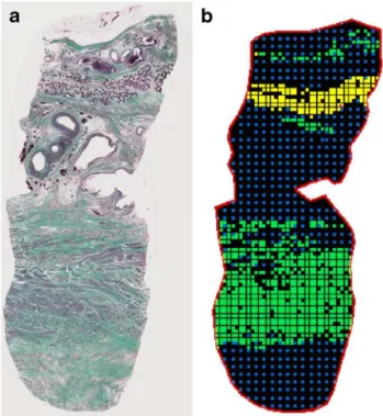
From February 2005 until March 2006, biopsies of the urogenital diaphragm of 22 formalin-fixed female cadavers were sampled at the Institute of Anatomy, University of Zurich, Switzerland. All 22 cadavers were female Cauca-sians. Due to the anonymity of the cadavers, no personal history except age was available. Mean age was 87 years (range, 74 to 101 years). Under the supervision of experienced senior anatomists, standardized biopsies were taken of the urogenital diaphragm 2 cm lateral of the introitus vaginae on the right side of the cadavers using a punch biopsy of 6-mm diameter (Fig.1). All samples were marked on the cranial side, where the fascia diaphragmatis urogenitalis superior is supposed to be. The specimens were refixed by immersion in 10% buffered formalin. To obtain exact transverse slices of the muscle tissue, the material was mounted in a vertical position. The muscle samples were embedded in paraffin, and the blocks were cut on a microtome in 10-µm serial sections. For histomorpholog-ical evaluation, all slides were stained with hematoxylin/ eosin (HE) and Goldner. For the morphometric analysis of the samples, the areas occupied by connective tissue, smooth muscle, and striated muscle were estimated using the Cavalieri estimator [2] (StereoInvestigator, MBF Bio-science, Williston, VT, USA) with a 300×300-μm point grid for connective tissue and a 150×150-μm point grid for smooth and striated muscle using a ×10 lens and a ×400 final magnification on-screen (Fig.2). Grid sizes aimed at counting on average 100 points in each of the tissue types
in each section, resulting in potential measurement errors within the sections of no more than 10% and typically much less [8]. Sectional area of tissue types will be directly proportional to their volume. Statistical analysis was done by Intercooled Stata 8.2. Data Analysis.
Results
The mean examined cross-sectional area of the urogenital diaphragm is 32.8 mm2 (range, 9.7–63.1 mm2, SD 12.2), corresponding to a thickness of 5.5 mm (range, 1.6 to 10.5 mm).
The main component of the urogenital diaphragm is the connective tissue. Eighty percent of the standardized biopsies of the urogenital diaphragm consists of connective tissue, including vessels, fat, and collagen fibers. Smooth muscle was found in all 22 biopsies. Striated muscle was present in 16 of the 22 samples. In these 16 biopsies, only four specimens showed more striated than smooth muscle (Fig. 3).
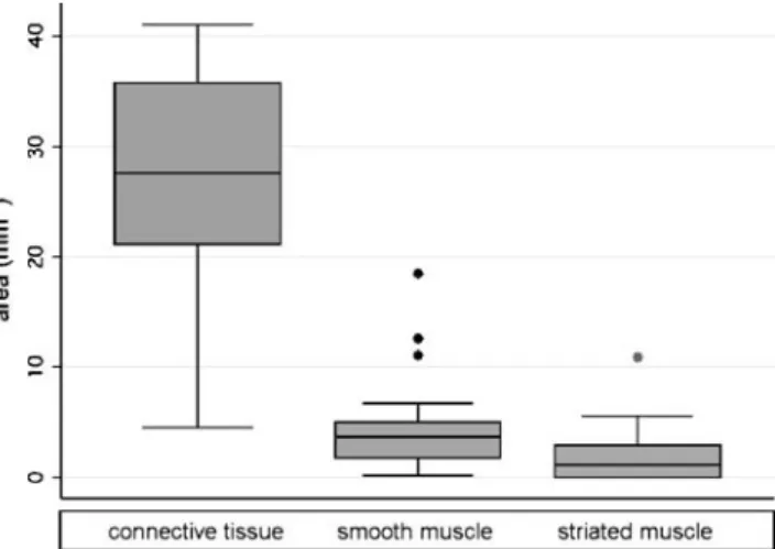
The mean area of smooth muscle was 4.55 mm2(range, 0.2–18.5 mm2; SD 4.4), of striated muscle 1.95 mm2 (range, 0–10.9 mm2
; SD 2.6), and of connective tissue 26.3 mm2(range, 4.5–41.1 mm2, SD 10.6) (Table1). The median area of smooth muscle was 3.65 mm2, of striated
Fig. 1 Biopsy taken from the urogenital diaphragm 2 cm lateral to the introitus vaginae on the right side. DTP deep transverse perineal muscle, STP superficial transverse perineal muscle
Fig. 2 a Histology of the urogenital diaphragm, ×10 magnification, Goldner staining. b Representation of the points counted using the Cavalieri estimator to estimate the sectional areas associated with connective tissue (dark gray/blue), smooth muscle (light gray/green), and striated muscle (white/yellow)
muscle 1.1 mm2, and of connective tissue 27.65 mm2 (Fig.4).
The average ratio of striated muscle to smooth muscle to connective tissue was 1:2.3:13.3 (Fig. 5). No correlation was found between age and striated muscle, smooth muscle, or connective tissue area (Spearman’s rho 0.099, 0.017, and 0.213, respectively; p<0.05). Both types of muscle fibers were dispersed in all parts of the specimens.
Discussion
In this histomorphological study of the urogenital dia-phragm, we expected a significant amount of striated muscle and found a surprisingly high proportion of connective tissue and smooth muscle. The literature gives quite different histomorphological descriptions of the urogenital diaphragm. In 1873, Henle [4] described the urogenital diaphragm containing a high proportion of smooth muscle, whereas“striated muscle fibers were rarely demonstrable or not at all.” In the anatomical textbook of Benninghoff, the thickness of the urogenital diaphragm is described to be 1 cm and to contain a “high amount of connective tissue and striated muscle” [9]. According to Benninghoff, sometimes, a 2- to 3-mm-thick layer of
smooth muscle cells is seen near the fascia diaphragmatica urogenitalis superior. Furthermore, the muscle is mentioned to be embedded in the fascia diaphragmatica urogenitalis superior and inferior. In our study, the muscle layers are dispersed all over the urogenital diaphragm. In all biopsies, no upper or inferior fascia could be demonstrated. Oelrich concluded in his histomorphological research of the perineal membrane that there is no superior fascia of the so-called urogenital diaphragm [10].
Our data show a higher ratio of smooth than striated muscle in the urogenital diaphragm. Similar results were recently reported for the levator ani muscle. Jundt et al. demonstrated that the levator ani muscle contains striated muscle in only 54% cases. These biopsies were obtained during pelvic floor surgery of 24 women with genital prolapse. The remaining biopsies consisted of smooth muscle and connective and fat tissue [11].
Disappearance of striated muscle in the female urethra has also been observed. Periurethral connective tissue
Fig. 4 Box plot of the tissue components with median and first and third quartile boundaries
Fig. 3 Descending proportion of the area of smooth muscle, striated muscle, and connective tissue in the urogenital diaphragm
Table 1 Mean area of the different tissue layers in square millimeters, standard deviation, minimum, and maximum
Smooth muscle (mm2) Striated muscle (mm2) Connective tissue (mm2) Mean 4.55 1.95 26.2 Standard deviation ±4.4 ±2.6 ±10.6 Minimum 0.2 0 4.5
Maximum 18.5 10.9 41.1 Fig. 5 Percentage of smooth muscle, striated muscle, and connective tissue of the urogenital diaphragmx
undergoes alteration in postmenopausal women with genital prolapse with and without stress incontinence [12]. Cadaver studies show that, in older women, all layers of striated urethral muscle become thinner; 2% of striated muscle in the anterior urethral wall is lost per year between the age of 20 and 80 [13, 14] and may be completely absent posteriorly [15]. Also, the study of Pandit et al. [16] supported the hypothesis that with increasing age the nerve density in the female striated urogenital sphincter muscle correlates with decreased muscular tissue. The nerve density in the urogenital diaphragm was not the focus of our interest; but, according to previously cited studies [14– 16], striated muscle loss in elderly women seems obvious. Striated muscle fibers were completely absent in six of our 22 samples.
The small number, the high age, and the anonymity of the female cadavers are limitations of our study.
The high age may explain why we could not find any correlation between age and muscle thickness in our study population. Nevertheless, the high quantity of connective tissue and the small amount of striated muscle in the urogenital diaphragm have never been demonstrated in a similar manner before. It will be the subject of further studies to compare our results with the urogenital dia-phragm of young parous or nulliparous women.
Due to the lack of data about parity and previous operations such as pelvic organ prolapse repair, which could increase the amount of connective tissue, we cannot interpret the high amount of connective tissue as a consequence of antecedent surgery in our sample. Never-theless, it is not likely that all 22 cadavers had undergone prolapse surgery.
Our biopsies were all taken at the same anatomical site. This was 2 cm lateral to the vaginal introitus, far enough from the urethra to avoid accidental biopsy of periurethral muscle. To minimize inter-individual variation, we took the biopsies always on the right side of the body. This is in accordance with Fischer’s histological–histochemical in-vestigation of the pubococcygeal muscle, which showed an intra-individual unambiguous right–left difference with regard to muscle quality, amount of muscle fibers, and connective tissue [17].
New operation techniques in cystocele surgery are based on strengthening the remnants of the urogenital diaphragm by mesh implants [18]. It is known that the success of surgery depends on the available tissue. The connective tissue of the subcutaneous layer is a good material for sutures in dog and cat [19]. The subcutaneous tissue supports the overlying structures and is important for further perfusion and wound healing. Removal of subcuta-neous connective tissue is accompanied by a higher risk of wound infections in dogs. This could encourage consider-ation that the connective tissue of the urogenital diaphragm
is important in wound healing of interposed meshes and prevents the patient from wound infection or mesh erosion, known as one of the major complications [1]. To support this hypothesis, further histomorphological studies before and after mesh implants must be performed.
An integrated action of various anatomical intra- and extraurethral structures are involved in the mechanism of female continence. The most important extraurethral struc-tures are the suburethral vaginal wall, the pubourethral ligaments, the pubococcygeus muscles, and the periurethral connective tissue [20]. The role of the urogenital diaphragm for the mechanism of maintaining continence is still unknown. Many studies investigating the histomorpholog-ical and histochemhistomorpholog-ical properties of striated muscles of the urethral sphincter muscle and the levator ani, regarding the control of continence, have been published [21–25]. Dimpfl et al. [23] have shown in histomorphological studies of the pelvic floor that both aging and vaginal birth may lead to myogenic changes. Chen and Creed [21] studied the levator ani and urethral striated muscle in dogs and sheep and found it to contain both slow type I fibers and fast type II fibers which are important in sustaining continence. Hanzal et al. [26] showed that the existence of striated muscle in the levator ani muscle has prognostic importance for the outcome of patients with stress incontinence undergoing antero-posterior vaginal repair. The high proportion of smooth muscle in the levator ani muscle and in the urogenital diaphragm could explain the limitation of pelvic floor re-education training. Whether the urogenital dia-phragm of elderly women contributes to the mechanism of continence remains still a matter of debate.
Controversial data have been published concerning the tissue composition and the existence or lack thereof of the urogenital diaphragm. Oelrich did not find a urogenital diaphragm in his monumental studies about the pelvic floor of embryos and female and male cadavers [10]. Also, Dorschner et al. [27], in their sectional and MRI studies, could not identify a urogenital diaphragm, consisting of the deep transverse perineal muscle. The term musculus trans-versus perineus profundus was attributed only to the male cadaver because this muscle is represented only by smooth muscle in the female cadaver. Mirilas and Skandalakis [7] gave a profound and controversial review of the anatomy of the perineal spaces over the last two centuries. The muscular layer between the two urogenital fascias was the object of a recent study of Bazhenov and Blinova [28]. In that study, the urogenital diaphragm of 36 female cadavers measured 0.7–0.9 mm and comprised two muscles, con-sisting of smooth muscle cells.
After the macroscopic study of the cadavers and histomorphological examination of the biopsies taken, we conclude that there is a triangularly shaped structure attached to the bony framework of the pubic arch. This
structure is oriented in the horizontal plane and has a free posterior margin. Based on our data, the idea of a musculus transversus perineus profundus and superficialis consisting mainly of striated muscle fibers appears to be exaggerated and must be refuted based on our samples of frail old women. The term “perineal membrane”, widely used in English and American textbooks, therefore seems to be much more appropriate for this anatomical structure, consisting mostly of connective tissue and smooth muscle.
Conflicts of interest None.
References
1. Dwyer PL, O’Reilly BA (2004) Transvaginal repair of anterior and posterior compartment prolapse with Atrium polypropylene mesh. BJOG 111:831–836
2. Gundersen HJG, Bendtsen TF, Korbo L, Marcussen N, Møller A, Nielsen K, Nyengaard JR, Pakkenberg B, Sørensen B, Vesterby A, West MJ (1988) Some new, simple and efficient stereological methods and their use in pathological research and diagnosis. Apmis 96:379–394
3. Luschka H (1864) Die Anatomie des Menschlichen Beckens. Laupp and Siebeck, Tübingen
4. Henle J (1873) Handbuch der Systematischen Anatomie des Menschen. Bd II. Friedrich Vieweg und Sohn, Braunschweig 5. Borley NR (2005) True pelvis, pelvic floor and perineum. In:
Standring S (ed) Gray’s anatomy. 39rd edn. Elsevier, Amsterdam, pp 1367–1369
6. Kalischer O (1900) Die Urogenitalmuskulatur des Dammes. Karger, Berlin
7. Mirilas P, Skandalakis J (2004) Urogenital diaphragm: an erroneous concept casting its shadow over the sphincter urethrae and deep perineal space. J Am Coll Surg 198:279–290
8. Gundersen HJG, Jensen EB (1987) The efficiency of systematic sampling in stereology and its prediction. J Microsc 147:229– 263
9. Benninghoff A, Drenckhahn D (2002) Makroskopische anatomie, histologie, embryologie, zellbiologie. Band 1: Harn- und Geni-talsystem. Urban und Fischer, München, Jena
10. Oelrich TM (1983) The striated urogenital sphincter muscle in the female. Anat Rec 2005(2):223–232
11. Jundt K, Kiening M, Fischer P, Bergauer F, Rauch E, Janni W, Peschers U, Dimpfl T (2005) Is the histomorphological concept of the female pelvic floor and its changes due to age and vaginal delivery correct? Neurourol Urodyn 24:44–50
12. Goepel C, Hefler L, Methfessel HD, Koelbl H (2003) Periurethral connective tissue status of postmenopausal women with genital
prolapse with and without stress incontinence. Acta Obstet Gynecol Scand 82:659–664
13. Perucchini D, DeLancey JO, Ashton-Miller JA, Peschers U, Kataria T (2002) Age effects on urethral striated muscle. I. Changes in number and diameter of striated muscle fibers in the ventral urethra. Am J Obstet Gynecol 186:351–355
14. Carlile A, Davies I, Rigby A, Brocklehurst JC (1988) Age changes in the human female urethra: a morphometric study. J Urol 139:532–535
15. Perucchini D, DeLancey JO, Ashton-Miller JA, Galecki A, Schaer GN (2002) Age effects on urethral striated muscle. II. Anatomic location of muscle loss. Am J Obstet Gynecol 186:356–360 16. Pandit M, DeLancey JO, Ashton-Miller JA, Iyengar J, Blaivas M,
Perucchini D (2000) Quantification of intramuscular nerves within the female striated urogenital sphincter muscle. Obstet Gynecol 96 (6):797–800
17. Fischer W, Pfister C, Tunn R (1992) Histomorphology of pelvic floor muscles in women with urinary incontinence. Zentralbl Gynaekol 114(4):189–194
18. Richter K (1988) Surgical anatomy of the bladder neck sphincter and its significance for vaginal surgery of stress incontinence. Geburtshilfe Frauenheilkd 48(8):541–550
19. Bohling MW, Henderson RA, Swaim SF, Kincaid SA, Wright JC (2006) Comparison of the role of the subcutaneous tissues in cutaneous wound healing in the dog and cat. Vet Surg 35(1):3–14 20. Ulmsten U, Falconer C (1999) Connective tissue in female urinary
incontinence. Curr Opin Obstet Gynecol 11(5):509–515 21. Chen X, Creed KE (2004) Histochemical and contractile
properties of striated muscles of urethra and levator ani of dogs and sheep. Neurourol Urodyn 23:702–708
22. DeLancey JO, Starr RA (1990) Histology of the connection between the vagina and levator ani muscles. Implications for urinary tract function. J Reprod Med 35:765–771
23. Dimpfl T, Jaeger C, Mueller-Felber W, Anthuber C, Hirsch A, Brandmaier R, Schuessler B (1998) Myogenic changes of the LAM in premenopausal women: the impact of vaginal delivery and age. Neurourol Urodyn 17:197–205
24. Gilpin SA, Gosling JA, Smith AR, Warrell DW (1989) The pathogenesis of genitourinary prolapse and stress incontinence of urine. A histological and histochemical study. Br J Obstet Gynaecol 96:15–23
25. Fritsch H, Pinggera GM, Lienemann A, Mitterberger M, Bartsch G, Strasser H (2006) What are the supportive structures of the female urethra? Neurourol Urodyn 25(2):128–134
26. Hanzal E, Berger E, Koelbl H (1993) Levator ani muscle morphology and recurrent genuine stress incontinence. Am Coll Obstet Gynecol 81:426–429
27. Dorschner W, Biesold M, Schmidt F, Stolzenburg J (1999) The dispute about the external sphincter and the urogenital diaphragm. J Urol 162:1942–1945
28. Bazhenov DV, Blinova NV (2002) Morphofunctional character-istics of the urogenital diaphragm in women. Morfologiia 122:64– 67