Sights and Signals
Ernst J. M. Speel and Paul Komminoth
Abstract
During the last decade, several strategies have been developed to improve the detection
sensitivity of
in situ
hybridization (ISH) by amplification of either target nucleic acid
sequences prior to ISH (e.g.,
in situ
PCR), or the detection signals after the hybridization
procedures (signal amplification). Here we outline the principles of tyramide signal
amplification using the catalyzed reporter deposition (CARD) technique, summarize
applications as well as possible limitations of CARD ISH, and discuss some future direc-
tions of
in situ
nucleic acid detection using this amplification strategy.
Key Words: In situ
hybridization; tyromide; signal amplification; mRNA; DNA; catalyzed
reporter deposition;
interphase cytogenetics.
Department of Pathology University of Zurich, Schmelzbergstrasse 12, CH-8091 Zurich, Switzerland Address correspondence to Ernst J. M. Speel. Ph.D. Department of Pathology, University of Zurich, Schmelzbergstrasse 12, CH-8091 Zurich, Switzerland. E-mail: [email protected] Endocrine Pathology, vol. 10, no. 3, 193-198, Fall 1999 9 Copyright 1999 by Humana Press Inc. All rights of any nature whatsoever reserved.
1046-3976/99/10:193-198/$11.50
Introduction
In situ
hybridization (ISH) is now an
established molecular tool in research and
diagnostics and has significantly advanced
the study ofgene structure and expression
at the level of individual cells. Currently,
the technique provides an optimal detec-
tion sensitivity of approx 1 kb of target
DNA in cell preparations using fluores-
cence approaches in combination with
charge-coupled device (CCD) recordings
and image analysis [1]. The ultimate
mRNA detection limit, however, is more
difficult to determine but may reach the
level of single mRNA molecules in the most
optimal test systems [2]. For detection
of nucleic acid sequences in routinely
processed tissue sections of paraffin-
embedded specimens, these sensitivities
may not be reached. As a consequence, ISH
detection limits on tissue sections are rather
in the range of 40 kb of target DNA and
10-20 copies ofmRNA or viral DNA per
cell [3-6].
In recent years, several strategies have
been developed to improve the sensitivity
oflSH. These include the use of increased
absolute amounts of hybridized probes
(cocktails of oligonucleotides or multiple
cRNA probes) [7] and the amplification
of either nucleic acid targets (target ampli-
fication) [8,9] or of (immuno) cytochemi-
cal detection signals (signal amplification)
in situ[10,11].
In general, target amplification methods
combine polymerase chain reaction
(PCR) and ISH to visualize specific ampli-
fied DNA and RNA sequences within cell
and tissue preparations. Theoretically
these
in situPCR techniques are straight-
forward, but in practice they are hampered
by several obstacles, such as low amplifi-
cation efficiency (restricted sensitivity),
poor reproducibility (restricted specific-
ity), and difficulties in quantification of
the results [12-15]. As a consequence,
other approaches to increase the sensitiv-
ity of ISH have been explored, of which
193
194
Endocrine Pathology Volume 10, Number 3 Fall 1999,'
illll
IIIIIIIIIIIIIIIIIIIIIIIIIIIIIIIIIIIIIIIIIIIIIIIIIIIIIIIIIIIII
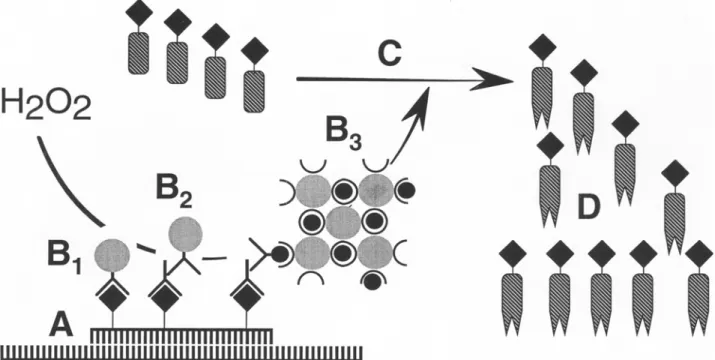
Fig. 1. Principles of CARD signal amplification for ISH. (A) Hybridization in situ with a hapten-labeled probe. (B) Application of a one-step (anti-hapten antibody conjugated to horseradish peroxidase; B~), two-step (anti-hapten antibody and a horseradish peroxidase labeled secondary antibody; B2), or three-step (avidin-biotin horseradish peroxidase complex; B3) probe detection system. (C) Production of haptenized tyramide radicals by horseradish peroxidase catalyzed CARD signal amplification. (D) Deposition of tyramide radicals to tyrosine moieties of proteins in situ in the vicinity of hybridization. Direct visualization of fluorochrome-labeled tyramides and indirect visualization of hapten- labeled tyramides with anti-hapten antibody or (strept)avidin conjugates labeled with fluorochromes or enzymes.
the CARD signal amplification method
appears to be the most promising.
Principles of CARD Signal
Amplification
The method of CARD signal amplifi-
cation has been developed by Bobrow et
al. [16] for use in immuno-blotting and
ELISA assays and is based on the deposi-
tion of a large number of haptenized
tyramide molecules promoted by peroxi-
dase activity (Fig. 1). Tyramine is a phenolic
compound and horseradish peroxidase
(HRP) can catalyze the dimerization of
such compounds when they are present at
high concentrations probably by the gen-
eration of free radical intermediates. If
applied in lower concentrations, such as
used in the signal amplification reaction,
the probability of tyramine dimerization
is reduced, whereas the binding of the
highly reactive intermediates to elec-
tron-rich moieties of proteins, such as tyro-
sine, at or near the site of the peroxidase
binding site is favored. In this way, many
hapten-labeled tyramine molecules (tyra-
mides) can be deposited at the hybridiza-
tion site
in situ.Visualization of deposited
tyramides can be performed either directly
after the CARD reaction with fluorescence
microscopy, if fluorochrome-labeled
tyramides are used, or indirectly with either
fluorescence or brightfield microscopy, if
biotin, digoxigenin, di- or trinitrophenyl
are used as haptens, which can act as fur-
ther binding sites for anti-hapten antibod-
ies or (strept)avidin conjugates (in the case
of biotinylated tyramides) [3]. Also fluo-
rescein and rhodamin can be used as hap-
tens, since specific antibodies against these
fluorochromes are commercially available
from several companies.
Applications and Limitations of
CARD Signal Amplification
CARD signal amplification with bioti-
nylated tyramides has been adapted for
immunohistochemistry by Adams [17],
allowing an increase in sensitivity of up to
1000-fold when compared with conven-
tional avidin biotinylated enzyme complex
(ABC) procedures [17-21 ]. In these stud-
ies, the amplification factor was assessed
by determining the maximal dilution of the
primary antibody leading still to identical
staining results as compared with standard
reactions. In most cases, however, the
increase in sensitivity seems to be rather in
the range of 50-100-fold, and sometimes
even less. As a consequence of this vari-
ability the optimal dilution for every pri-
mary antibody needs to be determined.
CARD signal amplification has also been
applied to visualize antigens or incorpo-
rated BrdU in fuorescence microscopy
[22,23] or electron
microscopy
[24,25],
and has further been used for double stain-
ing with two unconjugated primary anti-
sera raised in the same species [26].
Since 1995, CARD has further been
implemented in detection procedures of
both DNA and RNA ISH on cell prepara-
tions and tissue sections. With signal
amplification, the ISH sensitivity could be
improved in the range of 2- to 100-fold,
enabling the detection of up to three dif-
ferent repetitive and single-copy (1-5 kb)
DNA sequences in the same cell [22,27-
30] as well as low copy viral RNA [31,32]
and mRNA ranging from high to low
abundancy in cell and tissue preparations
[10,33-36].
As an example of the diagnostic poten-
tial of CARD ISH, we have implemented
CARD signal amplification for our diag-
nostic nonradioactive oligonucleotide ISH
procedure in order to increase the sensitiv-
ity of the assay and to shorten its overall
turnaround-time [10,33]. This approach
allows, e.g., the detection of peptide hor-
mone mRNA in tissue sections from rou-
tinely fixed, paraffin-embedded surgical
samples within one working day and makes
the assay suitable for routine diagnostic
purposes. Furthermore, it allows the use
of diaminobenzidine (DAB) as a chro-
mogen and, as a consequence, the applica-
tion of conventional counterstains and the
mounting of slides in xylene-based mount-
ing solutions, making the procedure more
acceptable to perform in a diagnostic setting.
To date, most of the protocols still use
biotinylated tyramides for the amplifica-
tion step, which can easily be obtained
commercially (e.g., NEN Life Science
Products, Boston, MA and Dako, Glost-
rup, Denmark) or synthesized in the labo-
ratory [16,17,27,28,30,37]. However,
similar to immunohistochemical proce-
dures, the use of biotin is associated with
significant disadvantages, especially when
working with tissue sections. Thus, in
tissues with high amounts of endogenous
biotin, such as liver or kidney, a low signal-
to-noise ratio due to high background
staining may be encountered. It is there-
fore desirable to be able to rely on differently
labeled tyramides, e.g., with digoxigenin,
di- or trinitriphenyl [10,30,33], or fluo-
rochromes
[10,22,30,33,34],
which is now
possible. These tyramide conjugates can also
be used in multiple-target ISH approaches
[22,29,30] or the combination of imm-
unohistochemistry and ISH with signal
amplification.
196
Endocrine Pathology
Volume 10, Number 3 Fall 1999Although the increase in ISH sensitivity
by using CARD signal amplification is
obvious from the literature, speculation
about the obtained amplification factor is
difficult. Moreover, since the tyramide
deposition reaction runs very quickly,
minor differences in amplification reaction
time may lead to variations in the final
signal intensities. Nevertheless, an ampli-
fication factor in the range of 5- to 10-fold,
or possibly higher, together with preserva-
tion of distinct localization of ISH signals
seems to be a realistic indication for both
DNA and mRNA ISH.
Since with CARD signal amplification
both specific and nonspecific (background)
ISH signals will be amplified, it is essential
that nonspecific probe binding and
detection have to be avoided in order
to successfully apply this procedure
[22,30,35,36]. Therefore, we recommend
that one should always optimize probe
hybridization and cytochemical probe
detection when applying CARD signal
amplification in order to achieve discretely
localized ISH signals of high intensity. In
our hands, the number of cytochemical
detection layers (e.g., one layer is sufficient
for repetitive, centromeric DNA detection
but minimal two layers are recommended
for DNA targets of 40 kb), the dilution of
detection conjugates (usually the first detec-
tion layer can be diluted 2-10-fold further
than in conventional detection systems),
the tyramide concentration in the CARD
amplification buffer (usually concentra-
tions in the range of 2-12 ~tM are used
[30]), and the reaction time (usually 5-15
min at room temperature or 37~ are the
most important parameters to consider.
Conclusions
CARD signal amplification using
labeled tyramides is an easy-to-perform,
fast, highly sensitive, and efficient proce-
dure to increase the detection sensitivity
of ISH and immunohistochemistry and
appears to become the method of choice
for diagnostic laboratories. It will not only
promote the detection of viral or mRNA
but also facilitate the evaluation of chro-
mosomal aberrations in cytological and
histological specimens. Furthermore, it
might also help to advance the develop-
ment of automated ISH spot-counting by
computer-assisted image generation and
analysis. The now available spectrum of
probe labels, detection systems, and
tyramide conjugates for CARD signal
amplification will further improve the
applicability and sensitivity oflSH as well
as promote multiple-target nucleic acid
detection in situ and procedures combin-
ing ISH and immunophenotyping [38].
Acknowledgments
The authors thank P. Saremaslani for
excellent technical assistance, N. Wey for
computer-assisted reproductions, and
A.H.N. Hopman, F.C.S Ramaekers,
J. Roth, and Ph.U. Heitz for continuous
support.
References
1. Lichter P, Bentz M, Joos S. Detection of chro- mosomal aberrations by means of molecular cytogenetics: Painting of chromosomes and chromosomal subregions and comparative genomic hybridization. In: Vogt P, IM Verma, eds. Oncogene techniques. San Diego, CA: Academic Press, 334-359, 1995.
2. Femino AM, Fay FS, Fogarty K, Singer RH. Visualization of single RNA transcripts in situ. Science 280:585-590, 1998.
3. Speel EJM, Ramaekers FC, Hopman AHN. Cytochemical detection systems for in situ hybridization, and the combination with immunocytochemistry. "Who is still afraid of red, green and blue?" Histochem J 27:833- 858, 1995.
4. H6fler H, Childers H, Montminy MR, Lechan RM, Goodmann RH, Wolfe HJ. In situ hybrid- ization methods for the detection of somatostatin mRNA in tissue sections using antisense RNA probes. Histochem J 18:597- 604, 1986.
5. Dirks RW. RNA molecules lighting up under the microscope. Histochem Cell Biol 106: 151-166, 1996.
6. McNicol AM, Farquharson MA. In situ hybridization and its diagnostic applications in pathology. J Pathol 182: 250-61, 1997. 7. Trembleau A, Bloom FE. Enhanced sensitiv-
ity for light and electron microscopic in situ hybridization with multiple simultaneous non-radioactive oligodeoxynucleotide probes. J Histochem Cytochem 43:829-841, 1995. 8. Komminoth P, Long AA. In situ polymerase
chain reaction and its applications to the study of endocrine diseases. Endocr Pathol 6:167-
171, 1995.
9. Long AA, Komminoth E In situ polymerase chain reaction: an overview. In: Gosden JR, eds. Methods in Molecular Biology Vol. 71. PRINS and in situ PCR Protocols. Totowa NJ: Humana Press Inc., 141-161, 1997. 10. Sped EJM, Hopman AHN, Komminoth E
Signal amplification for DNA and mRNA in situ hybridization. In: Darby J, eds. In situ hybridization protocols. Totowa: Humana Press, 2000.
11. Komminoth P, Werner M. Target and signal amplification: approaches to increase the sen- sitivity of in situ hybridization. Histochem Cell Biol 108: 325-333, 1997.
12. Long AA, Komminoth P, Lee E, Wolfe HJ. Comparison of indirect and direct in-situ polymerase chain reaction in cell preparations and tissue sections. Detection of viral DNA, gene rearrangements and chromosomal trans- locations. Histochemistry 99:151-162, 1993. 13. Komminoth P, Long AA. In-situ polymerase chain reaction. An overview of methods, applications and limitations of a new molecular technique. Virchows Arch B 64:67-73, 1993. 14. Komminoth P, Adams V, Long AA, Roth J, Saremaslani P, Flury R, Schmid M, Heitz PU. Evaluation of methods for hepatitis C virus (HCV) detection in liver biopsies: compari- son of histology, immunohistochemistry, in-situ hybridization, reverse transcriptase (RT) PCR and in-situ RT PCR. Path Res Pract 190:1017-1025, 1994.
15. HSfler H. In situ polymerase chain reaction: toy or tool? (Editorial). Histochemistry 99:103-104, 1993.
16. Bobrow MN, Harris TD, Shaughnessy KJ, Litt GJ. Catalyzed reporter deposition, a novel method of signal amplification. Application to immunoassays. J Immunol Methods 125:279-285, 1989.
17. Adams JC. Biotin amplification of biotin and horseradish peroxidase signals in histochemi- cal stains. J Histochem Cytochem 40:1457- 1463, 1992.
18. Berghorn KA, Bonnett JH, Hoffman GE. cFos immunoreactivity is enhanced with biotin amplification. J Histochem Cytochem 42:1635-1642, 1994.
19. Merz H, Malisius R, Mannweiler S, Zhou R, Hartmann W, Orscheschek K, Moubayed P, Feller AC. ImmunoMax. A maximized immunohistochemical method for the retrieval and enhancement of hidden antigens. Lab Invest 73:149-156, 1995.
20. Sanno N, Teramoto A, Sugiyama M, Itoh Y, Osamura RY. Application of catalyzed signal amplification in immunodetection of gona- dotropin subunits in clinically nonfunctioning pituitary adenomas. Am J Clin Pathol 106:16-21, 1996.
21. Werner M, von Waasielewski R, Komminoth P. Antigen retrieval, signal amplification and intensification in immunohistochemistry. Histochem Cell Biol 105:253-260, 1996. 22. van Gijlswijk RPM, Zijlmans HJ, Wiegant J,
Bobrow MN, Erickson TJ, Adler KE, Tanke HJ, Raap AK. Fluorochrome-labeled tyramides: use in immunocytochemistry and fluorescence in situ hybridization. J Histochem Cytochem 45:375-382, 1997.
23. Van Heusden J, de Jong P, Ramaekers F, Bruwiere H, Borgers M, Smets G. Fluores- cein-labeled tyramide strongly enhances the detection of low bromodeoxyuridine incorpo- ration levels. J Histochem Cytochem 45:315- 319, 1997.
24. Sch6fer C, Weipoltshammer K, Almeder M, Wachtler E Signal amplification at the ultra- structural level using biotinylated tyramides and immunogold detection. Histochem Cell Biol 108:313-319, 1997.
25. Mayer G, Bendayan M. Biotinyl-tyramide: a novel approach for electron microscopic immunocytochemistry. J Histochem Cyto- chem 45:1449-1454, 1997.
198
Endocrine Pathology
Volume 10, Number 3 Fall 199926. Shindler KS, Roth KA. Double immunofluo- rescent staining using two unconjugated pri- mary antisera raised in the same species. J Histochem Cytochem 44:1331-1335, 1996. 27. Kerstens HM, Poddighe PJ, Hanselaar AG. A
novel in situ hybridization signal amplifica- tion method based on the deposition of biotinylated tyramine. J Histochem Cytochem 43:347-352, 1995.
28. Raap AK, Van de Corput MPC, Vervenne RAW, van Gijlswijk RPM, Tanke HJ, Wiegant J. Ultra-sensitive FISH using peroxidase- mediated deposition of biotin- or fluorochrome tyramides. Hum Mol Genet 4:529-534, 1995. 29. Speel EJM, Ramaekers FCS, Hopman AHN. Sensitive multicolor fluorescence in situ hybridization using catalyzed reporter depo- sition (CARD) amplification. J Histochem Cytochem 45:1439-1446, 1997.
30. Hopman AHN, Ramaekers FCS, Speel EJM. Rapid synthesis of biotin-, digoxigenin-, trinitrophenyl-, and fluorochrome-labeled tyramides and their application for in situ hybridization using CARD-amplification. J Histochem Cytochem 46:771-777, 1998. 31. Adler K, Erickson T, Bobrow M. High sensi-
tivity detection of HPV-16 in SiHa and CaSki cells utilizing FISH enhanced by TSA. Histochem Cell Biol 108:321-324, 1997. 32. Reed JA, Nador RG, Spaulding D, Tani u
Cesarman E, Knowles DM. Demonstration of Kaposi's sarcoma-associated herpes virus cyclin D homolog in cutaneous Kaposi's sar- coma by colorimetric in situ hybridization using a catalyzed signal amplification system. Blood 91:3825-3832, 1998.
33. Speel EJM, Saremaslani P, Roth J, Hopman AHN, Komminoth P. Improved mRNA in situ hybridization on formaldehyde-fixed and paraffin-embedded tissue using signal ampli- fication with different haptenized tyramides. Histochem Cell Biol 110:571-577, 1998. 34. Schmidt BF, Chao J, Zhu Z, DeBiasio RL,
Fisher G. Signal amplification in the detection of single-copy DNA and RNA by enzyme-cata- lyzed deposition (CARD) of the novel fluores- cent reporter substrate Cy3.29-tyramide. J Histochem Cytochem 45:365-373, 1997. 35. Van de Corput MPC, Dirks RW, Van Gijlswijk
RPM, Van Binnendijk E, Hattinger CM, De Paus RA, Landegent JE, Raap AK. Sensitive mRNA detection by fluorescence in situ hybridization using horseradish peroxidase- labeled oligodeoxynucleotides and tyramide signal amplification. J Histochem Cytochem 46:1249-1259, 1998.
36. Yang H, Wanner IB, Roper SD, Chaudhari N. An optimized method for in situ hybrid- ization with signal amplification that allows the detection of rare mRNAs. J Histochem Cytochem 47:431-445, 1999
37. Jacobs W, Dhaene K, VanMarck E. Tyramine- amplified immunohistochemical testing using "homemade" biotinylated tyramine is highly sensitive and cost-effective. Archives of Pathology and Laboratory Medicine 122: 642-643, 1998.
38. Speel EJM. Detection and amplification sys- tems for sensitive, multiple-target DNA and RNA in situ hybridization: looking inside cells with a spectrum of colors. Histochem Cell Biol 112:89-113,1999.