Effect of 3-mercaptopyruvate Sulfurtransferase Deficiency
on the Development of Multiorgan Failure, Inflammation,
and Wound Healing in Mice Subjected to Burn Injury
Akbar Ahmad, PhD,* Nadiya Druzhyna, PhD,* and Csaba Szabo, MD, PhD, FPBS*,†,‡
The gaseous transmitter hydrogen sulfide (H2S) has been implicated in various forms of critical illness. Here, we have compared the outcome of scald burn injury in wild-type mice and in mice deficient in 3-mercaptopyruvate sulfurtransferase (3-MST), a mammalian H2S-generating enzyme. Outcome variables included indices of organ injury, clinical chemistry parameters, and plasma levels of inflammatory mediators. Plasma levels of H2S significantly increased in response to burn in wild-type mice, but remained unchanged in 3-MST-/- mice. The capacity of tissue
homogenates to produce H2S from 3-mercaptopyruvate was unaffected by burn injury. In 3-MST-/- mice, compared
to wild-type controls, there was a significant enhancement in the accumulation of polymorphonuclear cells (as assessed by the quantification of myeloperoxidase) in the liver (but not heart, lung, or skin) at 7 days postburn. Oxidative tissue damage (as assessed by malon dialdehyde content) was comparable between wild-type and 3-MST-deficient mice in all tissues studied. 3-MST-/- and wild-type mice exhibited comparable burn-induced elevations
in circulating plasma levels of hepatic injury; however, 3-MST-/- mice exhibited a higher degree of renal injury (as
reflected by elevated blood urea nitrogen levels) at 7 days postburn. Inflammatory mediators (eg, TNF-α, IL-1β, IL-2, IL-6, IL-10, and IL-12) increased in burn injury, but without significant differences between the 3-MST -/- and wild-type groups. The healing of the burn wound was also unaffected by 3-MST deficiency. In conclusion,
the absence of the H2S-producing enzyme 3-MST slightly exacerbates the development of multiorgan dysfunction but does not affect inflammatory mediator production or wound healing in a murine model of burn injury.
Hydrogen sulfide (H2S) is an endogenous gasotransmitter, which regulates multiple physiological and pathophysiologi-cal functions in the vascular, immune, and nervous system.1–6
H2S biogenesis in mammals is catalyzed by three enzymes:
cystathionine-gamma-lyase (CSE), cystathionine-beta-synthase (CBS), and 3-mercaptopyruvate sulfurtransferase (3-MST).1–6
The regulatory roles of H2S are emerging in various forms
of critical illness7–22 including burn injury.23–36 In some forms
of critical illness (eg, endotoxic shock, septic shock, as well as in burn injury), increased circulating H2S levels have been reported, and in many—but not all—published studies, phar-macological inhibition of H2S production, or genetic defi-ciency of H2S-producing enzymes exerted beneficial effects.
In other forms of critical illness (eg, ischemia-reperfusion, or hemorrhagic shock), H2S degradation appears to be increased,
resulting in local or systemic H2S deficiency; accordingly, in these conditions H2S donors (ie, pharmacological
supplemen-tation of H2S) was found to be therapeutically advantageous. However—and in line with the characteristic bell-shaped con-centration-response of H2S, where low vs high concentrations of this mediator can exert opposing biological effects in mod-ulating cell injury and inflammatory mediator production—in several preclinical models of critical illness, including murine burn models—both H2S donors and H2S synthesis inhibitors have been shown to exert beneficial effects.23–26
It was recently demonstrated that deficiency in CSE exerts beneficial effects in a mouse model of burn injury.36 However,
CSE is only one of the enzymes that contributes to the bio-synthesis of H2S; two other enzymes, CBS and 3-MST also
play important roles. So far, the pathophysiological role of 3-MST has been relatively less studied, in part because, until recently, specific experimental tools (eg, mice deficient in 3-MST or pharmacological inhibitors of 3-MST) have not been available.1 The goal of the current study, therefore, was
to elucidate the potential role of 3-MST on the development of multiorgan injury and inflammatory responses in a mouse model of burn injury.
METHODS
Materials
Unless indicated otherwise, all other all chemicals were obtained from Sigma-Aldrich (St. Louis, MO).
*Department of Anesthesiology, The University of Texas Medical Branch, Galveston; †Shriners Hospital for Children, Galveston, Texas; ‡Chair of Pharmacology, Faculty of Science and Medicine, University of Fribourg, Switzerland
Funding: This work was supported by a grant from the Shriners of North America (Grant #85800) and a grant from the Swiss National Foundation (31003A_179434) to C.S.
Conflict of interest statement. None declared.
Address correspondence to Csaba Szabo, MD, PhD, FPBS, Department of Anesthesiology, University of Texas Medical Branch, 601 Harborside Drive, Bldg. 21, Room 4.202J, Galveston, TX 77555-1102. Email: [email protected]
http://doc.rero.ch
Published in "Journal of Burn Care & Research 40(2): 148–156, 2019"
which should be cited to refer to this work.
Animals and Experimental Design
Male wild-type and 3-MST-/- mice37 (a kind gift of Dr. Noriyuki
Nagahara, Isotope Research Center, Nippon Medical School, 1-1-5 Sendagi, Bunkyo-ku, Tokyo 113–8602, Japan) (10–12 weeks of age) were housed at 24 to 26°C on a 12:12 light:dark cycle and subjected to burn injury as described.38,39 Briefly,
mice were anesthetized by isoflurane (3–5% inhalation). After an intraperitoneal (i.p.) injection of buprenorphine (0.1 mg/ kg), ~40% of the dorsum was shaved and approximately 1 ml Ringer’s lactate solution was injected under the skin along the spinal column. Subsequently, the dorsa were subjected to ~95°C water for 10 seconds. This produces a full thickness scald wound (~30% of the total BSA). The animals received 2 ml Ringer’s lactate for resuscitation. In order to minimize animal suffering, pain, or distress, animals were scored twice daily and evaluated based on an IACUC-approved Rodent Intervention Score Sheet. Buprenorphine was administered when needed indicated to reduce pain and distress.
N = 30 wild-type and N = 30 3-MST-/- animals were used
in the current study. Groups of animals (n = 10 wild-type and n = 10 3-MST-/-) were sacrificed at 24 hours, 7 days, or
21 days under anesthesia and blood, heart, lung, liver, and kidney tissues and skin tissue around the burn were harvested for analysis. Additional 10 wild-type animals were used as wild-type sham and additional 10 3-MST-/- animals were used
as 3-MST-/- sham animals (all killed at 24 hours).
The current investigation conforms to the Guide for the Care and Use of Laboratory Animals published by the National Institutes of Health (Eight Edition, 2011) and was performed in accordance with the IACUC, University of Texas Medical Branch, Galveston, TX, USA.
Measurement of Plasma H
2S Levels
Plasma H2S levels at 24 hours postburn were measured using
the fluorescent dye 7-azido-4-methylcoumarin (AzMC) as described.29 Plasma H
2S concentrations were calculated
against a Na2S standards reacted with AzMC and were expressed as µM.
Measurement of Tissue H
2S Production
H2S production in tissue homogenates was measured using
AzMC as described.33 Briefly, tissues were homogenized and
AzMc (10 μM), and either the CBS substrates L-cysteine and homocysteine (each at 2.5 mM final concentration) or the 3-MST substrate 3-mercaptopyruvate (2 mM final concentra-tion) was added to the homogenate and H2S fluorescence was measured at 3 hours. The L-cysteine/homocysteine-induced H2S production was used to estimate CBS activity and the 3-mercaptopyruvate-induced H2S production was used to
estimate 3-MST activity. H2S production was calculated against Na2S standards reacted with AzMC and expressed as
femtomoles of H2S produced/mg total tissue protein/min.
Myeloperoxidase Assay
Myeloperoxidase (MPO) activity, an indicator of tissue poly-morphonuclear leukocyte accumulation, was measured in heart, lung, liver, and kidney homogenates using a commer-cially available myeloperoxidase fluorometric detection kit (Enzo Life Sciences, , Farmingdale, NY) as described.39
Malondialdehyde Assay
Tissue malondialdehyde (MDA) levels, an index of cellular injury/oxidative stress, were measured in heart, lung, liver, and kidney homogenates using a fluorimetric MDA-specific lipid peroxidation assay kit (Enzo Life Sciences) as described.39
Measurement of Biochemical Parameters of Organ
Dysfunction
Blood samples were collected via cardiac puncture and were analyzed by using a Vetscan analyzer as described39 for various
biochemical parameters within 1 hour of collection.
Quantification of Plasma Cytokine Levels
Blood from all groups were collected into heparinized blood collection tubes and processed within 30 minutes of collection. Blood was centrifuged at 4°C for 10 minutes at 1000g. Plasma collected and stored at −80°C until analysis, using the Luminex system and Invitrogen’s Mouse Cytokine Magnetic 10-plex Panel kit, as described.39 Simultaneous quantification of the
fol-lowing analytes was conducted: tumor-necrosis factor α (TNF-α), interleukin (IL)-1β , IL-2, IL-4, IL-6, IL-10, IL-12 p70, interferon γ-induced protein 10 kDa (IP 10), chemokine (C-X-C motif) ligand 1/keratinocyte chemoattractant (KC), monocyte chemoattractant protein 1 (MCP-1), regulated on activation, normal T cell expressed and secreted/chemokine (C-C motif) ligand 5 (RANTES), vascular endothelial growth factor (VEGF), granulocyte-colony stimulating factor (G-CSF), and granulo-cyte-macrophage colony stimulating factor (GM-CSF).
Quantification of Wound Size
Wound areas at 24 hours after burn (to obtain a baseline value) and at 21 days were quantified as previously described,39 using
a transparent sheet and the NIS Elements Imaging Software (Nikon).
Statistical Analysis
The study was conducted in such a way that the person taking the measurements was blinded to the identity of the treatment groups. Numerical values in the text and figures are expressed as mean ± SEM for n observations. Student’s t-test, one-way and two-way analysis of variance with Tukey’s post hoc test were used to detect difference between groups using the Graph Pad software. P < .05 was considered statistically significant.
RESULTS
H
2S Production and Activity of H
2S-producing
Enzymes
Burn injury in wild-type mice resulted in an increase in circu-lating H2S levels, as assessed by the fluorescent dye AzMC.
However, no increase in circulating H2S was seen in the 3-MST-/- mice (Figure 1), suggesting that 3-MST contributes
to the burn-induced elevation in circulating H2S levels. Next, the H2S-producing capacity of liver and kidney
homogenates was measured. H2S production in response to cysteine/homocysteine was considered as an estimator of CBS activity; H2S production in response to 3-mercaptopyruvate was used as an estimator of 3-MST activity (Figure 2). In the liver,
maximal H2S production in response cysteine/homocysteine
(ie, CBS-dependent H2S production) was higher than maximal H2S production in response to 3-mercaptopyruvate (ie,
3-MST-dependent H2S production), while in the kidney, 3-MST activity was approximately twice as high as CBS activity (Figure 2). In 3-MST-/- liver homogenates, as expected,
3-mercaptopyruvate-induced H2S production was completely absent. However,
surprisingly, in 3-MST-/- tissues, the
cysteine/homocysteine-dependent H2S production was lower than the corresponding
levels in wild-type tissues, perhaps indicating an activating inter-action between the 3-MST and the CBS system in these tissues. Burn failed to affect either 3-mercaptopyruvate-catalyzed H2S production in liver and kidney homogenates; however,
cysteine+homocysteine-dependent H2S production appeared to increase over baseline at 7 days postburn (Figure 2).
Effect of CSE Deficiency on Multiorgan Injury and
Systemic Inflammatory Response in Burns
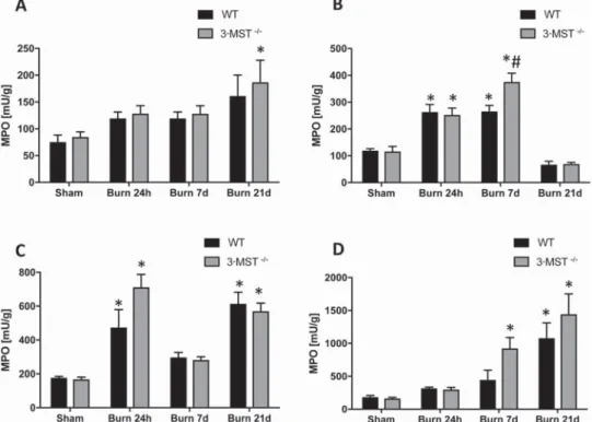
Burn induced marked increases in lung, liver, heart, and kidney MPO and MDA levels, indicating, respectively, the in-filtration of these tissues with polymorphonuclear cells, and increased oxidative burden of the tissues (Figures 3 and 4). The patterns and magnitudes of these burn-induced increases were, generally, comparable in wild-type mice and 3-MST
-/-mice, with the exception of the liver, where MPO levels 7 days postburn were higher in the 3-MST-/- mice than in the
wild-type control mice (Figure 3), possibly indicating a slight pro-tective role of endogenously produced, 3-MST-derived H2S.
Burn injury also induced marked changes in the clinical chemistry parameters and organ injury markers of the animals. There was an increase in the plasma levels of the hepatic/bone injury marker alkaline phosphatase, the hepatic injury marker alanine aminotransferase, the pancreatic injury marker amy-lase, and the renal dysfunction markers creatinine and blood urea nitrogen (Figures 5 and 6). Many of these parameters peaked at 24 hours, but alkaline phosphatase levels remained increased by 21 days postburn. Plasma blood urea nitrogen
levels tended to be higher in 3-MST-/- mice even under
base-line conditions, and they became significantly more elevated in 3-MST-/- mice than in wild-type mice 7 days postburn,
perhaps indicative of a slight protective role of endogenous, 3-MST-derived H2S against renal dysfunction (Figure 5).
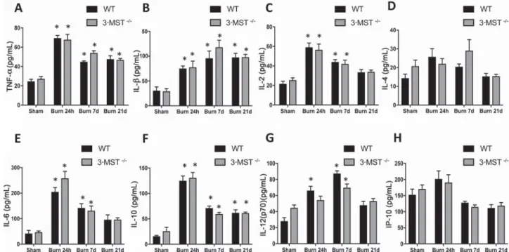
Consistent with the systemic inflammatory response syn-drome associated with burns, burn markedly increased the plasma levels of multiple chemokines, cytokines, and colony-stimulating and angiogenic hormones; however, plasma levels of these mediators were unaffected by the absence of 3-MST (Figures 7 and 8).
There were no differences in plasma troponin-I levels be-tween wild-type and 3-MST-deficient mice postburn, nor were any statistically significant differences in the rate of wound healing between the wild-type and the 3-MST-/-
ani-mals (Figure 9).
DISCUSSION
It been previously proposed that the role of H2S in burn may be time-dependent, with pro-inflammatory actions dominating in the early stages of the disease, and anti-inflammatory and wound-healing-modulating effects dominating in later stages.30
However, the relative role of the various H2S-producing enzymes in burns remains incompletely understood. Based on the beneficial effect of the CBS/CSE inhibitor aminooxyacetic acid,25 it was previously hypothesized that CBS-derived H
2S
plays a pathophysiological, deleterious role in burns. Moreover, based on the protection seen in CSE-deficient mice,36 it was
previously concluded that CSE-derived H2S also plays a path-ogenetic role in this model. Other studies have measured H2S-producing enzyme expression levels and circulating H2S concentrations, and demonstrated that in rodent models of burn, CSE mRNA expression is upregulated26 and both in
ro-dent models of burn as well as in patients with burn, circulating H2S levels are increased.26,29 Taken together, these experiments
indicate that CSE- and CBS-derived H2S both play
pathoge-netic (deleterious) roles in burns, by promoting inflamma-tory mediator production and exacerbating end-organ injury. However, the role of 3-MST in the pathogenesis of burn injury has not yet been elucidated, most likely because, until recently, neither mice deficient in 3-MST, nor sufficiently potent or se-lective pharmacological inhibitors of 3-MST were available.
The measurement of circulating H2S levels in the current study confirms and extends earlier studies25,29,36 showing that
burn induces an increase in circulating H2S levels and suggests that 3-MST is involved in this increase, because burn failed to increase circulating H2S levels in 3-MST-/- mice (Figure 1).
The tissue source of the increased 3-MST-derived H2S
pro-duction was not identified in the current study, as the liver and kidney tissues studied by us did not exhibit any increases in the activity of 3-MST, at least at the maximal (saturating) substrate (3-mercaptopyruvate) concentrations employed in this assay (Figure 2). These results do not, however, exclude the possibility that 3-MST (and/or other H2S-synthesizing
enzymes) become upregulated in response to burn in other cells or tissues that were not examined by us.
One interesting observation is that in tissues from 3-MST-/-
mice, the L-cysteine/homocysteine-induced H2S
produc-tion (which estimates CBS activity) was lower than the
Figure 1. Effect of burn on plasma H2S levels in wild-type and 3-MST-/- mice. Data show that H
2S plasma levels in sham mice and in wild-type (WT) and 3-MST-/- mice subjected to 24 hours of burn. H2S plasma levels increased in wild-type animals after burn; however, in 3-MST-/- mice, sham and postburn plasma H
2S levels are same. Data are shown as mean ± SEM of 10 animals for each group; *P < .05 shows significant increase in H2S levels in response to burn, com-pared to the sham group.
Figure 3. Burn injury increases in lung, liver, skin, and kidney myeloperoxidase (MPO) levels. Lung (A), liver (B), skin (C), and kidney (D) MPO levels (expressed as milliunits [mU]/g tissue) are shown in wild-type (WT) mice and in 3-MST-/- mice subjected to sham injury or to burn injury (24 hours, 7 days, and 21 days). Burn significantly increased MPO levels at 24 hours, 7 days, and 21 days (P < .05) compared to the values mea-sured in sham animals. MPO levels in 3-MST-/- burn mice were significantly increased as compared to wild-type burn mice in liver homogenates at 7 days. Data are shown as mean ± SEM of 10 animals for each group; *P < .05 shows significant increase in MPO in response to burn mice, compared to the sham group; #P < .05 shows significant increases in 3-MST-/- burn mice compared to wild-type burn mice.
Figure 2. Effect of burn on the enzymatic activity of cystathionine-beta-synthase (CBS) and 3-MST in wild-type and 3-MST-/- mice. Data show H2S production by liver homogenates in response to cysteine/homocysteine (an index of CBS activity) and in response to 3-mercatopyruvate (an index of 3-MST activity) in wild-type (WT) sham mice, in wild-type mice subjected to 24 hours and 7 days of burn, in 3-MST-/- sham mice and in 3-MST-/- mice subjected to 24 hours and 7 days of burn. Burn significantly increased CBS activity in liver (A) and kidney (C) samples from wild-type and 3-MST-/- mice at 7 days. 3-MST activities were comparable in liver (B) and kidney (D) samples from wild-type mice subjected to burn. Data are shown as mean ± SEM of 10 animals for each group; *P < .05 shows significant increase in CBS activity in response to burn, compared to the sham group.
Figure 4. Burn injury increases in lung, liver, skin, and kidney malondialdehyde (MDA) levels. Lung (A), liver (B), skin (C), and kidney (D) MDA levels are shown in wild-type (WT) mice and in 3-MST-/- mice subjected to sham injury or to burn injury (24 hours, 7 days, and 21 days). Burn significantly increased MDA levels at 24 hours, 7 days, and 21 days (P < .05) compared to the value measured in sham animals. Data are shown as mean ± SEM of 10 animals for each group; *P < .05 shows significant increase in MDA in response to burn, compared to the sham group.
Figure 5. Burn injury increases selected parameters of organ injury. Various physiological and organ injury marker levels (albumin (A), alkaline phosphatase (B), alanine aminotransferase (C), amylase (D), total bilirubin (E), plasma blood urea nitrogen (F), plasma calcium (G) and plasma phosphate (H)) measured by Vetscan analysis, are shown in wild-type (WT) mice and in 3-MST-/- mice subjected to sham injury or to burn injury (24 hours, 7 days, and 21 days). Burn significantly increased the indicated parameter at 24 hours, 7 days, and 21 days (P < .05) compared to the value measured in sham animals. The blood urea nitrogen level in 3-MST-/- burn mice was significantly higher than the corresponding values in wild-type burn mice at 7 days. Data are shown as mean ± SEM of 10 animals; *P < .05 shows significant changes in response to burn, compared to the sham group; #P < .05 shows a significantly higher increase in 3-MST-/- burn mice compared to wild-type burn mice.
corresponding H2S production in wild-type tissues. This is an unexpected finding, because the two enzymes have previ-ously not been linked functionally. Although the substrate of 3-MST (3-mercaptopyruvate) is produced from cysteine, and
cysteine is also a substrate of CBS, this commonality will also not explain the reduced tissue CBS activity in the 3-MST
-/-mice. The mechanism and the potential functional importance of these changes remain to be further explored.
Figure 7. Burn injury increases cytokine levels in plasma samples. Plasma cytokine levels, measured by Luminex analysis, are shown in wild-type (WT) mice and in 3-MST-/- mice subjected to sham injury or to burn injury (24 hours, 7 days, and 21 days). Burn markedly increased plasma TNF-α (A), IL-β (B), IL-2 (C), IL-4 (D), IL-6 (E), IL-10 (F), IL-12(p70) (G), and IP-10 (H) levels in wild-type and 3-MST-/- mice at 24 hours, 7 days, and 21 days. Data are shown as mean ± SEM of 10 animals; *P < .05 shows significant changes in response to burn, compared to the sham group. Figure 6. Burn injury increases in selected parameters of organ injury. Various physiological and organ injury marker levels (plasma creatinine (A), plasma glucose (B), plasma sodium (C), plasma potassium (D), plasma total protein (E) and plasma globulin (F)) measured by Vetscan analysis, are shown in wild-type (WT) mice and in 3-MST-/- mice subjected to sham injury or to burn injury (24 hours, 7 days, and 21 days). Burn significantly increased the indicated parameter at 24 hours, 7 days, and 21 days (P < .05) in wild-type and 3-MST-/- mice compared to the value measured in sham animals. Data are shown as mean ± SEM of 10 animals; *P < .05 shows significant changes in response to burn, compared to the sham group.
Based on prior studies focusing on CBS and CSE, our working hypothesis was that 3-MST deficiency will also exert protective effects in burns. However, unexpectedly, 3-MST deficiency did not provide any protective effects in burn in various tissues, nor did it affect circulating inflammatory mediator production. On a few selected parameters, at a few time points, we have even observed an exacerbation of the burn-induced pathophysiological changes in the 3-MST
-/-mice, although we must keep in mind that these differences are relatively minor, and, overall, probably the correct con-clusion should be that wild-type and 3-MST-deficient mice behave, in almost all respects, in the same manner when sub-jected to burn injury.
It should be noted that a recent study compared wild-type and 3-MST-deficient mice on hemodynamic or metabolic
parameters and mortality in an experimental model of trau-matic-hemorrhagic shock and observed no differences.40
Taken together, the available evidence so far suggests that 3-MST plays no major role in the pathogenesis of critical illness.
What, then, are the differences between 3-MST and CBS and CSE in terms of H2S production and related pathways that may explain these differences? First of all, 3-MST—in contrast to CBS and CSE—has a significant mitochondrial localization (although it is also present in the cytosol).41 Thus,
the intracellular localization of the H2S it produces may be different, and therefore, the cellular pathways/targets affected by H2S may also be different. Second, 3-MST—in contrast to CBS and CSE—produces a significant amount of polysulfides (rather than, or in addition to “free” H2S).41–43 Polysulfides
Figure 8. Burn injury increases cytokine levels in plasma samples. Plasma cytokine levels, measured by Luminex analysis, are shown in wild-type (WT) mice and in 3-MST-/- mice subjected to sham injury or to burn injury (24 hours, 7 days, and 21 days). Burn markedly increased plasma KC (A), MCP-1 (B), RANTES (C), VEGF (D), G-CSF (E), and GM-CSF (F) levels in wild-type and 3-MST-/- mice at 24 hours, 7 days, and 21 days. Data are shown as mean ± SEM of 10 animals; *P < .05 shows significant changes in response to burn, compared to the sham group.
Figure 9. Effect of burn injury on plasma troponin levels and burn wound area at Day 21. (A) Plasma troponin levels are shown in wild-type (WT) mice and in 3-MST-/- mice subjected to sham injury or to burn injury (24 hours, 7 days and 21 days). Burn markedly increased plasma troponin levels at 24 hours, 7 days, and 21 days; there was no statistically significant difference between the values measured in the wild-type and the 3-MST -/- burn group. *P < .05 shows significant increases in response to burn, compared to the sham group. (B) % wound area on Day 21 is shown in the wild-type and 3-MST-/- burn group. Wound healing was comparable in the two groups of mice. Data are shown as mean ± SEM of 10 animals.
have their own, very distinct regulatory roles in biological sys-tems, which are fairly different from the roles of H2S, due to the different array of chemical reactions polysulfides can catalyze (most importantly, a post-translational modification of proteins via S-sulfhydration).44 As far as the net output of
H2S from 3-MST vs CBS and CSE, no marked differences were noted (at least in the highly artificial conditions of tissue homogenates incubated with saturating concentrations of the respective substrates utilized in the current model). 3-MST is broadly expressed in all cells and tissues40,45 and therefore the
lack of effect on MDA or MPO levels in the various organs studied here cannot be due to the absence of 3-MST in those organs. It is also not the case that 3-MST, in general, is not biologically relevant: multiple studies, in multiple models, have already implicated functional roles of this enzyme in vari-ous biological systems ranging from neuronal roles to vascular and metabolic roles.41–53
Taken together, the data presented in the current paper do not find a major role of 3-MST in burn injury and support the concept that different H2S-producing enzymes (CBS, CSE, 3-MST) can play substantially different roles in the pathogen-esis of burn injury and other forms of critical illness.
ACKNOWLEDGEMENTS
The authors appreciate the editorial assistance of Dr. Anita Marton.
REFERENCES
1. Szabo C, Papapetropoulos A. International union of basic and clinical phar-macology. CII: pharmacological modulation of H2S levels: H2S donors
and H2S biosynthesis inhibitors. Pharmacol Rev. 2017;69:497–564.
2. Wang R. Physiological implications of hydrogen sulfide: a whiff
explora-tion that blossomed. Physiol Rev. 2012;92:791–896.
3. Predmore BL, Lefer DJ, Gojon G. Hydrogen sulfide in biochemistry and
medicine. Antioxid Redox Signal. 2012;17:119–40.
4. Vandiver M, Snyder SH. Hydrogen sulfide: a gasotransmitter of clinical
relevance. J Mol Med (Berl). 2012;90:255–63.
5. Kimura H. Signaling molecules: hydrogen sulfide and polysulfide.
Antioxid Redox Signal. 2015;22:362–76.
6. Huang CW, Moore PK. H2S synthesizing enzymes: biochemistry and
molecular aspects. Handb Exp Pharmacol. 2015;230:3–25.
7. Coletta C, Szabo C. Potential role of hydrogen sulfide in the
patho-genesis of vascular dysfunction in septic shock. Curr Vasc Pharmacol. 2013;11:208–21.
8. Szabo C, Ransy C, Módis K et al. Regulation of mitochondrial
bioener-getic function by hydrogen sulfide. Part I. Biochemical and physiological mechanisms. Br J Pharmacol. 2014;171:2099–122.
9. McCook O, Radermacher P, Volani C et al. H2S during circulatory shock:
some unresolved questions. Nitric Oxide. 2014;41:48–61.
10. Li L, Salto-Tellez M, Tan CH, Whiteman M, Moore PK. GYY4137, a
novel hydrogen sulfide-releasing molecule, protects against endotoxic shock in the rat. Free Radic Biol Med. 2009;47:103–13.
11. Tokuda K, Kida K, Marutani E et al. Inhaled hydrogen sulfide prevents
endotoxin-induced systemic inflammation and improves survival by alter-ing sulfide metabolism in mice. Antioxid Redox Signal. 2012;17:11–21. 12. Aslami H, Beurskens CJ, de Beer FM et al. A short course of infusion of a
hydrogen sulfide-donor attenuates endotoxemia induced organ injury via stimulation of anti-inflammatory pathways, with no additional protection from prolonged infusion. Cytokine. 2013;61:614–21.
13. Chen X, Xu W, Wang Y et al. Hydrogen sulfide reduces kidney injury
due to urinary-derived sepsis by inhibiting NF-κB expression, decreasing TNF-α levels and increasing IL-10 levels. Exp Ther Med. 2014;8:464–70.
14. Ferlito M, Wang Q, Fulton WB et al. Hydrogen sulfide [corrected]
increases survival during sepsis: protective effect of CHOP inhibition. J Immunol. 2014;192:1806–14.
15. Ahmad A, Druzhyna N, Szabo C. Delayed treatment with sodium
hydro-sulfide improves regional blood flow and alleviates cecal ligation and puncture (CLP)-induced septic shock. Shock. 2016;46:183–93.
16. Collin M, Anuar FB, Murch O, Bhatia M, Moore PK, Thiemermann C.
Inhibition of endogenous hydrogen sulfide formation reduces the organ injury caused by endotoxemia. Br J Pharmacol. 2005;146:498–505.
17. Li L, Bhatia M, Zhu YZ et al. Hydrogen sulfide is a novel mediator
of lipopolysaccharide-induced inflammation in the mouse. FASEB J. 2005;19:1196–8.
18. Zhang H, Zhi L, Moochhala S, Moore PK, Bhatia M. Hydrogen
sul-fide acts as an inflammatory mediator in cecal ligation and puncture-induced sepsis in mice by upregulating the production of cytokines and chemokines via NF-kappaB. Am J Physiol Lung Cell Mol Physiol. 2007;292:L960–L971.
19. Zhang H, Moochhala SM, Bhatia M. Endogenous hydrogen sulfide
regu-lates inflammatory response by activating the ERK pathway in polymicro-bial sepsis. J Immunol. 2008;181:4320–31.
20. Yan Y, Chen C, Zhou H et al. Endogenous hydrogen sulfide
for-mation mediates the liver damage in endotoxemic rats. Res Vet Sci. 2013;94:590–5.
21. Shirozu K, Tokuda K, Marutani E, Lefer D, Wang R, Ichinose F.
Cystathionine γ-lyase deficiency protects mice from galactosamine/
lipopolysaccharide-induced acute liver failure. Antioxid Redox Signal. 2014;20:204–16.
22. Badiei A, Chambers ST, Gaddam RR, Bhatia M. Cystathionine-γ-lyase
gene silencing with siRNA in monocytes/ macrophages attenuates in-flammation in cecal ligation and puncture-induced sepsis in the mouse. J Biosci. 2016;41:87–95.
23. Esechie A, Kiss L, Olah G et al. Protective effect of hydrogen sulfide in a murine model of acute lung injury induced by combined burn and smoke inhalation. Clin Sci (Lond). 2008;115:91–7.
24. Esechie A, Enkhbaatar P, Traber DL et al. Beneficial effect of a hydrogen sulphide donor (sodium sulphide) in an ovine model of burn- and smoke-induced acute lung injury. Br J Pharmacol. 2009;158:1442–53.
25. Ahmad A, Szabo C. Both the H2S biosynthesis inhibitor aminooxyacetic
acid and the mitochondrially targeted H2S donor AP39 exert protective
effects in a mouse model of burn injury. Pharmacol Res. 2016;113(Pt A):348–55.
26. Zhang J, Sio SW, Moochhala S, Bhatia M. Role of hydrogen
sul-fide in severe burn injury-induced inflammation in mice. Mol Med. 2010;16:417–24.
27. Li Y, Wang HJ, Song XF, Yang HL. Influence of hydrogen sulfide on
im-portant organs in rats with severe burn. Zhonghua Shao Shang Za Zhi. 2011;27:54–8.
28. Zeng J, Lin X, Fan H, Li C. Hydrogen sulfide attenuates the inflammatory
response in a mouse burn injury model. Mol Med Rep. 2013;8:1204–8.
29. Brunyanszki A, Erdelyi K, Szczesny B et al. Upregulation and
mito-chondrial sequestration of hemoglobin occur in circulating leukocytes during critical illness, conferring a cytoprotective phenotype. Mol Med. 2015;21:666–75.
30. Akter F. The role of hydrogen sulfide in burns. Burns. 2016;42:519–25.
31. Papapetropoulos A, Pyriochou A, Altaany Z et al. Hydrogen sulfide is
an endogenous stimulator of angiogenesis. Proc Natl Acad Sci USA. 2009;106:21972–7.
32. Módis K, Ju Y, Ahmad A et al. S-Sulfhydration of ATP synthase by
hy-drogen sulfide stimulates mitochondrial bioenergetics. Pharmacol Res. 2016;113(Pt A):116–24.
33. Chao C, Zatarain JR, Ding Y et al. Cystathionine-beta-synthase inhibition for colon cancer: enhancement of the efficacy of aminooxyacetic acid via the prodrug approach. Mol Med. 2016;33:361–79.
34. Ahmad A, Gerö D, Olah G, Szabo C. Effect of endotoxemia in mice
genetically deficient in cystathionine-γ-lyase, cystathionine-β-synthase or 3-mercaptopyruvate sulfurtransferase. Int J Mol Med. 2016;38:1683–92.
35. Szczesny B, Brunyánszki A, Ahmad A et al. Time-dependent and
organ-specific changes in mitochondrial function, mitochondrial DNA integrity, oxidative stress and mononuclear cell infiltration in a mouse model of burn injury. PLoS One. 2015;10:e0143730.
36. Ahmad A, Druzhyna N, Szabo C. Cystathionine-gamma-lyase deficient
mice are protected against the development of multiorgan failure and exhibit reduced inflammatory response during burn. Burns. 2017;43:1021–33.
37. Nagahara N, Nagano M, Ito T, Shimamura K, Akimoto T, Suzuki H.
Antioxidant enzyme, 3-mercaptopyruvate sulfurtransferase-knockout mice exhibit increased anxiety-like behaviors: a model for human mercap-tolactate-cysteine disulfiduria. Sci Rep. 2013;3:1986.
38. Toliver‐Kinsky TE, Cui W, Murphey ED, Lin C, Sherwood ER.
Enhancement of dendritic cell production by Fms‐like tyrosine kinase‐3 ligand increases the resistance of mice to a burn wound infection. J Immunol. 2005;174:404–10.
39. Ahmad A, Olah G, Herndon DN, Szabo C. The clinically used PARP
inhibitor olaparib improves organ function, suppresses inflammatory responses and accelerates wound healing in a murine model of third-degree burn injury. Br J Pharmacol. 2018;175:232–45.
40. Gröger M, Wepler M, Wachter U, Merz T, McCook O, Kress S,
Lukaschewski B, Hafner S, Huber-Lang M, Calzia E, Georgieff M, Nagahara N, Szabó C, Radermacher P, Hartmann C. The effects of
genetic 3-mercaptopyruvate sulfurtransferase deficiency in murine trau-matic-hemorrhagic shock. Shock. 2018. In press.
41. Nagahara N. Multiple role of 3-mercaptopyruvate sulfurtransferase: anti-oxidative function, H2 S and polysulfide production and possible SOx production. Br J Pharmacol. 2018;175:577–89.
42. Kimura Y, Toyofuku Y, Koike S et al. Identification of H2S3 and H2S
produced by 3-mercaptopyruvate sulfurtransferase in the brain. Sci Rep. 2015;5:14774.
43. Kimura Y, Koike S, Shibuya N, Lefer D, Ogasawara Y, Kimura H.
3-Mercaptopyruvate sulfurtransferase produces potential redox regulators cysteine- and glutathione-persulfide (Cys-SSH and GSSH) together with signaling molecules H2S2, H2S3 and H2S. Sci Rep. 2017;7:10459.
44. Kimura H. Hydrogen sulfide and polysulfide signaling. Antioxid Redox
Signal. 2017;27:619–21.
45. Tomita M, Yamada H, Adachi Y et al. Choroidal neovascularization is
provided by bone marrow cells. Stem Cells. 2004;22:21–6.
46. Miyamoto R, Otsuguro K, Yamaguchi S, Ito S. Contribution of cysteine
aminotransferase and mercaptopyruvate sulfurtransferase to hydrogen sul-fide production in peripheral neurons. J Neurochem. 2014;130:29–40.
47. Kuo MM, Kim DH, Jandu S et al. MPST but not CSE is the primary
regulator of hydrogen sulfide production and function in the coronary artery. Am J Physiol Heart Circ Physiol. 2016;310:H71–H79.
48. Tao B, Wang R, Sun C, Zhu Y. 3-Mercaptopyruvate sulfurtransferase,
not cystathionine β-synthase nor cystathionine γ-lyase, mediates hypoxia-induced migration of vascular endothelial cells. Front Pharmacol. 2017;8:657.
49. Mitidieri E, Tramontano T, Gurgone D et al. Mercaptopyruvate acts as
endogenous vasodilator independently of 3-mercaptopyruvate sulfur-transferase activity. Nitric Oxide. 2018;75:53–9.
50. Coletta C, Módis K, Szczesny B et al. Regulation of vascular tone, angio-genesis and cellular bioenergetics by the 3-mercaptopyruvate
sulfur-transferase/H2S pathway: functional impairment by hyperglycemia and
restoration by DL-α-lipoic acid. Mol Med. 2015;21:1–14.
51. Li M, Xu C, Shi J, Ding J, Wan X, Chen D, Gao J, Li C, Zhang J, Lin Y,
Tu Z, Kong X, Li Y, Yu C. Fatty acids promote fatty liver disease via the dysregulation of 3-mercaptopyruvate sulfurtransferase/hydrogen sulfide pathway. Gut. 2018;67:2169–80.
52. Szczesny B, Marcatti M, Zatarain JR et al. Inhibition of hydrogen sulfide biosynthesis sensitizes lung adenocarcinoma to chemotherapeutic drugs by inhibiting mitochondrial DNA repair and suppressing cellular bioener-getics. Sci Rep. 2016;6:36125.
53. Finnerty CC, McKenna CF, Cambias LA et al. Inducible satellite cell
depletion attenuates skeletal muscle regrowth following a scald-burn injury. J Physiol. 2017;595:6687–701.