REVIEW
Perinatal arrhythmias
Nicole Sekarski&Erik Jan Meijboom&
Stefano Di Bernardo&Tatiana Boulos Ksontini&
Yvan Mivelaz
Received: 13 December 2013 / Revised: 5 March 2014 / Accepted: 3 April 2014 / Published online: 17 April 2014 # Springer-Verlag Berlin Heidelberg 2014
Abstract Cardiac arrhythmias are very frequent in fetuses and newborns. The prognosis depends on the nature of the arrhythmias but is most often either spontaneously benign or following short-term medication administration. A correct diagnosis is essential for both management and prognosis. It is based on echocardiography during the fetal period and mainly on history, physical exam, and electrocardiogram after birth, but other modalities are available to record transient arrhythmic events. Irregular rhythms are mostly benign and rarely require therapy. In most fetuses and infants, tachyar-rhythmias resolve spontaneously or require short-term admin-istration of antiarrhythmics. Approximately one third of these may recur later on, especially during adolescence. Persistent bradyarrhythmias might require pacemaker implantation when associated with failure to thrive or with risk of sudden death. Conclusion: Arrhythmias in fetuses and infants are very common and mostly benign. History, physical exam, and recording of the arrhythmia are essential to make a correct diagnosis and establish an appropriate management for the rare potentially harmful arrhythmias.
Keywords Arrhythmia . Fetus . Newborn . Infant . Extrasystole . Tachycardia
Abbreviations
ABC Airway, breathing, circulation
AET Atrial ectopic tachycardia
APB Atrial premature beat
AV Atrioventricular
AVB Atrioventricular block
AVNRT Atrioventricular nodal reentrant tachycardia
AVRT Atrioventricular reentrant tachycardia
CHD Congenital heart disease
ECG Electrocardiogram
JPB Junctional premature beat
JET Junctional ectopic tachycardia
PALS Pediatric advanced life support
PJRT Permanent junctional reciprocating tachycardia
SVT Supraventricular tachycardia
VPB Ventricular premature beat
VT Ventricular tachycardia
WPW Wolff-Parkinson-White syndrome
Introduction
Arrhythmias occur as soon as the heart starts to beat and end on a final irreversible arrhythmia when we die. The same definition, classification, and therapies apply from fetuses to adulthood. They are noted in about 2 % of the pregnancies and account for 10 to 20 % of the referrals to fetal cardiology units
[13,20]. The prevalence of arrhythmias in normal infants
(child up to the age of 12 months) is difficult to estimate, ranging from 0.75 to 14 %, and is higher in children with
congenital heart disease (CHD) [7,32]. Arrhythmia is also a
frequent cause of pediatric cardiology referrals in order to rule out potentially life-threatening conditions. We review here the classification, the diagnosis, the management, and the Communicated by Patrick Van Reempts
N. Sekarski
:
E. J. Meijboom:
S. Di Bernardo:
T. B. Ksontini:
Y. Mivelaz (*)Cardiology Division, Pediatrics Department, Centre Hospitalier Universitaire Vaudois and University of Lausanne, Route du Bugnon 46, Bureau BH11/632, 1011 Lausanne, Switzerland
e-mail: [email protected] E. J. Meijboom
Current affiliation: Pediatric Cardiology, University of Twente, Enschede, Netherlands
outcome of arrhythmias in fetuses and newborns (child under 28 days of age).
Conduction system development
The first heartbeat occurs by 3 weeks post-conception when the heart is only a primitive tubular structure. Major morpho-logical remodeling occurs simultaneously with the develop-ment of the cardiac conduction system, which results by 7 weeks of gestation in a four-chambered heart with synchro-nous contraction of the atrial and ventricular chambers at a rate of approximately 110 bpm. Progressively, the sinus node acts as the primary pacemaker and the heart rate reaches 170 bpm by 9 to 10 weeks. Later on in gestation, heart rate slowly decreases. Between 20 and 40 weeks of gestation, the heart rate is regular, with a range from 110 to 180 bpm and a
maximal beat-to-beat variation of 15 bpm [20,44]. After birth,
heart rate slowly decreases and normal data for all electrocar-diogram (ECG) time intervals including heart rate and wave
axis have been published (Table1) [9].
Rhythm analysis
During fetal life, a real-time fetal ECG is not obtainable due to the parasitic electrical field generated by the maternal heart and abdominal muscles. Magnetocardiography allows record-ing of the fetal heart magnetic field instead of the traditional electric field recorded by ECG. It is actually the best modality
to analyze the fetal heart rhythm [36,48]. However, this
tech-nology is restricted to select centers due to its high cost. Therefore, the analysis of arrhythmias is routinely based on the ultrasound assessment of the temporal relationship of atrial and ventricular contractions. Different ultrasound modalities (M-mode, Doppler, or tissue Doppler) allow the classification
of the arrhythmias (Fig.1) [6,13,40]. In some cases, it may
even be superior to ECG for arrhythmia assessment, as atrial contraction can be identified even when the ventricle is
contracting or during the repolarization phase [37]. A
system-atic analysis of the ultrasound tracing is mandatory. Stepwise interpretation of the fetal heart rhythm is based on the deter-mination of rhythm origin, regularity, and relationship be-tween atrial and ventricular events and rate. Normal fetal rhythm includes an atrial origin, a regular atrial and ventricular contraction in a 1:1 fashion, and a normocardic heart rate.
In newborn and infants, analysis of the rhythm relies es-sentially on the surface ECG. ECG interpretation should fol-low a similar initial approach with assessment of rhythm origin, regularity, relationship, and rate (mnemonic: 4R). In addition, a careful analysis of each wave (P, QRS, and T) and segment (PR, QT) in each lead will add information on the
type of arrhythmia. For both diagnosis and management, it is Ta
b le 1 Hea rt rat e and ess enti al p ar amet er s: normal v al ues b y ag e Age g ro up He ar t ra te a(bpm) F rontal plane Q RS axi s a(de g re es) P w ave amplitude b (m m) PR inte rva l a(s) Q RS dur ati on V 5 a(s) R V1 c(m m) R V 6 c(m m) S V 1 c(m m) S V 6 c(mm) Le ss th an 1 d ay 93 –1 5 4 (123) +59 to + 192 (13 5 ) 2 .8 0.08 –0.16 (0.1 1) 0.02 –0.08 (0.05) 5.2 –26 .1 0– 11 .1 0– 22.7 0– 9.6 1– 2d ay s 9 1– 1 5 9 (123) +64 to + 197 (13 4 ) 2 .8 0.08 –0.14 (0.1 1) 0.02 –0.07 (0.05) 5.3 –26 .9 0– 12.2 0– 20.7 0– 9.4 3– 7d ay s 9 0– 1 6 6 (129) +77 to + 187 (13 2 ) 2 .9 0.08 –0.14 (0.10) 0.02 –0.07 (0.05) 2.8 –24 .2 0.3 –12.1 0– 16.8 0– 9.8 1– 4 w eeks 107 –182 (149) +65 to + 160 (1 10) 3.0 0 .07 –0.14 (0.10) 0.02 –0.08 (0.05) 3.2 –20 .8 2.6 –16.4 0– 10.8 0– 9.8 1– 3 m onths 121 –179 (150) +31 to + 1 1 4 (75) 2.6 0 .07 –0.13 (0.10) 0.02 –0.08 (0.05) 3.3 –18 .4 5.2 –21.4 0– 12.4 0– 6.4 3– 6 m onths 106 –186 (141) +1 to +104 (60) 2.5 0 .07 –0.15 (0.1 1) 0.02 –0.08 (0.05) 2.7 –19 .8 6.4 –22.4 0– 17.1 0– 9.9 6– 12 months 109 –169 (134) +1 to +99 (56) 2.5 0 .07 –0.16 (0.1 1) 0.02 –0.08 (0.05) 1.4 –20 .3 5.8 –22.7 0– 18.1 0– 7.2 Ada p te d from [ 9 ] a Sec ond and 98th p er ce ntil es (mea n) bNine ty-eigh th p er cent ile s (1 m m = 0.1 m V) cSec ond and 98th p er ce ntil es (1 mm = 0 .1 mV)
essential to distinguish a wide or a narrow QRS. Preexcitation (delta wave) or prolonged QT interval should also be ruled out.
Irregular rhythm—premature beats
Definition
A premature beat, also called premature contraction, ectopic beat, or extrasystole can originate from the atria, the atrioven-tricular junction, or the ventricle, and bypasses the sinus node. It is by far the most common type of arrhythmia encountered during the perinatal period, and their incidence in healthy patients varies greatly depending of the duration of the heart rhythm assessment from 1 %, when based on a single surface ECG, up to more than 50 % when based on a 24-h heart rate
monitoring [17,46].
Atrial premature beats (APB) Incidence
APB account for about 90 % of irregular rhythm in fetuses and
infants [17,22].
Diagnosis
On a Doppler or an ECG recording, APB is defined as a premature beat originating from the atria, which might be either conducted (generating a ventricular contraction) or blocked (not conducted through the atrioventricular (AV)
node) (Fig.2). Prenatally, the demonstration of the atrial origin
of the extra beat is based on the analysis of the timing of the atrial contraction and the resetting phenomenon of the sinus node with APB resulting in less than full compensatory pause. Postnatally, on ECG, P waves resulting from an APB
(called P′) usually have a different morphology than the sinus
P wave. The morphology of the QRS is also helpful since most of the time a narrow QRS with similar morphology than
sinus QRS is noted following an APB. A P′ wave is
some-times visible in the preceding T wave (Fig.2).
Management
As arrhythmias can be associated with cardiac malformations
[50], it is important to ensure that a routine mid-trimester fetal
screening scan for cardiac anomalies is performed. If the APBs are very frequent (more than 5 per minute), persisting for more than 3 weeks or associated with signs of cardiac failure or extracardiac anomalies, it is recommended to
per-form a detailed fetal cardiac ultrasound [13]. A weekly
mon-itoring of the fetal heart rate is recommended as long as the arrhythmia persists to detect the onset of supraventricular tachycardia (SVT). Indeed, the susceptibility to cardiac failure in fetuses with SVT and the inability to observe clinical manifestations justify this strategy. After birth, if the medical history and the physical exam are free of any cardiac sign or symptom and the ECG is normal for age except for the premature beat, monitoring or further investigation are not recommended. Exceptions are frequent APB and APB arising from multiple foci seen as P waves with different
morphol-ogies on surface ECG [52].
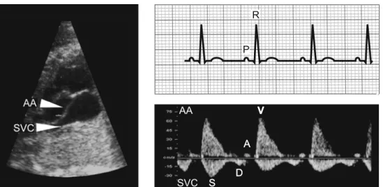
Fig. 1 Left, Two-dimensional ultrasound image showing the anatomical proximity of the SVC and the AA. The sample gate can easily be placed so as to record both proximal parts of SVC and AA. Right, Fetal virtual ECG (upper panel) and SVC/AA Doppler recording (lower panel, Dopp-ler tracing turned upside down in order to illustrate analogies with the ECG tracing) in normal sinus rhythm. Ventricular ejection (V) in the AA
appears above baseline. Venous flow is typical with systolic (S) and diastolic (E) waves below baseline and the retrograde flow wave (A) due to the atrial contraction above. A late diastolic wave due to atrial contraction, AA ascending aorta, AV atrioventricular interval, D early diastolic wave, ECG electrocardiogram, P P wave, R R wave, S systolic wave, SVC superior vena cava, V ventricular ejection
Outcome
APBs are mostly benign and remain self-limited with a spon-taneous resolution after the diagnosis of the arrhythmia before birth in 95 % of fetuses and by 1 year of age in 95 % of
children [46]. No association with sudden infant death or any
other serious clinical problems has ever been described. The immaturity of conduction pathways in fetuses is characterized by an increased number of accessory pathways connecting the atrial and ventricular myocardium and a high rate of APB
[31]. Both phenomena are responsible for the peak incidence
of SVT by an atrioventricular (AV) reentrant mechanism during fetal life, which occurs in approximately 1 to 3 % of
fetuses with APB [13,20]. After birth, with the maturation of
the conduction pathways, both APBs and SVTs tend to be
self-limited [45]. Occasionally, APB can be associated with
cardiac malformations, such as atrial septal defect. In sick babies, other causes should be ruled out: mechanical problems (central venous line, atrial distension), metabolic anomalies (hypo- or hyperkaliemia, hypercalcemia, hypoglycemia, hypoxia), pharmacological causes (antiarrhythmics, inotropes), or previous cardiac surgery (atrial scar).
Ventricular premature beats (VPB) Incidence
VPB in fetuses are rare and represent less than 2 % of fetal premature beats. After birth, the incidence of VPB in infants is highest in the neonatal period and decreases rapidly thereafter (incidence of VPBs on 24 h ambulatory ECG decreasing from
18 to 6 %, respectively) [17]. Therefore, some authors
con-sider neonatal isolated monomorphic VPB a variation of
normal rhythm [53].
Diagnosis
AVPB is diagnosed when, on a Doppler or an ECG recording, a premature ventricular beat is observed without any alteration of the atrial rate, giving rise to a full compensatory pause after
the extra beat (Fig. 3). On ECG, a premature wide QRS,
usually with a different morphology than the preceding QRS, makes the diagnosis easier postnatally.
Management
Prenatally, a careful exam of the cardiac anatomy and function should be performed to detect malformation or signs of car-diac failure. Weekly monitoring of the fetal heart rate is
recommended as long as the arrhythmia persists [13,20]. After
birth, besides a cardiovascular-oriented medical history and physical exam, an ECG should be obtained. But there is no agreement on the management of VPB diagnosed postnatally. Proposed managements are as follows: no further investiga-tion, repetition of ECG at 1- or 2-month intervals, perfor-mance of a 24-h ambulatory electrocardiography monitoring to look for higher grade ectopic activity (nonisolated or poly-morphic premature beats), and referral to a pediatric
cardiol-ogist for a cardiovascular work-up [27,52]. We believe that if
medical history (personal and familial) and physical exam are free of any cardiovascular symptoms or signs, and if a pedi-atric cardiologist has ruled out abnormalities suggesting an Fig. 2 Upper panel, ECG
showing one nonconducted premature atrial beat (black arrowheads) occurring during T wave after every conducted atrial beat called bigeminy. This results in a ventricular bradycardia with a ventricular rate of 70 bpm. Lower panel, SVC/AA Doppler recording showing the same phenomenon than on the upper panel with an APB (white arrowheads) occurring at the end of the ventricular contraction which did not result in a ventricular contraction. A atrial contraction, AA ascending aorta, P P wave, R R wave, SVC superior vena cava, V ventricular contraction
underlying myocardial disease on ECG, it is reasonable to repeat the ECG beyond the neonatal period and perform a cardiovascular work-up for those who show persistent irregu-lar rhythm at auscultation or ECG.
Outcome
The prognosis of VPB is also benign in most cases. However, it may be a manifestation of a serious under-lying myocardial disease such as cardiac malformations, tumors, or cardiomyopathies. In most instances, those could be ruled out by medical history, physical exami-nation, and ECG.
Junctional premature beats (JPB)
JPB describe premature beats originating from the AV node area. They are rarely described in fetuses and infants. The prenatal diagnosis is challenging because differentiation from VPB might be impossible. On ECG, an early narrow QRS not preceded by any P wave is diagnostic. Prognosis and
man-agement are similar to those with APBs [52].
Tachycardia Definition
Tachycardia is defined as an area of the heart that depolarizes faster than the normal range for age for at least three consec-utive beats, above 180 bpm in the fetus between 20 and 40 weeks of gestation and above 200 bpm in infants. As for extrasystoles, supraventricular origin is by far the most fre-quent cause, with AV nodal and ventricular origins being infrequent in healthy fetuses and infants. In fetuses, 70 % of the tachyarrhythmias are paroxysmal AV reentry tachycardia, 24 % primary atrial tachycardias (mostly atrial flutter), and
6 % sinus tachycardia [22]. During infancy, 80 % of nonsinus
tachycardia are AV reentry tachycardia, 15 % are primary atrial tachycardia (mostly atrial flutter), and 5 % are AV nodal
reentry SVT [32]. The electrophysiological mechanism of all
SVT is described in Table2. During the perinatal period and
infancy, a reentrant circuit is by far the most common mech-anism. The reentrant circuit is either involving both the atria and ventricles, with the abnormal pathway crossing the AV groove in AV reentry tachycardia and within or just next to the AV node in AV nodal reentry tachycardia, or limited to the atria in atrial flutter (AF).
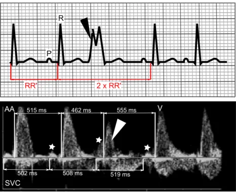
Fig. 3 Upper panel, surface ECG: VPB is diagnosed when a wide premature QRS complex (black arrowhead) is observed on surface ECG. Since in rare instances a wide QRS could result from an APB, another feature of VPB is that the interval between the preceding and the following sinus beat is equal to twice the time between two regular sinus beats. This phenomenon called“full compensatory pause” is due, in contrast to APB, to the absence of resetting of the sinus node in VPB. Lower panel, SVC/AA Doppler recording: prenatally, since ECG is not
obtainable, this phenomenon is essential to the diagnosis. RR′ interval before the VPB (white arrowhead) is 515 ms. The interval from the preceding and the following regular ventricular beat equals 1,017 ms, approximately twice the RR′ interval (=1,030 ms). Another feature is the identification of regular atrial contractions (white stars), independent of the premature ventricular contraction. AA ascending aorta, P P wave, R R wave, SVC superior vena cava, V ventricular contraction
Ta b le 2 C h ara ct eri sti cs o f n ar ro w Q RS ta chyc ar dia S inu s A VR T (co nc ea le d) A V R T (WP W ) A VNR T A F A ET / M A T JE T P JR T Onse t/t er min ati on W ar m -up/ co ol-down A br up t A b ru p t A bru p t A br up t W ar m -up /c ool-d o w n W ar m-up/ co ol-down Ince ss ant Me ch an ism A u tom ati c R ee n tr y R ee n try Re en tr y R eentry Automatic or tr ig ger A u tom ati c Ree n tr y P w av e axis N or ma l R et ro gr ad e R etr o g ra d e R etro g ra d e E cto p ic E ct o p ic N or ma l o r re tro gr ad e R et ro gr ad e Atr ial ra te in SVT V ar ia ble F ixe d F ix ed F ix ed V ar iab le V aria b le No rm al o r va ri ab le F ixe d A:V ratio/rate in S VT A = V A = V A = V A = V A ≥ VA ≥ VA ≤ VA = V V A in te rva l Lo ng Shor t, V A >70 m s S ho rt, V A > 7 0 ms Sh ort , V A <7 0 m s L on g L ong V ar ia b le Lon g Resp ons e to v ag al sti m ul ati on/a d en o sine Mild decr rat e None or termin ation N one o r terminati on N one or ter m in at ion T ra nsi ent AV b lo ck T ran sie n t A V b lo ck No ne Non e o r tr an sie n t termination Resp ons e to D C ca rd iov ers ion None T ermination T erminati on T erminat ion T ermination N one None T ransient terminati on Pr ev al en ce in fe tu se s N /A C o mm on C o m m on Ra re In te rme d ia te Rar e Ra re Rar e Pr ev al en ce in in fa nts N /A C o mm on C o m m on Ra re Oc ca sio n al R ar e M o st ly po sto p . R ar e 1st line chronic therapy in: Fe tuses [ 26 ] N one D igoxin/sotal o l/ fle cain ide N/ A D igoxin/ sotalol/ fl ec ain ide Digoxin/sotal o l/ flec ain ide Digo xin /sot alo l/ fl ec ai nid e Di gox in/s ota lol / fl ec ainid e Digoxin/sotal o l/ fle cain ide In fa n ts [ 4 , 8 , 45 ] N o n e D ig oxin /pr opr anolo l p ro p ra no lol D igo x in/ p ro pr an olol N/A D igo x in /pr opr an olo l/ sotal o l/flecaini d e Am iod ar one Dig oxin /pr opr anolo l Ada p te d from [ 52 ], exc ept fo r fe tal th er apy [ 21 ] and infant th erapy [ 4 , 8 , 45 ] AE T atrial ectopic tachycardia, AF at ri al fl ut te r, AV N R T atrioventricular nodal reentrant tachycardia, AV R T atrioventricular ree ntrant tachycardia, JE T ju nctional ectopic tachycardia, MA T mul tipl e atr ial ta chyc ard ia, PJ RT permanent junctio nal reciprocating tachy cardia, VT ventricular tachycardia
For the purpose of clarity, we will use a common terminol-ogy based on the depolarized cardiac chamber to characterize SVT on Doppler recordings and ECG. This means that instead of the traditional P, QRS waves, and PR interval, we will use the terms A (corresponding to atrial activity or event), V (corresponding to ventricular activity or event), and AV
inter-val [52]. This allows to differentiate tachycardias based on the
ratio of atrial versus ventricular events: =1, >1, or <1 and on the time interval between those events: VA interval < AV interval, called short VA SVT; or VA interval > AV interval,
called long VA SVT (Table2; Fig.4).
Supraventricular tachycardia Incidence
The incidence of fetal SVT is about 1:3.700 pregnancies [50].
It accounts for about 5 to 10 % of all fetal arrhythmias, but for
more than 50 % of the clinically significant ones [22].
Post-natally, the incidence ranges between 1:250 and 1:1,000
chil-dren, with a peak incidence in the neonatal period [17].
Diagnosis
The determination of the type of SVT is based on the assess-ment of the AV relationship and other specific characteristics
(see Table2and Figs4and5). Based on that, sinus
tachycar-dia and ventricular tachycartachycar-dia should be ruled out since their management and prognosis differ considerably from nonsinus SVT. Provoking factors for nonsinus SVT in the fetus have to be looked for: co-existing CHD, hyperthyroidism, or maternal caffeine, alcohol, or nicotine consumption. These last causes
are among the most frequent ones [49]. Sinus tachycardia is
the most common cause of long VA tachycardia and the heart rate is usually lower than in other SVT with frequent gradual acceleration and slowing. Atrial contractions remain always clearly visible on Doppler recording prenatally, but on ECG, P waves might be transiently hidden in the T waves and visible only when the heart rate decelerates. Maternal fever, drug use, or hyperthyroidism and hypoxemia are the most frequent cause in fetuses. During infancy, the same etiologies are encountered, but hypovolemia due to dehydration or shock
is the leading cause [18].
Management
Acute therapy Classification of SVT is the main step to es-tablish a prognosis and guide the therapy. Prenatally, due to the risk of a rapid progression to cardiac failure and the difficulty to convert SVT in a hydropic fetus, all in utero sustained SVT should be treated when delivery is not an
option [13,20]. Antiarrhythmic drugs have been used for years
now and adverse events for the mother and the fetus remain rare. However, any drug administration to a healthy individu-al, the mother, is a great concern and careful introduction and monitoring is mandatory. The choice of the drug will depend mostly on the state of the fetus (signs of heart failure, fetal hydrops) as well as on the type of SVT. Since no large prospective randomized controlled trial (RCT) has been un-dertaken, there is to date no agreement on the best antiarrhyth-mic. This should be solved in the near future with the multicentric international RCT planned by Jaeggi et al. Be-cause of their efficacy and safety, digoxin, sotalol, and flecainide are the first-line agents most commonly used
(Table 2). All allow monitoring of the dosage based on
their maternal serum level (serum level of sotalol and
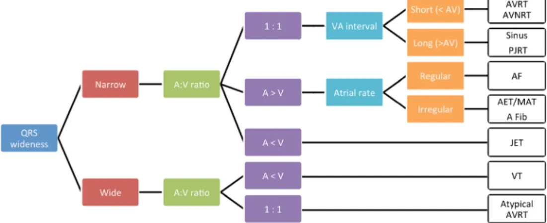
Fig. 4 SVT classification. After birth, QRS wideness allows to differen-tiate SVT from VT. The positive predictive value of a wide QRS for the diagnosis of VT is very high. Rare false positive cases are due to SVT with a bundle branch block or with antegrade conduction through an accessory pathway. Prenatally, without the availability of an ECG, tachy-cardia could still be differentiated based on the A:V ratio and the AV
interval, except for differentiating JET from VT, both rare entities in healthy fetuses and newborn. AET atrial ectopic tachycardia, AF atrial flutter, A Fib atrial fibrillation, AVRT atrioventricular reentrant tachycar-dia, AVNRT atrioventricular nodal reentrant tachycartachycar-dia, JET junctional ectopic tachycardia, PJRT permanent junctional reciprocating tachycar-dia, Sinus sinus tachycartachycar-dia, VT ventricular tachycardia
flecainide might not be available in some centers) and the corrected QT measurement on maternal ECG. Com-bination of antiarrhythmics or second-line antiarrhyth-mics (propafenone, amiodarone) might be used in
hydropic fetuses (Table 2). Indeed, in this situation,
the conversion rate decreases to less than 25 % with digoxin alone and usually a median of 2 medications
are needed to convert to sinus rhythm [20]. Currently,
the combinations of flecainide + digoxin and sotalol + digoxin are most commonly used. In severe cases, when all transplacental therapies have failed, direct adminis-tration of antiarrhythmic agents either through fetal in-tramuscular or transumbilical intravenous administration may be attempted.
The acute management of SVT in infants is based on pediatric advanced life support (PALS) guidelines from the
American Heart Association [29]. Probable SVT with pulses
and poor perfusion should be managed as follows: ABC status (i.e., airways, breathing, and circulation), oxygen delivery, cardiac monitoring, and IV access. If ECG recording confirms SVT and the patient is unstable, electrical synchronized direct current cardioversion 0.5 to 1 J/kg has to be performed im-mediately. If the patient is hemodynamically stable, vagal maneuvers trial can be performed as long as adenosine ad-ministration is not delayed. Vagal maneuvers include diving
reflex and ice bag placement on the face of infants. A mix of crushed ice with water in a plastic bag or glove should be applied on the infant face for 10 to 15 s, without compromis-ing his breathcompromis-ing. In a stable patient, this might be repeated once. Carotid sinus massage and ocular pressure are not recommended due to prolonged asystole and potential retinal detachment, respectively. If not successful, adenosine should be given at a starting dose of 0.1 mg/kg (maximum 6 mg) intravenously by rapid bolus followed by saline flush admin-istered as close to the patient as possible in the largest intra-venous access. Conversion to sinus rhythm with the starting dose is frequently not obtained, especially in infants with less
than 25 % conversion with 0.1 mg/kg [11]. Therefore, the
dose should progressively be increased up to 0.3 mg/kg
(max-imum 12 mg) [39]. Administration through umbilical artery
catheter is the only contraindicated route due to the rapid metabolization of adenosine prior to its reaching the
myocar-dium [30]. Adenosine acts by blocking the AV node and,
therefore, only terminates arrhythmias involving the AV node such as AV reentrant tachycardia (AVRT) or AV nodal reen-trant tachycardia (AVNRT). It will not terminate primary atrial tachycardias such as AF, but it will slow AV conduction and often unmask the underlying arrhythmia. It is therefore essen-tial to record the ECG tracing during its administration. Aden-osine is very effective for terminating SVT and also safe due to Fig. 5 Typical SVT with a short VA interval. On the surface ECG (upper
panel), a narrow QRS tachycardia is observed. If we look carefully at the T wave, a little deflation (black arrowhead) can be observed sometimes corresponding to the retrograde depolarization of the atria. Prenatally (lower panel), a tachycardia is also noted on the Doppler recording based on the ventricular rate, but the atrial contractions are very clearly identi-fiable following shortly every ventricular depolarization. This phenome-non called caphenome-non A wave is due to the occurrence of the atrial contraction
before the opening of the tricuspid valve (the ventricle is still contracting and ejecting blood). The VA interval is much smaller than the AV interval, 83 and 171 ms, respectively, for a heart rate of 240 bpm. A late diastolic wave due to atrial contraction, AA ascending aorta, AV atrioventricular interval, D early diastolic wave, R R wave, S systolic wave, SVC superior vena cava, V ventricular ejection, VA ventriculoatrial interval
its short half life (less than 10 s). Major side effects are infrequent, and they include severe bradycardia, asystole, atrial fibrillation, AV blocks, ventricular arrhythmias, and bronchospasm. Therefore, during its administration, a resus-citation cart has to be available at the bedside. In cases when adenosine is not recommended or unsuccessful, a pediatric cardiologist should be consulted for further management, either electrical cardioversion, overdrive pacing via esopha-geal leads or antiarrhythmic drugs administration. Acute heart failure is sometimes encountered in long-lasting tachycardia. In this situation, rapid restoration of sinus rhythm is crucial and vagal maneuvers and negative inotropes should be avoided. In this setting, digoxin is a valuable option for acute and long-term therapy.
AF is managed prior to birth by transplacental
antiarrhyth-mic administration (Table 2, Fig. 4). Conversion to sinus
rhythm is very effective with the administration of sotalol
[34]. Postnatally, electrical cardioversion is the first-choice
therapy either by atrial overdrive pacing or synchronized
direct current cardioversion [3].
Long-term therapy Duration of therapy depends widely on the type of tachycardia and the difficulties encountered to convert it. Very little data exists on the postnatal management for fetuses diagnosed with SVT. Two strategies are mainly used either to discontinue all therapies at birth, or to continue antiarrhythmics prophylactically for 6 to 12 months. The first option is favored for short VA SVT easily converted with a single drug due to low recurrence rate and the second one for all other cases (long VA SVT, short VA SVT requiring more
than one antiarrhythmic) [24]. Due to a very low recurrence
rate after successful atrial flutter conversion, a chronic antiar-rhythmic treatment is recommended neither in fetuses nor in
infants [24,34]. However, it is important to teach parents the
signs that might reflect a recurrent SVT: irritability, poor feeding, pallor, diaphoresis, and tachypnea. Since those signs might be subtle and also because of parental anxiety, some propose to teach parents to check the heart rate of their infant during sleep or feeding by either placing their hand on the
child’s chest or by using a stethoscope [28].
Postnatally, if a short VA SVT recurred or is newly diag-nosed, a chronic therapy is usually initiated in infants to prevent further episodes. Drugs most used are digoxin and
propranolol with similar efficiency [45]. However, since
di-goxin accelerates conduction through accessory pathways (AP), it is nowadays mostly avoided in children with WPW syndrome due to the risk of ventricular fibrillation in case of
atrial fibrillation [16,32]. Other antiarrhythmics frequently
used are sotalol, flecainide, propafenone, and amiodarone. Long-term therapy is usually discontinued after 6 to 12 months. In case of recurrency of SVT after discontinuation of medical therapy, the antiarrhythmic therapy is maintained for a longer period.
Other rare forms of SVT, like atrial ectopic dia (AET), permanent junctional reciprocating tachycar-dia (PJRT), and junctional ectopic tachycartachycar-dia (JET), are often more difficult to treat medically than short VA SVT. They respond poorly to acute management with adenosine administration, atrial overdrive pacing, or di-rect current cardioversion. They often require multiple therapies directed at controlling at least the rate of the tachycardia when converting to sinus rhythm is not achievable.
Until recently, radiofrequency ablation in children less than 5 years of age and weighing less than 15 kg was restricted to malignant or poorly tolerated
arrhyth-mias [16]. With improving technologies and expertise, it
is now a safe and effective alternative in infants [5].
Catheter ablation should be considered at any age for SVT associated with aborted sudden death ,arrhythmia-related syncopal episodes, ,ventricular dysfunction, re-fractoriness to multidrug therapy, or severe drug-related
side effects [3,5]. This procedure should be performed
only in experienced hands.
Outcome
Studies show that the recurrence rate of paroxysmal SVT is the lowest for prenatal cases and then inversely related to age
at initial diagnosis [41]. Patients with initial SVT during
infancy have a freedom from SVT at 1 year of age of 90 %, but approximately one third of them may have recurrences later in childhood. The reason for the low recurrence rate in prenatal cases lies probably with two phenomena: the pro-gressive disappearance of the muscular accessory pathways
connecting the atria and ventricles as the infant’s heart grows
and the low rate of premature beats as initiating events during
infancy [19].
In the recent era, fetal SVT in hydropic fetuses is still associated with a significant mortality (as high as 17 %) and morbidity rate (neurological abnormalities reported in 10 to
20 % of cases even after successful treatment) [23,38].
Ventricular tachycardia Incidence
VT is very rare in fetuses and infants, accounting for less than 1 % of all tachyarrhythmias and with an incidence
of 0.3 episodes/100,000 infants [16,42]. An underlying
structural heart disease (hypertrophic cardiomyopathy, long QT syndrome, right ventricular dysplasia, left ven-tricle noncompaction, congenital cardiac malformation) is present in approximately half of the pediatric cases.
Diagnosis
Prenatally, the diagnosis is challenging, but should be suspected if AV dissociation is observed with more ventricular contractions than atrial ones on fetal echo. After birth, the diagnosis is made if, on ECG, there are more than three consecutive wide QRS beats of ventricular origin occurring at a faster rate than the underlying rhythm. A rare benign cause of broad QRS tachycardia in neonates is accelerated idioventricular rhythm, characterized by a broad QRS on ECG with a frequency rate slightly higher than the prevailing sinus rhythm in a perfectly asymptomatic patient. Spontane-ous conversion to sinus rhythm might be observed when heart rate rises due to the infant agitation. Etiology is unknown, and it is always self-limited, benign, and never evolves toward a
VT [14,42].
Management
Prenatal treatment includes beta-blockers, flecainide, sotalol, lidocaine, and amiodarone, but due to the very limited number of cases, success rate of treatment is not clearly established and a first-line agent remains to be established. Amiodarone and sotalol have proven to be successful, but there are con-cerns about their proarrhythmic side effects in case of associ-ated LQTS. Therefore, some advocate the use of flecainide or propranolol, which decreases the QT interval and thus the risk of inducing torsades de pointes. Postnatally, management of sustained VT also follows the American Heart Association PALS guidelines: ABC status, oxygen delivery, cardiac mon-itoring, and IV access and ECG recording for rhythm assess-ment and electrical synchronized cardioversion 0.5 to 1 J/kg. Hemodynamically stable infants with VT might be managed
by drug administration with lidocaine or amiodarone [10,33].
In the setting of prolonged QT and torsades de pointes (poly-morphic ventricular tachycardia), intravenous magnesium sul-fate is the treatment of choice. The long-term management still has to be established since, without the occurrence of life-threatening events or death, VT resolution occurs with and
without outpatient antiarrhythmic medication [33].
Outcome
The outcome of VT depends on its etiology. Congenital heart disease, cardiomyopathies (hypertrophic, arrhythmogenic right ventricular cardiomyopathy), long QT syndrome, and cardiac tumors are associated with a poor outcome.
The mortality reported is 36 % in this population [42].
Inversely, in half of fetuses and infants, VT is observed in apparently healthy individuals and is associated with
a good prognosis [10,33,42].
Bradycardia Definition
Bradycardia is defined as an area of the heart that depolarizes slower than the normal range for age for at least three succes-sive beats. A ventricular rate in fetuses and infants below 110 bpm and below 100 bpm, respectively, is usually
consid-ered bradycardia [9,13].
Incidence
Bradycardia represents less than 5 % of arrhythmia referral in
fetuses and infants [13].
Etiology
The most common cause is sinus bradycardia. Transient sinus bradycardia is often secondary to vagal stimulation, frequently occurring during fetal scan as a result of the pressure applied to the maternal abdomen and is also noted during deep sleep,
vomiting, and defecation in infants [47]. Persistent sinus
bra-dycardia is often related to cardiac failure and hydrops in fetuses, but might also be caused by either maternal hypother-mia or long QT syndrome. In infants, sustained sinus brady-cardia is mostly related to secondary sinus node dysfunction
(Table3). Nonsinus bradycardia consists of sinus node
dys-function either idiopathic or related to a CHD pre- or
post-repair, blocked atrial bigeminy (Fig. 2), 2:1 second-degree
atrioventricular block (AVB), or complete AVB. Atrioventric-ular blocks will be discussed separately in the next section. Diagnosis
In utero atrial and ventricular contraction rate and relationship allows identification of the type of bradycardia, either sinus bradycardia, blocked atrial bigeminy, 2:1 AVB or complete
AVB (Fig. 2). Postnatally, the diagnosis is easily made on
surface ECG. Management
For all bradycardias, management depends on etiology and severity, but a close follow-up is always necessary in order to detect the occurrence of cardiac failure.
Outcome
Usually, blocked atrial bigeminy is not associated with the development of cardiac failure and converts spontaneously to sinus rhythm. The outcome of sinus bradycardia depends highly on the underlying cause.
Atrioventricular conduction disorders Complete AV block
Definition
Complete AVB is the complete dissociation between atrial and ventricular contractions. Atrial and ventricular impulses are generated independently from each other. Complete AVB occurring prenatally are equally related to either CHD (left isomerism, atrioventricular septal defect, corrected transposi-tion of the great arteries) or maternal autoantibodies (associ-ated with autoimmune diseases such as systemic lupus
ery-thematosus) [35,47]. Postnatally, the majority is acquired after
surgery for CHD, but carditis (viral myocarditis and Lyme diseases carditis essentially) and idiopathic causes might be
encountered [35,52].
Diagnosis
The diagnosis is easily made prenatally and postnatally by identifying atrial and ventricular events, on Doppler or ECG recording, occurring independently from each other at their own pace.
Management
When delivery is not an option, therapeutic choices are limit-ed. It has been suggested that immune-related second-degree and complete AVB might benefit from transplacental steroid administration to prevent AV node and fetal myocardial
dam-age, but this is still controversial [21,26,35,43]. Significant in
utero morbidity and mortality has been reported related main-ly to cardiac failure and hydrops, which occurs in 9 to 27 % of the pregnancies with a higher rate in those fetuses with
asso-ciated CHD [25,35]. Since with a heart rate below 55 bpm the
prognosis seems very poor, therapies aiming at increasing heart rate have been proposed. Transplacental salbutamol showed some effects in increasing the fetal ventricular rate and case reports describe the use of in utero pacing, but the clinical benefit of these therapies remains to be demonstrated
[2,21,26,43].
In contrary to prenatal management, postnatal management is more clearly established due to the possibility of pacemaker implantation. Advanced second- or third-degree AVB follow-ing surgery and persistfollow-ing more than 7 days requires pace-maker implantation. Nonsurgical advanced second- or third-degree AVB might be followed conservatively. Criteria for pacemaker implantation in those patients include the follow-ing: failure to thrive, cardiovascular symptoms related to the bradycardia, low cardiac output, and resting heart rate during
infancy less than 55 bpm [5]. Finally, any infant with
symp-tomatic sinus bradycardia or chronotropic incompetence
should be referred for pacemaker implantation [5].
Outcome
Complete AVB with co-existing CHD carries the worst
prog-nosis with only 20–40 % survival beyond the neonatal period
[35]. One-year survival for isolated perinatal complete AVB is
between 80 and 90 %. Dilated cardiomyopathy is observed in
approximately 25 % of the survivors [43,51].
Second-degree AV block
Definition-diagnosis-management-outcome
In second-degree AV block, not every atrial beat is conducted to the ventricles. The diagnosis is made similarly for all AV blocks by identifying the atrial and ventricular beats and the blocked atrial beat, and measuring the AV delay. In Mobitz type I subtype (Wenckebach), there is a progressive increase in AV conduction delay ending in a nonconducted atrial beat. This condition shares the same etiologies and prognosis as first-degree AVB. It is also a physiologic condition, frequently observed when vagal tone is more prominent, especially dur-ing sleep. However, progression to higher degrees of AV block is possible, especially if it is related to maternal Table 3 Causes of sinus bradycardia in fetuses or infants
• Primary sinus node dysfunction
– Certain myopathies and inflammatory diseases – Following cardiac surgery
– Heterotaxy syndrome associated with absent SA node (left isomerism) • Autonomic mediated
–Hypervagotonia
– Pallid breathholding spells – Long QT syndrome • Medication (antiarrhythmics) • Situational
– Vasovagal syncope – Carotid sinus pressure – Eye surgery • Central nervous system
– Tumors
– Increased intracranial pressure – Meningitis • Metabolic – Hypoxia – Hypothermia – Hypothyroidism – Acidosis • Sepsis Adapted from [52]
autoantibodies exposition. Those have to be searched for prenatally and appropriate treatment should be established. An underlying cardiac condition is also possible and has to be excluded during the prenatal ultrasound and after birth by history and physical examination. Patient should be referred to a pediatric cardiologist and a 24-h ambulatory heart rate monitoring is indicated to observe if the condition is only transient and related with periods of high vagal tone. If so, follow-up is not necessary. For the remaining cases, etiologies should be looked for and a yearly follow-up is suggested.
In Mobitz type II second-degree AV block, there is no progressive lengthening of the AV conduction, but rather a sudden interruption of AV conduction. This is a rare but more serious condition than type I, often related either to inflamma-tion (maternal autoantibodies) or traumatic injury. There is a higher risk for progression to complete AVB, especially for advanced second-degree AV block (defined as the blocking of two or more consecutive P waves). Therefore, pacemaker implantation is indicated when such block occurs following
surgery [5]. Its incidental finding in an asymptomatic patient
can be managed conservatively, but deserves careful follow-up. First-degree AV block
Definition-diagnosis-management-outcome
In first-degree AV block, every atrial beat is conducted to the ventricles with some delay. The diagnosis is based on the measurement of a prolonged AV or PR interval for age on Doppler recording or ECG. AV interval increases slightly through gestation and normal data varies depending on the
technique used [1,12]. Normal PR interval is less than 160 ms
the first day of life and decreases progressively down to
130 ms at 3 months of age [1,9]. Then its upper limit rises
up to 150 ms and to 160 ms from 3 to 6 and 6 to 12 months,
respectively (Table1).
First-degree AV block is a benign condition and is well tolerated. It does not require any therapy, but it might reflect damage from maternal autoantibodies to the conduction path-way. This should be looked for prenatally and followed
appro-priately [15]. A careful medical history and physical exam has
to be performed to rule out other underlying cardiac diseases. This is mostly a benign condition, physiological in most instances, encountered in infants with high levels of vagal tone, especially during sleep, but it can also be associated with CHD, antiarrhythmic medication, hypothyroidism,
myocardi-al inflammation, or surgicmyocardi-al trauma [52].
Conclusion
Arrhythmias in fetuses and infants are very common. History, physical exam, and recording of the arrhythmia are the
cornerstones of the management. Doppler echocardiography and ECG are the preferred methods for the prenatal and postnatal periods, respectively. Premature beats are almost always benign. The most common SVTs, AV re-entrant tachy-cardia and atrial flutter, carry an excellent prognosis. Acute and chronic antiarrhythmic medications are available to treat and prevent recurrent SVT until spontaneous resolution of the tachyarrhythmia occur, as is the case in up to 90 % of the treated fetuses and infants. If therapy fails, which is more common for rarer forms of SVT, electrophysiological study and catheter ablation could be an option. Due to the increased risks of such procedures in infants, it has to be undertaken in experienced hands. Complete AVB is a serious condition with significant morbidity and mortality, especially when associat-ed with CHD and may neassociat-ed pacemaker implantation. Acknowledgments Some Doppler recording tracings were provided courtesy of Jean-Claude Fouron MD, Pediatric Cardiology, Sainte Justine Hospital, Montreal, Canada.
Conflict of interest The authors do not have any conflict of interest regarding this publication.
References
1. Andelfinger G, Fouron JC, Sonesson SE, Proulx F (2001) Reference values for time intervals between atrial and ventricular contractions of the fetal heart measured by two Doppler techniques. AJC 88:1433– 1436– A8
2. Assad RS, Zielinsky P, Kalil R et al (2003) New lead for in utero pacing for fetal congenital heart block. J Thorac Cardiovasc Surg 126:300–302. doi:10.1016/S0022-5223(03)00220-4
3. Bauersfeld U, Pfammatter JP, Jaeggi E (2001) Treatment of supra-ventricular tachycardias in the new millennium—drugs or radiofre-quency catheter ablation? Eur J Pediatr 160:1–9
4. Bonney WJ, Shah MJ (2013) Incessant SVT in children: ectopic atrial tachycardia and permanent junctional reciprocating tachycar-dia. Progress in Pediatric Cardiology 1–8. doi:10.1016/j.ppedcard. 2012.11.005
5. Brugada J, Blom N, Sarquella-Brugada G et al (2013) Pharmacological and non-pharmacological therapy for arrhyth-mias in the pediatric population: EHRA and AEPC-Arrhythmia Working Group joint consensus statement. Europace 15:1337–1382. doi:10.1093/europace/eut082
6. Carvalho JS, Prefumo F, Ciardelli V et al (2007) Evaluation of fetal arrhythmias from simultaneous pulsed wave Doppler in pulmonary artery and vein. Heart 93:1448–1453. doi:10.1136/hrt.2006.101659
7. Chiu S-N, Wang J-K, Wu M-H et al (2008) Cardiac conduction disturbance detected in a pediatric population. J Pediatr 152:85–89. doi:10.1016/j.jpeds.2007.05.044
8. Collins KK, Van Hare GF, Kertesz NJ et al (2009) Pediatric nonpost-operative junctional ectopic tachycardia. JAC 53:690–697. doi:10. 1016/j.jacc.2008.11.019
9. Davignon A, Rautaharju P, Boisselle E et al (1980) Normal ECG standards for infants and children. Pediatr Cardiol 1:123–152 10. Davis AM, Gow RM, McCrindle BW, Hamilton RM (1996) Clinical
spectrum, therapeutic management, and follow-up of ventricular tachycardia in infants and young children. Am Heart J 131:186–191
11. Dixon J, Foster K, Wyllie J, Wren C (2005) Guidelines and adenosine dosing in supraventricular tachycardia. Arch Dis Child 90:1190– 1191. doi:10.1136/adc.2005.077636
12. Epstein AE, DiMarco JP, Ellenbogen KA et al (2008) ACC/AHA/ HRS 2008 guidelines for device-based therapy of cardiac rhythm abnormalities: a report of the American College of Cardiology/ American Heart Association Task Force on Practice Guidelines (Writing Committee to Revise the ACC/AHA/NASPE 2002 Guideline Update for Implantation of Cardiac Pacemakers and Antiarrhythmia Devices): developed in collaboration with the American Association for Thoracic Surgery and Society of Thoracic Surgeons. Circulation 117:e350–408. doi:10.1161/ CIRCUALTIONAHA.108.189742
13. Fouron J-C (2004) Fetal arrhythmias: the Saint-Justine hospital ex-perience. Prenat Diagn 24:1068–1080. doi:10.1002/pd.1064
14. Freire G, Dubrow I (2008) Accelerated idioventricular rhythm in new-borns: a worrisome but benign entity with or without congenital heart disease. Pediatr Cardiol 29:457–462. doi:10.1007/s00246-007-9024-z
15. Friedman DM, Kim MY, Copel JA et al (2008) Utility of cardiac monitoring in fetuses at risk for congenital heart block: the PR Interval and Dexamethasone Evaluation (PRIDE) prospective study. Circulation 117:485–493. doi:10.1161/CIRCULATIONAHA.107. 707661
16. Friedman RA, Walsh EP, Silka MJ, et al. (2002) NASPE Expert Consensus Conference: radiofrequency catheter ablation in children with and without congenital heart disease. Report of the Writing C o m m i t t e e . N o r t h A m e r i c a n S o c i e t y o f P a c i n g a n d Electrophysiology. In: Pacing Clin Electrophysiol. pp 1000–1017 17. Garson AJ, Gillette PC, McNamara DG (1981) Supraventricular
tachycardia in children: clinical features, response to treatment, and long-term follow-up in 217 patients. J Pediatr 98:875–882 18. Gimovsky ML, Nazir M, Hashemi E, Polcaro J (2004) Fetal/neonatal
supraventricular tachycardia. J Perinatol 24:191–193. doi:10.1038/sj. jp.7211036
19. Hahurij ND, Gittenberger-De Groot AC, Kolditz DP et al (2008) Accessory atrioventricular myocardial connections in the developing human heart: relevance for perinatal supraventricular tachycardias. Circulation 117:2850–2858. doi:10.1161/CIRCULATIONAHA.107. 756288
20. Hornberger LK, Sahn DJ (2007) Rhythm abnormalities of the fetus. Heart 93:1294–1300. doi:10.1136/hrt.2005.069369
21. Jaeggi ET (2004) Transplacental fetal treatment improves the out-come of prenatally diagnosed complete atrioventricular block without structural heart disease. Circulation 110:1542–1548. doi:10.1161/01. CIR.0000142046.58632.3A
22. Jaeggi E (2009) Electrophysiology for the perinatologist. In: Yagel S, Silverman NH, Gembruch U (eds) Fetal cardiology, 2nd edn. Informa Healthcare London, New York, pp 435–447
23. Jaeggi ET, Carvalho JS, De Groot E et al (2011) Comparison of transplacental treatment of fetal supraventricular tachyarrhythmias with digoxin, flecainide, and sotalol: results of a nonrandomized multicenter study. Circulation 124:1747–1754. doi:10.1161/ CIRCULATIONAHA.111.026120
24. Jaeggi E, Fouron JC, Drblik SP (1998) Fetal atrial flutter: diagnosis, clinical features, treatment, and outcome. J Pediatr 132:335–339 25. Jaeggi ET, Hornberger LK, Smallhorn JF, Fouron JC (2005) Prenatal
diagnosis of complete atrioventricular block associated with structur-al heart disease: combined experience of two tertiary care centers and review of the literature. Ultrasound Obstet Gynecol 26:16–21. doi:10. 1002/uog.1919
26. Jaeggi ET, Silverman ED, Laskin C et al (2011) Prolongation of the atrioventricular conduction in fetuses exposed to maternal anti-Ro/ SSA and anti-La/SSB antibodies did not predict progressive heart block. JAC 57:1487–1492. doi:10.1016/j.jacc.2010.12.014
27. Kaltman J, Shah M (2004) Evaluation of the child with an arrhythmia. Pediatr Clin N Am 51:1537–51– viii. doi:10.1016/j.pcl.2004.08.002
28. Kantoch MJ (2005) Supraventricular tachycardia in children. Indian J Pediatr 72:609–619
29. Kleinman ME, Chameides L, Schexnayder SM et al (2010) Part 14: pediatric advanced life support: 2010 American Heart Association Guidelines for Cardiopulmonary Resuscitation and Emergency Cardiovascular Care. Circulation 122:S876–908. doi:10.1161/ CIRCULATIONAHA.110.971101
30. Knick BJ, Saul PJ (2001) Immediate arrhythmia management. In: Zeigler VL, Gillette PC (eds) Practical management of pediatric cardiac arrhythmias. Futura, Armonk, NY, pp 161–230
31. Ko JK, Deal BJ, Strasburger JF, Benson DW (1992) Supraventricular tachycardia mechanisms and their age distribution in pediatric pa-tients. AJC 69:1028–1032
32. Lee KW, Badhwar N, Scheinman MM (2008) Supraventricular tachycardia—part I. Curr Probl Cardiol 33:467–546. doi:10.1016/j. cpcardiol.2008.06.002
33. Levin MD, Stephens P, Tanel RE et al (2010) Ventricular tachycardia in infants with structurally normal heart: a benign disorder. Cardiol Young 20:641–647. doi:10.1017/S1047951110000867
34. Lisowski LA, Verheijen PM, Benatar AA et al (2000) Atrial flutter in the perinatal age group: diagnosis, management and outcome. JAC 35:771–777
35. Lopes LM, Tavares GMP, Damiano AP et al (2008) Perinatal out-come of fetal atrioventricular block: one-hundred-sixteen cases from a single institution. Circulation 118:1268–1275. doi:10.1161/ CIRCULATIONAHA.107.735118
36. Mivelaz Y (2014) Electrophysiology of the fetal heart assessed by magnetocardiography: a hidden area revealed but with some limita-tions. Heart Rhythm. doi:10.1016/j.hrthm.2013.12.040
37. Mivelaz Y, Sarquella-Brugada G, Fournier A, Fouron J-C (2009) The underestimated potential of Doppler ultrasound to assess fetal arrhyth-mia: first report of a prenatal, transient, atypical atrioventricular block. Heart Rhythm 6:1226–1228. doi:10.1016/j.hrthm.2009.03.049
38. Oudijk MA, Gooskens RHJM, Stoutenbeek P et al (2004) Neurological outcome of children who were treated for fetal tachy-cardia complicated by hydrops. Ultrasound Obstet Gynecol 24:154– 158. doi:10.1002/uog.1106
39. RCPCH (2003) Medicines for children, 2nd edn. RCPCH Publications Limited, London
40. Rein AJJT (2002) Use of tissue velocity imaging in the diagnosis of fetal cardiac arrhythmias. Circulation 106:1827–1833. doi:10.1161/ 01.CIR.0000031571.92807.CC
41. Riggs TW, Byrd JA, Weinhouse E (1999) Recurrence risk of supra-ventricular tachycardia in pediatric patients. Cardiology 91:25–30 42. Roggen A, Pavlovic M, Pfammatter J-P (2008) Frequency of
spon-taneous ventricular tachycardia in a pediatric population. AJC 101: 852–854. doi:10.1016/j.amjcard.2007.10.047
43. Rosenthal E, Gordon PA, Simpson JM, Sharland GK (2005) Letter regarding article by Jaeggi et al,“Transplacental fetal treatment im-proves the outcome of prenatally diagnosed complete atrioventricular block without structural heart disease”. Circulation 111:e287–e288. doi:10.1161/01.CIR.0000164275.69617.B2, author reply e287–8 44. Sadovsky G, Nicolaides KH (1989) Reference ranges for fetal heart
rate patterns in normoxaemic nonanaemic fetuses. Fetal Ther 4:61–68
45. Sanatani S, Potts JE, Reed JH et al (2012) The study of antiarrhyth-mic medications in infancy (SAMIS): a multicenter, randomized controlled trial comparing the efficacy and safety of digoxin versus propranolol for prophylaxis of supraventricular tachycardia in in-fants. Circ: Arrhythmia Electrophysiol 5:984–991. doi:10.1161/ CIRCEP.112.972620
46. Schwartz PJ, Salice P (1984) Cardiac arrhythmias in infancy: preva-lence, significance and need for treatment. Eur Heart J 5 Suppl B:43–50 47. Southall DP, Johnson AM, Shinebourne EA et al (1981) Frequency and outcome of disorders of cardiac rhythm and conduction in a population of newborn infants. PEDIATRICS 68:58–66
48. Strasburger JF, Cheulkar B, Wakai RT (2008) Magnetocardiography for fetal arrhythmias. Heart Rhythm 5:1073–1076. doi:10.1016/j. hrthm.2008.02.035
49. Tan HL, Lie KI (2001) Treatment of tachyarrhythmias during preg-nancy and lactation. Eur Heart J 22:458–464. doi:10.1053/euhj.2000. 2130
50. Vergani P, Mariani E, Ciriello E et al (2005) Fetal arrhythmias: natural history and management. Ultrasound Med Biol 31:1–6. doi:10.1016/ j.ultrasmedbio.2004.10.001
51. Villain E, Coastedoat-Chalumeau N, Marijon E et al (2006) Presentation and prognosis of complete atrioventricular block in childhood, according to maternal antibody status. J Am Coll Cardiol 48:1682–1687. doi:10.1016/j.jacc.2006.07.034
52. Walsh EP, Triedman JK, Berul CI (1992) Cardiac arrhythmias. In: Keane JF, Lock JE, Fyler DC (eds) Nadas’ pediatric cardiology, 2nd edn. Saunders Elsevier, Philadelphia, pp 377–433
53. Yabek SM (1991) Ventricular arrhythmias in children with an appar-ently normal heart. J Pediatr 119:1–11