ORIGINAL ARTICLE
Effects of Er:YAG laser on bacteria associated with titanium
surfaces and cellular response in vitro
Irmgard Hauser-Gerspach&Corinna Mauth&
Tuomas Waltimo&Jürg Meyer&Stefan Stübinger
Received: 7 November 2012 / Accepted: 5 March 2013 / Published online: 19 March 2013 # Springer-Verlag London 2013
Abstract This in vitro study examined (a) the anti-bacterial efficacy of a pulsed erbium-doped yttrium aluminum garnet (Er:YAG) laser applied to Streptococcus sanguinis or Porphyromonas gingivalis adhered to either polished or microstructured titanium implant surfaces, (b) the response of osteoblast-like cells and (c) adhesion of oral bacteria to titanium surfaces after laser irradiation. Thereto, (a) bacteria adhered to titanium disks were irradiated with a pulsed Er: YAG laser (1=2,940 nm) at two different power settings: a
lower mode (12.74 J/cm2calculated energy density) and a
higher mode (63.69 J/cm2). (b) After laser irradiation with
both settings of sterile titanium, disks were seeded with 104
MG-63 cells/cm2. Adhesion and proliferation were
deter-mined after 1, 4, and 24 h by fluorescence microscopy and scanning electron microscopy. (c) Bacterial adhesion was also studied on irradiated (test) and non-irradiated (control) surfaces. Adhered P. gingivalis were effectively killed, even
at the lower laser setting, independent of the material’s
surface. S. sanguinis cells adhered were effectively killed
only at the higher setting of 63.69 J/cm2. Laser irradiation of
titanium surfaces had no significant effects on (b) adhesion or proliferation of osteoblast-like MG-63 cells or (c) adhesion of both oral bacterial species in comparison to untreated surfaces. An effective decontamination of polished and rough
titanium implant surfaces with a Er:YAG laser could only be
achieved with a fluence of 63.69 J/cm2. Even though this
setting may lead to certain surface alterations, no significant adverse effect on subsequent colonization and proliferation of MG-63 cells or increased bacterial adhesion was found in comparison to untreated control surfaces.
Keywords Titanium surfaces . Er:YAG laser . Decontamination . Oral bacteria . Osteoblast-like cells
Introduction
Rehabilitation and reconstruction of missing or displaced teeth by means of dental implants or orthodontic treatment has become an essential part of state-of-the-art dentistry to
improve life quality of patients [1,2]. However, a serious
complication and risk factor for the long-term success of any intraoral biomedical device is that of bacterial infec-tion. The high affinity and adhesion of microorganisms to implant surfaces impede normal physiologic or
mechani-cal cleaning by means of saliva or oral hygiene [3].
Subsequent bacterial colonization and biofilm formation can lead to a tremendous loss of dental hard and soft
tissue structures [4,5]. Following this, the onset of severe
inflammatory reactions like gingivitis and periodontitis around teeth or mucositis and peri-implantitis around dental
implants is often inevitable [6].
Periodontitis as well as peri-implantitis are diseases char-acterized by inflammation, swelling, and bleeding of a soft tissue lesion; the severity and extent of these diseases are influenced by the microbial composition of the individual’s
indigenous oral flora and periodontal pathogens [7]. In the
pathogenesis of peri-implant disease bacterial adhesion and colonization are considered to play a key role. These pro-cesses are initiated by early colonizers, like Streptococcus sanguinis, whenever an implant surface is exposed to the I. Hauser-Gerspach (*)
:
T. Waltimo:
J. MeyerInstitute of Preventive Dentistry and Oral Microbiology, School of Dental Medicine, University of Basel, Hebelstrasse 3, 4056 Basel, Switzerland
e-mail: [email protected] C. Mauth
Institute Straumann AG, Peter Merian-Weg 12, 4002 Basel, Switzerland
S. Stübinger
Competence Center for Applied Biotechnology and Molecular Medicine, University of Zürich, Winterthurerstrasse 190, 8057 Zürich, Switzerland
oral environment. Bacteria adhering to the implant usually
grow in a biofilm which make them difficult to treat [8].
Although various different therapeutic options have been advocated for the treatment and prevention especially for peri-implantitis, until now, no generally approved treatment
con-cept could be established [8]. The initial infection rate of dental
implants is still reported to be 5–8 % [9]. Moreover, a recent
consensus meeting has concluded that peri-implantitis occurs at 12–40 % of sites of implants which makes peri-implantitis a
true threat for the long-term success of dental implants [10].
The use of antimicrobial agents like chlorhexidine or antibi-otics does not provide an effective countermeasure to peri-implantitis particularly over the long term. A crucial factor is that bacteria in biofilms are less sensitive against many anti-microbials, which make these infections difficult to treat. Besides classical mechanical removal strategies of the biofilm or novel approaches like application of gaseous ozone, differ-ent laser irradiation regimes have been commonly proposed as
a promising treatment option [11]. Several clinical and
exper-imental studies could demonstrate the advantages summarized
in [10,12] of laser irradiation on decontamination of implant
surfaces. Beneath comfortable clinical handling, especially pulsed Er:YAG lasers have gained a growing popularity due to scientific evidence provided by animal studies in a canine
model [13–15]. However, recent own experiments with a
pulsed Er:YAG laser revealed certain surface alterations of polished and microstructured titanium implant surfaces by
using clinically accepted laser settings [16].
Respecting the 3R principle (reduce, replace, refine) of animal welfare it is consequently desirable to evaluate such basic laser applications in an in vitro system to draw any possible conclusions. To obtain persistent success, the laser irradiation should (1) eliminate bacteria effectively, (2) enhance re-osseointegration, and (3) impede bacterial re-colonization of the surface.
Therefore, it was the aim of the present in vitro study to analyze the biological and microbiological potential of a pulsed Er:YAG laser, on S. sanguinis and Porphyromonas gingivalis adhered to either a polished or a sandblasted, large-grit, acid-etched (SLA) titanium surface in an approved
disk model. The stated H0 hypothesis was that irradiated
implant surfaces do neither impede the proliferation of osteoblast-like cells nor advocate adhesion of S. sanguinis and P. gingivalis in comparison to non-irradiated surfaces.
Materials and methods
Test specimens and laser system
Disks (5.0 mm in diameter) of titanium (commercial pure, ASTM grade II) either sandblasted, large grit, acid etched (SLA), or polished (POL) (Straumann AG Basel, Switzerland)
were sterilized by gamma irradiation. For the bacterial exper-iments, a total of 108 disks was coated by exposure to a sterile
saliva–serum mixture (10:1) during 15 min at 35 °C [17].
Treatment of bacteria, adhered to the materials’surface, was carried out with an OpusDuo laser system (OpusDuo ECTM, Lumenis GmbH, Dreieich, Germany) with a fiber-optic
deliv-ery system. The Er:YAG laser, with a wavelength of 2.94μm,
allows variations of pulse energy up to 1,000 mJ, a pulse frequency of 7 to 20 Hz, and pulse duration of 250 to
400μs. A 1,000-μm fiber tip (HPX Conical Sapphire Contact
Tips) was used for application. The pulse frequency of 10 Hz
and pulse durations between 250 to 400μs were chosen as
recommended by the manufacturer.
All titanium specimens were laser irradiated by hand-guiding the application tip at a constant distance of 0.5 to
1 mm. According to the findings of a previous study [16],
the following two laser settings were chosen:
1. 100 mJ, 10 Hz, 10 s, calculated energy density 12.
74 J/cm2, which causes no surface alterations and
2. 500 mJ, 10 Hz, 10 s, calculated energy density 63.
69 J/cm2, which may cause certain surface alterations.
Microbiological experiments
The two bacteria examined were S. sanguinis (DSM 20068), an early colonizer, and P. gingivalis (ATCC 33277), fre-quently associated with peri-implantitis. Microbiological
procedures were as previously described [17–19]. Bacteria
used were suspended in the 10:1 saliva–serum mixture at a
concentration of 108–109colony-forming unit CFU/mL, to
simulate in vivo conditions.
To irradiate adhered bacteria, the coated disks were placed on the bottom of 24-well plates (Becton Dickinson, Basel, Switzerland) and exposed to the bacterial suspensions for 2 h at 35 ° C. The disks were then placed for 1 min on a pad saturated with 70 % ethanol to inactivate bacteria ad-hered to the bottom side of the disks. Bacteria on the other side were then irradiated as described. Afterwards, the disks were suspended in 3 mL 0.9 % NaCl, vortexed for 60 s and sonicated for 15 s (30 W, 20 kHz; VibracellTM, Ultrasonic Processor, Sonics, Newtown, USA). Viable bacteria were determined by culture on blood agar plates and anaerobic incubation. For one bacterial suspension, three disks were used, one for the untreated control, and two for the laser applications. Each material with either S. sanguinis or P. gingivalis was tested in five independent experiments.
Sterilized uncoated disks were treated with the Er:YAG laser as above, then coated with the saliva–serum mixture and ex-posed to the bacterial suspensions for 2 h. Each material with either S. sanguinis or P. gingivalis was tested in four indepen-dent experiments. Viable bacteria adhered were enumerated by
Cellular experiments
In vitro experiments were performed by incubation of hu-man osteosarcoma cells (MG-63, ATCC) on each material surface. Cells were maintained as subconfluent monolayers in Minimal Essential Medium supplemented with 10 % fetal calf serum and 1 % penicillin–streptomycin (all Sigma-Aldrich, Buchs, Switzerland) as described by
Hauser-Gerspach et al. [18]. Cells were used for experiments no
later than passage 4 and seeded on disks placed in 96-well culture plates (Techno Plastic Products AG, Trasadingen,
Switzerland) at a density of 10,000 cells/cm2. Determination
of cell morphology, attachment, spreading, and proliferation was performed after incubation for 1, 4, and 24 h. All experiments were repeated in duplicates with n=5 for an independent experiment.
For the evaluation whether the substrate type affected cell morphology, the specimens (n=2) were examined by scanning electron microscopy (Philips XL-30, Eindhoven, Netherland). Images were recorded at ×500 magnification.
Cell attachment and spreading was examined by immuno-cytochemical staining of fixed cells (n=3). Nuclei staining was performed using 4′,6 – diamidin-2phenylindol (DAPI), and actin cytoskeleton was stained with Alexa Fluor® 488 phalloidin (both Sigma-Aldrich). Each five images were recorded of all material samples at ×10 magnification using a fluorescent microscope (Nikon, 90i, Egg, Switzerland), whereby areas were chosen randomly at five regions in the center of the disks, to the right, left, bottom, and top of it. Calculation of the area of attached cells as well as counts of nuclei was performed using the Visiopharm software Version 7 (Hoersholm, Denmark).
One-way ANOVA was used to determine whether there was a statistically significant difference between titanium SLA and polished surface. P values of <0.05 were considered to be statistically significant.
Results
Decontamination efficiency of Er:YAG laser
Overall, the Er:YAG laser had a superior decontaminating effect on sandblasted, large-grit, acid-etched (Ti-SLA) sur-faces to polished (Ti-POL) titanium sursur-faces which had
been seeded with S. sanguinis (Table 1). At the higher
power setting of 63.69 J/cm2, laser irradiation killed S.
sanguinis adhered to Ti-SLA disks to below detection limit (>4 logs reduction) while bacteria adhered to polished sur-faces (Ti-POL) were incompletely eliminated. Similarly, at
the lower setting of 12.74 J/cm2, laser irradiation caused more
efficient killing of S. sanguinis on Ti-SLA (1 % survivors) than on Ti-POL surfaces (about 10 % survivors).
On both titanium surfaces, Er:YAG laser irradiation of
energy density 63.69 J/cm2was more strongly bactericidal
to S. sanguinis than at the setting of 12.74 J/cm2. Adhered P.
gingivalis cells were very sensitive to laser applications: The bacteria were inactivated to below detection limit on either
titanium surface by either laser setting (Table1).
Cell spreading and proliferation on surfaces irradiated before
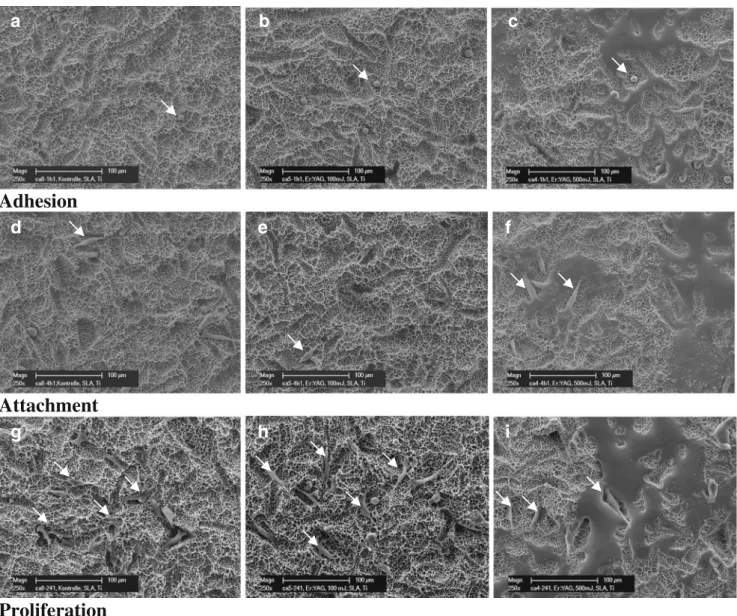
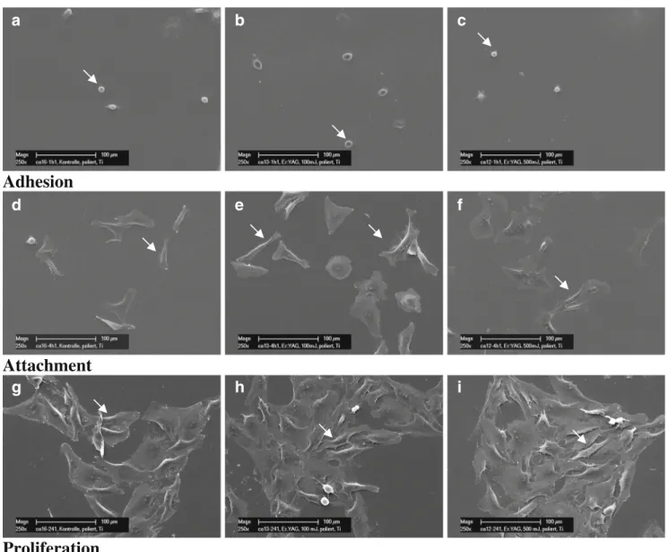
The MG-63 cells seeded on the Ti-SLA and Ti-POL sur-faces showed a spherical morphology after 1 h, became gradually flattened after 4 h and were uniformly flattened
after 24 h (Figs.1and2). Cell spreading was quantified as
percentage covered (Fig.3a, b). The increase over 24 h was
similar on both titanium surfaces without a significant effect of laser irradiations.
Proliferation of MG-63 cells as measured by average nuclei count was slightly higher on Ti-SLA surface and was not affected by the laser treatment in a significant way
(Fig.3c, d).
Attachment of bacteria to surfaces irradiated before
The other question addressed was whether Er:YAG laser irradiation would affect subsequent adherence of bacteria. Interestingly, adhesion of both bacterial species S. sanguinis and P. gingivalis to both titanium Ti-SLA and Ti-POL was statistically not significantly changed after irradiation of the
surface in comparison to the untreated control (Table2).
Discussion
Treatment of peri-implantitis aims at eliminating or substan-tially reducing the bacterial load on the implant in order to Table 1 Bactericidal effects of Er:YAG laser on bacteria adhered to titanium surfaces. Log reduction of adhered viable bacterial cells in relation to surface alterations observed after laser irradiation Er:YAG laser
parameters
Ti-SLA Ti-POL
S. sanguinis P. gingivalis S. sanguinis P. gingivalis 12.74 J/cm2 2.0 logsa ≥3.5 logsa 0.9 logsa ≥3.6 logsa 63.59 J/cm2 ≥4.3 logsb ≥3.5 logsb 2.6 logsb ≥3.6 logsb Mean and S.D. are given (n=5)
Surface alterations as reported by Stübinger et al. [16] are indicated by superscripted letters
Ti-SLA titanium sandblasted, large grit, acid-etched; Ti-POL titanium polished
a
No visible alteration
b
arrest disease progression. Therefore the first aim of this in vitro study was to investigate the bactericidal potential of Er: YAG laser on bacteria adhered to titanium surfaces. Indeed, differences in the anti-bacterial activity were detected between the two laser settings using S. sanguinis. Thus, for maximal decontamination the higher laser dose was required. However,
this dose may lead to alterations of the surface [16].
The results also showed that not all bacterial species are eliminated with equal efficiency: Attached P. gingivalis was more sensitive to the Er:YAG laser tested than S. sanguinis, as there were marked differences at the lower laser settings. The decontaminating potential of lasers has been explored in vitro using a range of different titanium implant or other surfaces, bacterial or fungal species, and laser systems
[19–28]. Investigations using Er:YAG laser irradiation regimes
are listed in Table3. The data illustrate the enormous
hetero-geneity of the studies. The energy densities applied varied in a
wide range of 0.04 to 63.69 J/cm2 in the different studies.
Therefore, any direct comparisons of results are hardly possi-ble. Some of the surfaces materials irradiated with the higher doses underwent structural changes. The bacteria used repre-sented different segments of the oral microbiota (Gram posi-tive or negaposi-tive, aerobic, microaerophilic, or anaerobic) and even bacteria of the same species could be of different strains (type strains from collections vs recent clinical isolates). Among the support/implant materials tested, titanium domi-nated, including different surface qualities, but glass and hy-droxyapatite were also used. Some surfaces were conditioned with serum or saliva before microbial adhesion which was allowed to proceed for one to several hours. Alternatively,
Adhesion
Attachment
Proliferation
a
d
g
b
e
h
c
f
i
Fig. 1 Representative SEM imagines of osteoblastic cells (arrows) seeded on Ti-SLA (a, d, g, untreated; b, e, h, after laser treatment Er: YAG-1, 12.74 J/cm2; c, f, i, Er:YAG-2, 63.69 J/cm2) and incubated for
1 h (a–c), demonstrating the initial cell adhesion; 4 h (d–f), demon-strating cell attachment; 24 h (g–i), demonstrating cell growth
incubation continued to yield a biofilm. Analyses of the laser effects involved light or electron microscopy (measuring dead and live cells), cultural techniques (measuring CFU) or en-zyme assays (measuring total cell metabolic state). The results are difficult to compare because of the very diverse parame-ters, even though P. gingivalis and S. sanguinis are often among the bacteria tested. From these studies, it is difficult to draw general conclusions with respect to predictable clinical Er:YAG laser applications.
Another important point is the evaluation of the measure-ments documenting the antimicrobial efficacy of the laser system. Reduction of bacterial counts by laser irradiation in the order of 90 to 99 % (1 to 2 logs) are most likely
statistically significant (Table3). However, the relevant
bio-logical question remains unanswered: is a short-term drastic
bacterial reduction (of e.g. 2 logs P. gingivalis) in the peri-implant region achieved by low laser energy sufficient for stable clinical improvements. A threshold level of P. gingivalis or any other peri-implant bacteria to obtain predictable clinical
success is not known [29]. In periodontitis therapy, a
statisti-cally significant reduction of periodontopathogenic bacteria does not guarantee clinical success because reinfection from other oral sites may occur or bacteria may survive
intracellu-larly at the treated site [30].
From previous studies, it is known that laser irradiation of
titanium surfaces may lead to detectable alterations [16,
31–33]. Even after application of these higher laser dose
conditions, neither initial cell adhesion nor spreading and proliferation of osteoblast-like MG-63 cells was altered on rough (Ti-SLA) and smooth (Ti-POL) surfaces. A slight
Adhesion
Attachment
Proliferation
b
a
f
e
d
i
h
g
c
Fig. 2 Representative SEM imagines of osteoblastic cells (arrows) cultured on Ti-POL (a, d, g, untreated; b, e, h, after laser treatment Er: YAG-1, 12.74 J/cm2; c,f,i, Er:YAG-2, 63.69 J/cm2) and incubated for
1 h (a–c), demonstrating the initial cell adhesion; 4 h (d–f), demon-strating cell attachment; 24 h (g–i), demondemon-strating cell growth
decrease of initial cell adhesion could be assumed due to the surface energy alteration of untreated versus laser treated surfaces. However, a significant effect on MG-63 cells has not been determined. Similar results have also been reported in human osteoblastic Saos-2 cells on machined and sandblasted acid-etched surfaces following Er:YAG laser treatment observing no differences in cell proliferation after 24 h. This study also did not demonstrate any effect on the
cell differentiation [34].
Similarly, the altered surfaces did not interfere with subse-quent bacterial adhesion. No inhibition effect which would be desirable and no positive stimulation were observed. To date,
there are only few studies evaluating the effects on bacterial adhesion on surfaces after Er:YAG laser treatment. Also
Duarte et al. [31] did not find higher levels of S. sanguinis
adhesion on titanium surfaces in vitro after Er:YAG laser
treatment at 8.4 J/cm2.
In the present in vitro study, effective decontamination of polished and rough titanium implant surfaces with a pulsed Er:YAG laser could be only achieved with an energy density
of 63.69 J/cm2. Even though these settings may lead to
certain surface alterations, no significant adverse effect on subsequent colonization and proliferation of MG-63 cells or increased bacterial adhesion was found in comparison to
2 -G A Y : r E 1 -G A Y : r E d e t a e r t n u
b
a
c
d
Fig. 3 Quantification of cell spreading per surface by actin staining. MG-63 cells cultured for 1, 4, and 24 h on different surfaces. a Ti-SLA and b Ti-POL without and after laser treatment. Quantification of
nuclei per image area. MG-63 cultured for 1, 4, and 24 h on different surfaces. c Ti-SLA and d Ti-POL without and after laser treatment. Er: YAG-1, 12.74 J/cm2; Er:YAG-2, 63.69 J/cm2
Table 2 Adhesion of S. sanguinis or P. gingivalis on titanium surfaces after laser irradiation of the surfaces (log)
Mean and SD are given (n=4) Ti-SLA titanium sandblasted, large grit, acid-etched; Ti-POL titanium polished
Control Er:YAG laser parameters
12.74 J/cm2 63.69 J/cm2 Ti-SLA
S. sanguinis log CFU per disk 5.9±0.07 5.8±0.14 5.8±0.12 P. gingivalis log CFU per disk 5.6±0.19 5.4±0.15 5.5±0.21 Ti-POL
S. sanguinis log CFU per disk 5.1±0.20 5.1±0.22 5.2±0.24 P. gingivalis log CFU per disk 4.9±0.07 4.9±0.08 4.9±0.07
T able 3 Antimicrobial activities of Er:Y AG laser application in vitro Auth ors Er:Y AG laser Bacte ria/fungi Results C o mment Calculated ener gy density Spec ies Support surfa ce Measureme nts Reduct ion in viabil ity Ando et al. [ 24 ] Single pulse at 0.04 –10.6 J/cm 2 P . ging ivalis , Aggr egatibac ter (Actinobac illus ) actinomy cetemcomitans Nutrient agar plates Growth inhibi tion zones; survival in laser -irradiated colo nies Growth inhibition zones at ≥ 0.3 Jcm − 2for P . gingivalis + Aggr egatibacter (Actinobacillus ) ac tinomyc etemcom itans 17 % surv ivors P . gingivalis at 10.6 J/cm 2 Kreisler et al. [ 21 ] 2 6 o r 52.2 J/cm 2 pulse S . san guinis Bacteria in PBS adhere d 1 h , 37° to titanium disks (3 surfaces) CFU by conve ntional culture tec hnique (after son ication) 1.8 –2.4 logs at 26 J/cm 2 Incom p lete bact erial k il ling 2.8 –3.2 logs at 52.2-J/ cm 2 val ues depe ndent on surfa ce Noiri et al. [ 25 ] 0.38 or 0.71 or 0.98 J/cm 2 pulse 4 aerobic G + and 3 anaerob ic G − species a 7-day aerobic, 14-day anae robic mono species biofil m o n hydrox yapatite disks CFU by conve ntional culture tec hnique (after sc raping of f+ so nication) G + :0 –3 logs at 0.71 J/cm 2 ; 0– 4 logs at 0.98 J/cm 2 V iabil ity speci es-depende nt Lactobac illus casei most res istant G − : > 3 logs at 0.71 and 0.9 8 J/cm 2 Quaran ta et al. [ 27 ] 10.7 J/cm 2 pulse P . ging ivalis 48-h anaerobic incubation on dif ferent titanium impl ants Cou nting bacterial cel ls by SEM 76.2 –98. 3 % decontamination val ues depe ndent on surfa ce Live or dead bacteria? Senn henn-Kirchner et al. [ 26 ] 12.0 or 15.2 J/cm 2 pulse Can dida albican s (clinical isolates) 5-day biofilm on glass and ti tanium disks in BHI Colorimetric assay to determine mito chondri al dehydrog enase activity (metabolic state) Significan tly reduc ed enzy me act ivity Schw arz et al. [ 28 ] 12.7 J/cm 2 24-h ex vivo biofilm on titan ium disks Residua l plaque area measured o n microscopic images 97 % clean impla nt surface Hauser -Gerspach et al. this study 12.74 J/cm 2 or 63.6 9 J/cm 2pulse S . san guinis Bacteria in human saliva/ se rum adhered 2 h , 37° to ti tanium disks (2 surfa ces) CFU by conve ntional culture tec hnique (after son ication) S. sanguin is : 0.9 –2.0 logs at 12. 74 J/cm 2 P . ging ivalis P . gingivalis :≥ 3.5 logs at 12.74 J/cm 2 S. sanguin is : 2.6 –≥ 4.3 logs at 63.69 J/cm 2 J/cm 2P . ging ivalis :≥ 3.5 log s at 63.69 J/cm 2 V alu es depen dent on surface a Gram positive: Actinomyces naeslundii , Enter ococcus faecalis , L. casei , Pr oprionibacterium acnes ; Gram negative: Fusobacterium nucleatum , P . gingivalis , Pr evotella nigr escens
untreated control surfaces. Further detailed analyses of bio-molecular and microbiological interactions and processes on laser-irradiated implant surfaces are necessary to draw any further conclusions on re-osseointegration and biofilm formation of dental titanium implants in vivo.
Acknowledgments We thank the ITI Foundation (grant no. 518/ 2007) and the SSO Funds (grant no.: 248–09) for financial support. The authors also thank Catiana Gass for doing some of the in vitro cell experiments at Straumann laboratories.
References
1. Wennerberg A, Albrektsson T (2011) Current challenges in successful rehabilitation with oral implants. J Oral Rehabil 38: 286–294
2. Preston CB, Maggard MB, Lampasso J, Chalabi O (2008) Long-term effectiveness of the continuous and the sectional archwire techniques in leveling the curve of Spee. Am J Orthod Dentofacial Orthop 133:550–555. doi:10.1016/j.ajodo. 2006.02.039
3. Furst MM, Salvi GE, Lang NP, Persson GR (2007) Bacterial colonization immediately after installation on oral titanium im-plants. Clin Oral Implants Res 18:501–508
4. Lang NP, Berglundh T (2011) Periimplant diseases: where are we now?—consensus of the Seventh European Workshop on Peri-odontology. J Clin Periodontol 38(Suppl 11):178–181
5. Lee A, Wang HL (2010) Biofilm related to dental implants. Im-plant Dent 19:387–393
6. Berglundh T, Zitzmann NU, Donati M (2011) Are peri-implantitis lesions different from periodontitis lesions? J Clin Periodontol 38(Suppl 11):188–202
7. Persson LG, Berglundh T, Lindhe J, Sennerby L (2001) Re-osseointegration after treatment of peri-implantitis at different im-plant surfaces. An experimental study in the dog. Clin Oral Im-plants Res 12:595–603
8. Renvert S, Roos-Jansåker AM, Claffey N (2008) Non-surgical treatment of peri-implant mucositis and peri-implantitis: a litera-ture review. J Clin Periodontol 35(8 Suppl):305–315
9. Pye AD, Lockhart DE, Dawson MP, Murray CA, Smith AJ (2009) A review of dental implants and infection. J Hosp Infect 72:104–110
10. Lindhe J, Meyle J (2008) Peri-implant diseases: consensus report of the sixth European Workshop on Periodontology. J Clin Periodontol 35(8 Suppl):282–285
11. Deppe H, Horch HH (2007) Laser applications in oral surgery and implant dentistry. Lasers Med Sci 22:217–221
12. Kotsovilis S, Karoussis IK, Trianti M, Fourmousis I (2008) Ther-apy of peri-implantitis: a systematic review. J Clin Periodontol 35:621–629
13. Schwarz HP, Dorner F, Mitterer A, Mundt W, Schlokat U, Pichler L, Turecek PL (1999) Preclinical evaluation of recombinant von Willebrand factor in a canine model of von Willebrand disease. Wien Klin Wochenschr 111:181–191
14. Schwarz HP, Dorner F, Mitterer A, Mundt W, Schlokat U, Pichler L, Turecek PL (1998) Evaluation of recombinant von Willebrand factor in a canine model of von Willebrand disease. Haemophilia Off J World Fed Hemophilia 4(Suppl 3):53–62
15. Schwarz F, Bieling K, Nuesry E, Sculean A, Becker J (2006) Clinical and histological healing pattern of peri-implantitis lesions following non-surgical treatment with an Er:YAG laser. Lasers Surg Med 38:663–671
16. Stübinger S, Etter C, Miskiewicz M, Homann F, Saldamli B, Wieland M, Sader R (2010) Surface alterations of polished and sandblasted and acid-etched titanium implants after Er:YAG, car-bon dioxide, and diode laser irradiation. Int J Oral Maxillofac Implants 25:104–111
17. Hauser-Gerspach I, Kulik EM, Weiger R, Decker E-M, Von Ohle C, Meyer J (2007) Adhesion of Streptococcus sanguinis to dental implant and restorative materials. Dent Mater J 26:361–366 18. Hauser-Gerspach I, Vadaszan J, Deronjic I, Gass C, Meyer J, Dard
M, Waltimo T, Stübinger S, Mauth C (2012) Influence of gaseous ozone in peri-implantitis: bactericidal efficacy and cellular re-sponse. An in vitro study using titanium and zirconia. Clin Oral Investig 16:1049–1059
19. Hauser-Gerspach I, Stübinger S, Meyer J (2010) Bactericidal ef-fects of different laser systems on bacteria adhered to dental implant surfaces: an in vitro study comparing zirconia with titani-um. Clin Oral Implants Res 21:277–283
20. Kato T, Kusakari H, Hoshino E (1998) Bactericidal efficacy of carbon dioxide laser against bacteria-contaminated titanium im-plant and subsequent cellular adhesion to irradiated area. Lasers Surg Med 23:299–309
21. Kreisler M, Kohnen W, Marinello C, Götz H, Duschner H, Jansen B, d’Hoedt B (2002) Bactericidal effect of the Er:YAG laser on dental implant surfaces: an in vitro study. J Periodontol 73: 1292–1298
22. Prates RA, Yamada AMJ, Suzuki LC, Eiko Hashimoto MC, Cai S, Gouw-Soares S, Gomes L, Riberio MS (2007) Bactericidal effect of malachite green and red laser on Actinobacillus actinomycetemcomitans. J Photochem Photobiol B 86:70–76
23. Kojima T, Shimada K, Iwasaki H, Ito K (2005) Inhibitory effects of a super pulsed carbon dioxide laser at low energy density on periodontopathic bacteria and lipopolysaccharide in vitro. J Periodont Res 40:469–473
24. Ando Y, Aoki A, Watanabe H, Ishikawa I (1996) Bactericidal effect of erbium YAG laser on periodontopathic bacteria. Lasers Surg Med 19:190–200
25. Noiri Y, Katsumoto T, Azakami H, Ebisu S (2008) Effects of Er: YAG laser irradiation on biofilm-forming bacteria associated with endodontic pathogens in vitro. J Endod 34:826–839
26. Sennhenn-Kirchner S, Schwarz P, Schliephake H, Konietschke F, Brunner E, Borg-von Zepelin M (2009) Decontamination efficacy of erbium:yttrium-aluminium-garnet and diode laser light on oral Candida albicans isolates of a 5-day in vitro biofilm model. Lasers Med Sci 24:313–320
27. Quaranta A, Maida C, Scrascia A, Campus G, Quaranta M (2009) Er:YAG laser application on titanium implant surfaces contami-nated by Porphyromonas gingivalis: an histomorphometric evalu-ation. Minerva Stomatol 58:317–330
28. Schwarz F, Sculean A, Romanos G, Herten M, Horn N, Scherbaum W, Becker J (2005) Influence of different treatment approaches on the removal of early plaque biofilms and the viability of SAOS2 osteoblasts grown on titanium implants. Clin Oral Investig 9:111–117
29. Romeo E, Ghisolfi M, Carmagnola D (2004) Peri-implant dis-eases. a systematic review of the literature. Minerva Stomatol 53:215–230
30. Johnson JD, Chen R, Lenton PA, Zhang G, Hinrichs JE, Rudney JD (2008) Persistence of extracrevicular bacterial reservoirs after treatment of aggressive periodontitis. J Periodontol 79:2305–2312
31. Duarte PM, Reis AF, de Freitas PM, Ota-Tsuzuki C (2009) Bacterial adhesion on smooth and rough titanium surfaces after treatment with different instruments. J Periodontol 80:1824–1832
32. Shin SI, Min HK, Park BH, Kwon YH, Park JB, Herr Y, Heo SJ, Chung JH (2011) The effect of Er:YAG laser irradiation on the scanning electron microscopic structure and surface roughness of various implant surfaces: an in vitro study. Lasers Med Sci 26:767–776
33. Kim JH, Herr Y, Chung JH, Shin SI, Kwon YH (2011) The effect of erbium-doped: yttrium, aluminium and garnet laser irradiation on the surface microstructure and roughness of double acid-etched implants. J Periodont Imlant Sci 41:234–241
34. Galli C, Macaluso GM, Elezi E, Ravanetti F, Cacchioli A, Gualini G, Passeri G (2011) The effects of Er:YAG laser treatment on titanium surface profile and osteoblastic cell activity: an in vitro study. J Periodontol 82:1169–1177