HAL Id: halshs-00957270
https://halshs.archives-ouvertes.fr/halshs-00957270
Submitted on 19 Mar 2018
HAL is a multi-disciplinary open access
archive for the deposit and dissemination of
sci-entific research documents, whether they are
pub-lished or not. The documents may come from
teaching and research institutions in France or
abroad, or from public or private research centers.
L’archive ouverte pluridisciplinaire HAL, est
destinée au dépôt et à la diffusion de documents
scientifiques de niveau recherche, publiés ou non,
émanant des établissements d’enseignement et de
recherche français ou étrangers, des laboratoires
publics ou privés.
Towards producing pure phytolith concentrates from
plants that are suitable for carbon isotopic analysis
Rémi Corbineau, Paul Reyerson, Anne Alexandre, Guaciara Santos
To cite this version:
Rémi Corbineau, Paul Reyerson, Anne Alexandre, Guaciara Santos. Towards producing pure phytolith
concentrates from plants that are suitable for carbon isotopic analysis. Review of Palaeobotany and
Palynology, Elsevier, 2013, pp.179-185. �10.1016/j.revpalbo.2013.06.001�. �halshs-00957270�
Research paper
Towards producing pure phytolith concentrates from plants that are
suitable for carbon isotopic analysis
Rémi Corbineau
a,b, Paul E. Reyerson
c, Anne Alexandre
a,⁎
, Guaciara M. Santos
da
Centre Européen de Recherche et d'Enseignement des Géosciences de l'Environnement (UMR 7330), CNRS, Aix-Marseille Université, Europôle méditerranéen de l'Arbois BP 80, 13545 Aix en Provence cedex 04, France
b
Laboratoire d'Archéologie Médiévale et Moderne en Méditerranée (UMR 7298), CNRS, Aix-Marseille Université, MMSH, 5 rue du Château de l'Horloge, BP 647, 13094 Aix-en-Provence, France
c
Department of Geography, University of Wisconsin-Madison, 550 North Park Street, Madison, WI 53706, USA d
Earth System Science, University of California, Irvine, B321 Croul Hall, Irvine, CA 92697-3100, USA
a b s t r a c t
a r t i c l e i n f o
Article history: Received 28 June 2012
Received in revised form 6 June 2013 Accepted 9 June 2013
Available online 19 June 2013 Keywords:
Phytolith
AMS radiocarbon dating Extraction protocol Carbon isotopes
Phytoliths are micrometric particles of amorphous silica that form inside or between the cells of higher plant tis-sues throughout the life of a plant. Phytolith morphological assemblages extracted from sediments and buried soils are increasingly used as proxies of grassland diversity and tree cover density. When found in significant amounts in archeological sites they can be used for identifying food habits, cultural and agricultural practices. Phytoliths can contain small amounts of C occluded in their structure (phytC). It is generally assumed that the source of this phytC is atmospheric CO2that wasfixed by the plant via photosynthesis. Isotopic analyses of
phytoliths (δ13C,14C) were thus expected to inform respectively on the photosynthetic pathway or on the age
of the mineralized host plants. However recent14C analyses of phytC from phytolith concentrates extracted
from soils and harvested grasses yielded unexpected14C ages of several hundreds to kyr old. These14C phytC
re-sults raised the question of a possible source of refractory/old soil organic matter component taken up by roots, which can be attached or occluded in phytoliths. Simultaneously these results highlighted the need for setting standardized protocols leading to concentrates entirely devoid of organic residues, as well as for a robust method for checking phytolith purity. The goal of this work was thus to develop protocols for extracting phytoliths from plants, leading to 100% phytolith purity, as required for phytC analyses. Protocol 1 utilizes a multi-step process of dry ashing and acid digestion, while protocol 2 also uses acid digestion as well as a separate alkali immersion step which removes surface layers. Phytolith concentrate purity was gauged in a semi-quantitative fashion through the use of SEM–EDS analysis. This quality check for phytolith purity can reveal small C particulate contamination of phytolith concentrates that may considerably bias isotopic and quantitative analyses of phytC. Results indicate that the two protocols were able to entirely remove small C particulate contamination. Protocol 1 produced phy-tolith concentrates with well defined morphologies suitable for both morphological and isotopic analyses. How-ever measurement of C yields showed that protocol 1 probably induced C leakage, leading to lower recovery. Protocol 2 is faster, leads to higher C yield but may lead to a beginning of dissolution. With these protocols on hand, sources of phytC can be properly investigated.
© 2013 Elsevier B.V. All rights reserved.
1. Introduction
Phytoliths are micrometric particles of amorphous silica (ASi) that form inside or between the cells of higher plant tissues throughout the life of a plant. Silicon (Si) is taken up by the roots in its dissolved form, translocated in the sap, and deposited in the cells where it can take the shape of the host cell. The concentration of phytoliths ranges from less than 0.01% of dry weight in many gymnosperms and dicot-yledon angiosperms to more than 8% of dry weight in Poaceae,
Arecaceae, and Equisetaceae (e.g.Geis, 1973; Bozarth, 1992; Webb
and Longstaffe, 2002). With plant decay, phytoliths that are preserved
in oxidizing environments are either incorporated into soils or exported to sediments via regional watersheds. Phytolith morphological assem-blages extracted from sediments and buried soils are increasingly used as proxies of grassland diversity and tree cover density (e.g.Blinnikov et al., 2002; Strömberg, 2002; Boyd et al., 2005; Bremond et al., 2005a, b; 2008a,b; Piperno, 2006; Lentfer and Torrence, 2007; Neumann
et al., 2009; Messager et al., 2010; Prasad et al., 2011). When found in
significant amounts in archeological sites they can be used for identify-ing food habits, cultural and agricultural practices (e.g.Delhon et al.,
2008; Li et al., 2010; Yost and Blinnikov, 2011). In parallel, phytoliths
in plants, soils, and rivers were quantified for investigating the
⁎ Corresponding author. Fax: +33 4 42 97 15 42.
E-mail addresses:corbineau@mmsh.univ-aix.fr(R. Corbineau),reyerson@wisc.edu
(P.E. Reyerson),alexandre@cerege.fr(A. Alexandre),gdossant@uci.edu(G.M. Santos).
0034-6667/$– see front matter © 2013 Elsevier B.V. All rights reserved.
http://dx.doi.org/10.1016/j.revpalbo.2013.06.001
Contents lists available atSciVerse ScienceDirect
Review of Palaeobotany and Palynology
biogeochemical cycle of Si, which itself is coupled to the global C cycle (e.g.Blecker et al., 2006; Struyf et al., 2009; Alexandre et al., 2011; Cornelis et al., 2011).
Phytoliths can contain small amounts of C occluded in their struc-ture (phytC), which is thought to range from 0.1 to 2% of phytolith dry weight (Wilding, 1967; Prychid et al., 2003). Raman spectroscopy and GC–MS analyses of phytC evidenced the presence of aliphatic com-pounds and lignins (Perry et al., 1987; Smith and Anderson, 2001), as well as aromatic hydrocarbon and graphite or coal when phytoliths were burnt (Pironon et al., 2001). PCR associated with protein staining detected the presence of glycoproteins but could notfind evidence of any DNA (Elbaum et al., 2009).
It is generally assumed that the source of this phytC is atmospheric CO2that wasfixed by the plant via photosynthesis (Wilding, 1967;
Kelly et al., 1991; Raven et al., 1999; Piperno, 2006; Carter, 2009).
From this basis, the assumption that phytC may be a terrestrial sink of C in the global C cycle was recently suggested (Parr and Sullivan,
2005; Jansson et al., 2010). In parallel, carbon isotopic studies have
investigated the potential of phytCδ13C signatures for providing
infor-mation about photosynthetic pathways (Kelly et al., 1991; Smith and White, 2004; Webb and Longstaffe, 2010; Strömberg and McInerney, 2011) or deriving a paleo-atmospheric CO2 record (Carter, 2009).
However, isotopic calibration studies of phytoliths from grasses showed that the difference betweenδ13C
tissueandδ13CphytCvalues is not
con-stant from a plant to another (Smith and White, 2004; Webb and
Longstaffe, 2010) and that changes inδ13C
phytCvalues are not related
to expected variation in theδ13C values of atmospheric CO 2(Webb
and Longstaffe, 2010).
Few studies have used14C ages of phytolith concentrates from soils
and archeological sediments as chronological indicators (Piperno and
Becker, 1996; Piperno and Stothert, 2003; McMichael et al., 2012). One
of them found a modern or post-bomb age for phytolith concentrates
extracted from superficial soil (Piperno and Becker, 1996). Thousand-year ages were also reported for topsoil phytoliths (McMichael et al., 2012). This was justified by to the long mean residence time of phytoliths in soils. Other studies failed in matching14C phytC values
with expected or independent chronologies (Wilding, 1967; Kelly et al., 1991; McClaran and Umlauf, 2000; Prior et al., 2005; Rieser
et al., 2007; Boaretto, 2009). Encountered difficulties were thought to
be associated with stratigraphic inversions, preferential oxidation of younger phytoliths or mostly with ineffectiveness of phytolith extrac-tion procedures (Wilding, 1967; Kelly et al., 1991; Prior et al., 2005;
Rieser et al., 2007; Boaretto, 2009). Neither the phytolith chemical
pro-cedural blank assessment nor the reproducibility and accuracy checks on14C of large pools of phytC were ever attempted to corroborate or
explain the14C results obtained. A recent study (Santos et al., 2010a)
evaluated the background of phytolith chemical extractions and the reproducibility and accuracy of14C phytC on phytolith concentrates
extracted from soils and harvested grasses. Surprisingly, the phytC from harvested grasses yielded unexpected14C ages of several kyr old
(though bulk material from the same plants gave contemporary14C
values), when using an established protocol (Kelly, 1990; Kelly et al., 1991). This case is supported by phytC14C-AMS data obtained from
harvested bamboo leaves and underlying litter layers that were expected to reproduce contemporaneous atmospheric14C values (Sullivan and
Parr, 2008, 2013). As recently discussed inSantos et al. (2012a,b)the
dataset shows varying depletions of at least 5 pMC (percent modern carbon) relative to the values expected when taking into account the southern hemisphere bomb radiocarbon peak and its recent decreasing trend in the atmosphere (Santos et al., 2012b). The minimum 5 pMC off-set is equivalent to 400 years, reflecting incorporation of a substantial amount of“old” carbon in the phytolith concentrates (Santos et al.,
2012b). The offset is maximum for the harvested leaves (which yielded
an age of 3.5 ka BP), and undisturbed green litter with minimal contact
Table 1
Main steps of published protocols originally set up for phytolith extraction from plants. These protocols are commonly used for morphological identification purposes; some of them have been used for phytC analyses.
Protocols Original references Used for phytC analyses
Wet oxidation Main oxidizing agent: H2SO4/H2O2
Rinsing of plant material with HCl.
Boiling samples in 70% ethanol, washing and drying. Oxidation with concentrated H2SO4 at 70°C.
Addition of H2O2 until solution is clear at room temperature. Rinsing with distilled water.
Geis,1973, 1978 Kelly, 1990
Pironon et al., 2001 Smith and Anderson, 2001 Krull et al., 2003 Smith and White, 2004 Carter, 2009 Santos et al., 2010a Webb and Longstaffe, 2010 Main oxidizing agent: HNO3/KClO3
Rinsing of plant material.
Oxidation with concentrated HNO3 and KClO3 at 100°C. Removal of carbonates using HCl at room temperature. Rinsing with distilled water.
Rovner, 1972 Pearsall, 1989 Piperno, 2006 Main oxidizing agent: HNO3/HClO4
Rinsing of plant material with HCl.
Two oxidation steps with a 1:1 HNO3-HClO4 mixture at 80°C. Addition of H2O2 at 80°C.
Rinsing with distilled water.
Rovner, 1971 Elbaum et al., 2009
Santos et al., 2010a
Microwave digestion Oxidation with HNO3, H2O2 and HCl in closed digestion tubes. Microwave irradiation for 30 min.
Sieving at 250 µm. Rinsing in ethanol.
Parr et al., 2001b
Parr, 2002 Krull et al., 2003Parr et al., 2010
Parr and Sullivan, 2005, 2011 Ashing
Rinsing of plant material with distilled water. Heating crucibles in muffle furnace at 500°C for 6h. Remove from crucibles to test tubes.
Oxidation with HCl at 70°C for 20 min/rinsing. Oxidation with H2O2 or HNO3 at 70°C for 20 min. Rinsing with distilled water.
with soil contaminants (1.8 ka BP). These14C results in addition to
Santos et al.'s (2010a)findings raised the question of a possible source
of refractory/old soil organic matter (SOM) component in soils taken up by roots, through nitrogen assimilation (amino acids or proteins) or in dissolved form (Santos et al., 2012aand references within), which can be attached to or occluded in phytoliths. Scanning Electron Micro-scope images coupled with Energy Dispersive Spectrometer analyses (SEM–EDS) from splits of the phytoliths of living grasses dated by
Santos et al. (2010a)revealed the presence of particulate organic matter
(OM) residues (Santos et al., 2012a) that may have contributed to bias in phytC ages. This fact emphasizes the need for setting standardized proto-cols for phytC analysis that can dissolve or oxidize all external organic remains.
Methods commonly used for extracting phytoliths from modern plant materials can be grouped into three categories: wet oxidation,
dry ashing and microwave digestion (Table 1). Although they were originally set up for phytolith morphological identification which does not require 100% purity, a survey of the published literature shows that some of them were used for plant phytC isotopic analyses
(Table 1). Very few reports are available to evaluate phytolith
concen-trate purity (Parr et al., 2001a,b). This type of assessment is mostly performed by optical microscope evaluations, sometimes including cross-polarized light (Parr et al., 2010; Parr and Sullivan, 2011) which does not allow for the identification of non-polarizing organic remains (other than cellulose). FT-IR spectroscopy can be used on phytolith con-centrates to check mineral purity (Cabanes et al., 2011), however, it has a low detection limit of organic compounds (1%).
Here we used SEM–EDS analyses to check for OM remains in phyto-lith concentrates. Moreover, we developed two protocols for extracting phytoliths from plants, leading to 100% phytolith purity, as required for
60
µm
b
C
O
Al
Si
Au Pd
C
O
Al
Si
Au
Pd
10
µm
c
C:Si = 0.21
C:Si = 0.06
2
.
c
1
.
c
60
µm
a
C
O
Al
Si
Au
Pd
10
µm
d
C:Si = 1.35
C
O
Al
Si
Au
Pd
20
µm
f
C:Si = 0.58
C
O
Al
Si
Au
Pd
40 µm
e
C:Si = 1.13
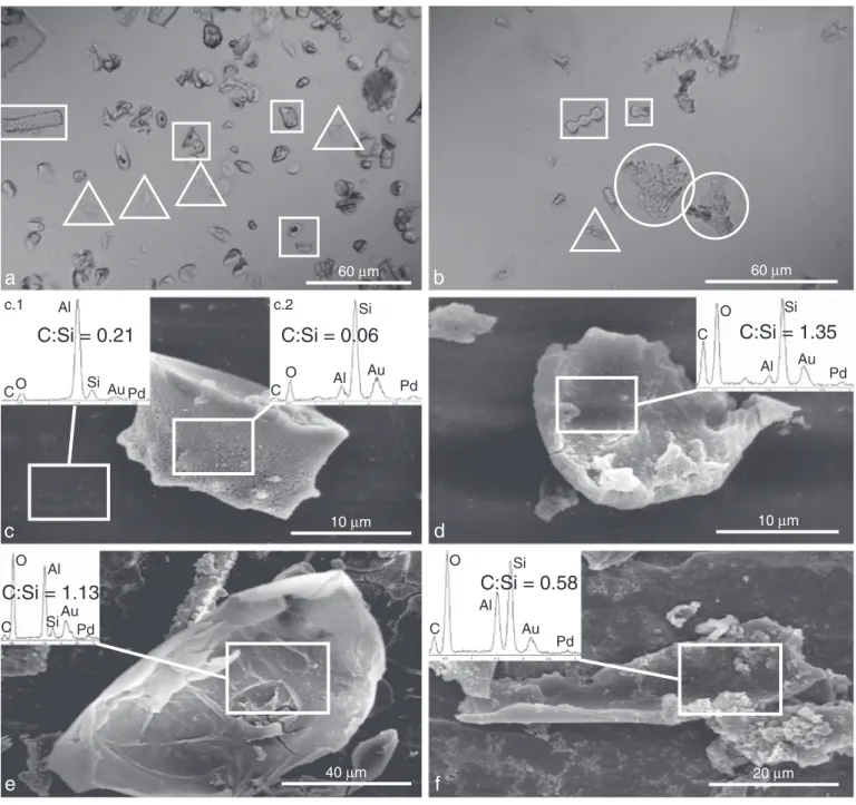
Fig. 1. Images of non-pure phytolith concentrates from Grass 1 and MN samples extracted followingKelly (1990). Optical microscopic images from (a) Grass 1, and (b) MN, showing highly refractive phytolith particles (white rectangles), poorly refractive particles without characteristic shape (white triangles) and tissue-like particles (white circles:). SEM images and EDS spectra of (c.1) Al sample holder, (c.2) rondel type phytolith, (d), (e) and (f) organic particles. 1-c, 1-d and 1-e are reproduced fromSantos et al. (2012a)for the purpose of the comparison between pure and non-pure phytolith concentrates.
phytC analyses. Regarding phytolith extraction from soils and sediments, we already have in hand a high purity protocol commonly used for phy-tolith oxygen isotopic analyses (Crespin et al., 2010; Alexandre et al., 2012).
2. Material and methods
Several combinations of the protocols listed inTable 1were tested on random grasses of no relevance until consistently satisfactory results
were achieved. The twofinal protocols were applied to 50 g of leaves of harvested sorghum (Sorghum bicolor), in preparation for ulterior
14C-AMS analyses of phytC. The protocols were devoid of any organic
chemicals that can sorb to the surface of clean silica (Schlechtriem et al., 2003; Santos et al., 2010a).
Final phytolith concentrates were observed under optical microscopy and analyzed using a SEM (Philips XL30) associated with an EDS system (Energy-dispersive X-ray spectroscopy, Oxford) according to the follow-ing steps: 1) coatfollow-ing with gold and palladium of the samples placed on
Table 2
Two protocols of phytolith extraction from plants assessed at CEREGE and UWM as leading to pure phytolith concentrates.
Protocol 1—wet digestion/dry ashing Protocol 2—wet digestion
(1) Dry sample at 110 °C for 24 h then cut into cm-sized pieces. (2) Grind samples.
(3) Place sample with DW⁎in ultrasonic bath for 15 min to remove particulate matter from the surface.
(4) Place sample in a porcelain crucible and oven dry at 110 °C for 24 h. (5) Place the crucible in a muffle furnace and increase temperature
incrementally from 300 °C to 500 °C (300 °C–325 °C–350 °C–375 °C–500 °C) over 4 h. Hold for 4 h at 500 °C.
(6) Place sample in a glass beaker and rinse/soak with 10% HCl for 30 min to remove any carbonates. Rinse with DW⁎3 times.
(7) Add a 65% HNO3/70% HClO4mixture (2:1) and place on sand bath at 80 °C for 16 h.
(8) Rinse with DW three times. Place sample in a porcelain crucible and oven dry at 110 °C for 24 h.
(9) Repeat steps (5) and (7).
(10) Add H2O2and place on sand bath at 80 °C for 16 h. (11) Rinse with DW 3 times.
(12) Check purity by SEM/EDS. If some OM⁎still remains, repeat steps 2 to 6.
(1) Dry sample at 110 °C for 24 h then cut into cm-sized pieces. (2) Place sample with DW⁎in ultrasonic bath to remove particulate matter
from the surface.
(3) Immerse in 10% HCl to remove any carbonates and agitate for 30 min. Rinse with DW 3 times.
(4) Place samples in glass beaker and add concentrated H2SO4. Allow to react for 2 h at 70 °C then allow to sit unheated overnight.
(5) Reheat to 70 °C then slowly add 30% H2O2while stirring until the supernatant turns clear. Keep under heat for 2 h, stirring occasionally. Allow to sit, heated, for another hour. Pour off the supernatant then rinse the sample 3 times with DW. (6) Add concentrated HNO3and heat at 70 °C for 2 h. Add a pinch of KClO3to facilitate
digestion. Allow to sit unheated overnight. Decant then repeat this step to maximize organic matter oxidation. Rinse 3 times with DW.
(7) Immerse the phytoliths in 0.001 M KOH solution (pH = 11) and heat at 70 °C for 15 min to remove any alkali-soluble forms of OM⁎.
(8) Extract material using PPfilters. Rinse with DW, transfer to glass vials then dry at 110 °C overnight.
(9) Check purity by SEM/EDS. ⁎ DW: distilled water; OM: organic matter.
a
50
µm
40
µm
C
O
Al
Si
Au
Pd
C
O
Al
Si
Au
Pd
C:Si = 0.08
C:Si = 0.08
b
40
µm
C
O
Si
Au
Al
Pd
C
O
Al
Si
Au
Pd
C:Si = 0.08
C:Si = 0.05
c
Fig. 2. (a) Optical microscopy images of phytoliths from Sorghum bicolor leaves, extracted following Protocol 1, showing highly refractive phytolith-like particles (white rectangles) and tissue-like particles (white circle). SEM images and EDS spectra of (b) phytoliths and (c) tissue-like particles.
an Al holder; 2) SEM spotting of organic-like particles showing tissue-like or non-phytolith morphologies; 3) EDS semi-quantitative analyses of C, Si and Ca (indicative of the presence of C-bearing Ca-carbonates). Although SEM–EDS data are semi-quantitative and little accurate for C determination, C:Si % mass ratios, obtained from 10 to 30μm spots were distinct enough to allow detection of silica particles and organic remains (Fig. 1).
For the purpose of comparison, two samples obtained by Santos and co-workers (Grass 1 and MN;Santos et al., 2010a) using the H2SO4/H2O2 protocol from which organic remains were previously
evidenced (Santos et al., 2012a) were re-investigated using optical microscopy and SEM–EDS (Fig. 1).
In order to determine the carbon percentage of the phytC obtained by the optimized protocols, 100% pure phytolith concen-trate samples were combusted at 900 °C in evacuated sealed 6 mm OD pre-baked quartz tubes in the presence of cupric oxide (Santos
et al., 2010a), and the cryogenically cleaned CO2yield was measured
manometrically at room temperature on a vacuum line (Santos et al., 2004).
3. Results and discussions
When observed in optical microscopy, non-pure phytolith concen-trates obtained from Grass 1 and MN samples evidenced three catego-ries of particles (Fig. 1): 1) highly refractive phytoliths; 2) poorly refractive particles without characteristic shape; and 3) tissue-like par-ticles. SEM–EDS analyses (Fig. 1) allowed us to clearly categorize the observed particles into: 1) silica particles (or phytoliths) with values of C:Si % mass ratios equal or lower than values obtained for the Al sam-ple holder (b0.5); 2) organic remains with values of C:Si % mass ratios higher than 0.5. Most of the tissue-like particles were silica (C:Si %
massb0.5). Organic remains (C:Si % mass >0.5) were mainly unshaped particles. No Ca was found by EDS analysis.
After several trial experiments to refine phytolith extraction protocols, two protocols werefinally kept. They are described inTable 2. Protocol 1
(Table 2) combines methods described inRovner (1971), Parr et al.
(2001a)andPiperno (2006). It includes multi-step wet oxidation using
HNO3and HClO4along with dry ashing. Protocol 2 combines methods
de-scribed inGeis (1973),Pearsall (1989),Prior et al. (2005)andPiperno (2006). It is a wet digestion using H2O2, HNO3, KClO3, and KOH,
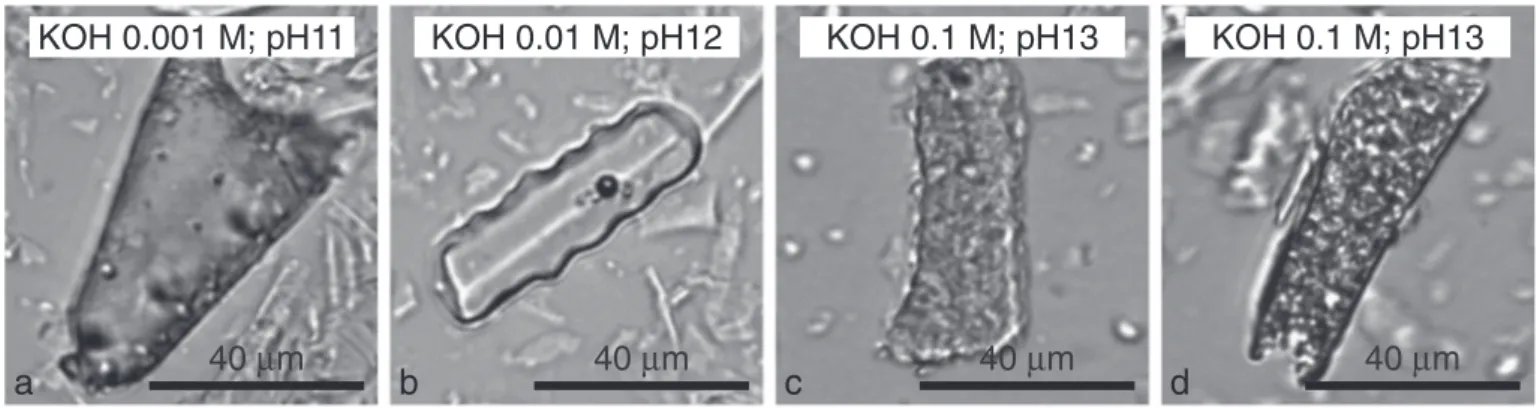
some-times used for soil phytolith extraction (Piperno, 2006). Several tests were made with three concentrations of KOH (0.001 M @ pH 11; 0.01 M @ pH 12 and 0.1 M @ pH 13). Methodic checking for carbon par-ticulate contamination, using SEM–EDS analysis, revealed the absence of Ca and only the presence of particles with C:Si peak area ratios lower than 0.1 (Figs. 2 and 3). Thus, both protocols led to phytolith concentrates pure enough for phytC isotopic analyses. Protocol 1 produced phytolith concentrates including tissue-like Si particles with well preserved orna-mentation (Madella et al., 2005) (Fig. 2). Ornamentation features were well preserved when Protocol 2 included the use of KOH @ pH 11 and 12 but started dissolve when KOH was used @ pH 13 (Fig. 4).
The carbon yields of phytolith concentrates from protocols 1 and 2 were 0.053 and 0.21%, respectively, and were reproducible among dupli-cates. From SEM–EDS analyses and phtyC CO2yields, we can conclude
that both of these protocols show advantages and disadvantages: Protocol 1 produces phytoliths with preserved ornamentation excellent for morphological counting prior to phytC analyses. However repeated ashing up to 500 °C may lead to C leakage as evidenced by the low CO2
yield measured herein. This necessitates that a large amount of phytoliths (ca. 800 mg) need to be extracted from plant material (ca. 800 g assum-ing a phytolith content of 0.1% dry weight of bulk plant tissue) for obtaining large graphite targets of ~0.4 mg of C. However several AMS
10
µm
b
C
O
Al
Si
Au
Pd
C:Si = 0.05
20
µm
3
µm
a
Fig. 3. (a) and (b) SEM images and EDS spectra of phytoliths particles from Sorghum bicolor leaves extracted following Protocol 2, including the use of KOH @ pH 13. Pits scattered on the phytolith surfaces evidence dissolution.
40
µm
40
µm
40
µm
40
µm
KOH 0.01 M; pH12
KOH 0.001 M; pH11
KOH 0.1 M; pH13
KOH 0.1 M; pH13
c
a
b
d
Fig. 4. Optical microscopy images of phytoliths obtained after Protocol 2 using three concentrations of KOH: (a) 0.001 M @ pH11; (b) 0.01 M @ pH 12; (c) and (d) 0.1 M @ pH 13. Pits scattered on the phytolith surfaces evidence dissolution @ pH 13.
facilities are today able to measure samples as small as 0.010 mg C with 1 sigma errors of just 1% on close-to-modern targets (Santos et al., 2007; de Rooij et al., 2010; Fahrni et al., 2010; Ruff et al., 2010) which brings the re-quired amount of phytolith to ca. 20 mg (using protocol 1) or 5 mg (using protocol 2). However, one should note that14C-AMS samples produced
from some chemical extractions bear higher blanks (Santos et al., 2010a, b), and therefore slightly larger amounts of biosilica concentrates in order to produce larger graphite samples are recommended. Another drawback of protocol 1 is the use of HClO4, which is dangerous to handle
and requires the use of a specialized fume hood. Protocol 2, which does not utilize dry ashing, is faster and avoids the problem of C leakage. We were able to extract approximately four times more phytC with this ap-proach, as compared to protocol 1. However the use of KOH concentra-tions above pH 12 should be avoided as it induces fast silica dissolution which needs to be carefully monitored. Finally, these protocols are both suitable for phytolith extraction prior to phytC isotopic analyses. The choice of one or another will depend on the amount of plant material, on available time and equipment, and on the amount of pure phytoliths required for accurate and reproducible analyses.
4. Conclusion
This study highlights the need of a robust method for checking phyto-lith purity prior to phytC quantification and isotopic analyses. SEM–EDS analysis of Ca, C and Si, although yielding semi-quantitative estimations, is suitable to be used as a routine method for phytolith concentrate purity evaluation. Organic remains, that may not be accounted for under light microscopy, can be clearly individualized from their high C:Si % mass ra-tios. This quality check for phytolith purity may reveal small C particulate contamination of phytolith concentrates that may considerably bias isoto-pic and quantitative analyses of phytC.
With the aim of phytC isotopic analyses, we have assessed two plant phytolith extraction protocols which yielded phytolith concentrates de-void of organic remains. Methodological advantages and drawbacks of both protocols were presented. With these protocols on hand, sources and concentration of phytC and possibly organic carbon involvement in biosilica synthesis of higher plants can be now properly investigated. Acknowledgments
We thank Dr. Bruce A. Kimball and Dr. Michael J. Ottman from the U.S. Arid-Land Agricultural Research Center, Maricopa, AZ for providing samples of Sorghum bicolor plant tissue used in this work. Aspects of this work were partially supported by the NSF grant DEB-1144888, FIR 2010 (Aix-Marseille Université), ECCOREV 2011 and AIR Archéométrie 2011 (CNRS). We also thank two anonymous reviewers for their suggestions. References
Alexandre, A., Bouvet, M., Abbadie, L., 2011.The role of savannas in the terrestrial Si cycle: a case-study from Lamto, Ivory Coast. Global and Planetary Change 78, 162–169.
Alexandre, A., Crespin, J., Sonzogni, C., Sylvestre, F., Hilbert, D.W., 2012.The oxygen isoto-pic composition of phytoliths from troisoto-pical rainforest soils (Queensland, Australia): application of a new paleoenvironmental tool. Climate of the Past 8, 307–324.
Blecker, S.W., McCulley, R.L., Chadwick, O.A., Kelly, E.F., 2006.Biologic cycling of silica across a grassland bioclimosequence. Global Biogeochemical Cycles 20 (3), GB3023.1–GB3023.11.
Blinnikov, M., Busacca, A., Whitlock, C., 2002.Reconstruction of the late Pleistocene grassland of the Columbia basin, Washington, USA, based on phytolith records in loess. Palaeogeography, Palaeoclimatology, Palaeoecology 177 (1–2), 77–101.
Boaretto, E., 2009.Dating materials in good archaeological contexts: the next challenge for radiocarbon analysis. Radiocarbon 51, 275–281.
Boyd, W.E., Lentfer, C.J., Parr, J.F., 2005.Interactions between human activity, volcanic eruptions and vegetation during the Holocene at Garua and Numundo, West New Britain, PNG. Quaternary Research 64 (3), 384–398.
Bozarth, S.R., 1992.Classification of opal phytoliths formed in selected dicotyledons native to the great plains. In: Rapp, G., Mulholland, S.C. (Eds.), Phytolith Systematics. Plenum, New York, pp. 193–214.
Bremond, L., Alexandre, A., Hély, C., Guiot, J., 2005a.A phytolith index as a proxy of tree cover density in tropical areas: calibration with Leaf Area Index along a forest–
savanna transect in southeastern Cameroon. Global and Planetary Change 45 (4), 277–293.
Bremond, L., Alexandre, A., Peyron, O., Guiot, J., 2005b.Grass water stress estimated from phytoliths in West Africa. Journal of Biogeography 32 (2), 311–327.
Bremond, L., Alexandre, A., Peyron, O., Guiot, J., 2008a.Definition of grassland biomes from phytoliths in West Africa. Journal of Biogeography 35 (11), 2039–2048.
Bremond, L., Alexandre, A., Wooller, M.J., Hely, C., Williamson, D., Schafer, P.A., Majule, A., Guiot, J., 2008b.Phytolith indices as proxies of grass subfamilies on East African tropical mountains. Global and Planetary Change 61, 209–224.
Cabanes, D., Weiner, S., Shahack-Gross, R., 2011.Stability of phytoliths in the archaeological record: a dissolution study of modern and fossil phytoliths. Journal of Archaeological Science 38 (9), 2480–2490.
Carter, J.A., 2009.Atmospheric carbon signatures in phytolith-occluded carbon. Quaternary International 193 (1–2), 20–29.
Cornelis, J.-T., Delvaux, B., Georg, R.B., Lucas, Y., Ranger, J., Opfergelt, S., 2011.Tracing the origin of dissolved silicon transferred from various soil–plant systems towards rivers: a review. Biogeosciences 8, 89–112.
Crespin, J., Sylvestre, F., Alexandre, A., Sonzogni, C., Paillès, C., 2010.Re-examination of the thermo-dependent relationship betweenδ18O diatoms andδ18O lake water.
Implications for palaeoclimatic applications. Journal of Paleolimnology 44, 547–557.
de Rooij, M., van der Plicht, J., Meijer, H.A.J., 2010.Porous iron pellets for AMS14C analysis
of small samples down to ultra-microscale size (10–25 μgC). Nuclear Instruments and Methods in Physics Research Section B 268, 947–951.
Delhon, C., Martin, L., Argant, J., Thiébault, S., 2008.Shepherds and plants in the Alps: multi-proxy archaeobotanical analysis of neolithic dung from“La Grande Rivoire” (Isère, France). Journal of Archaeological Science 35 (11), 2937–2952.
Elbaum, R., Melamed-Bessudo, C., Tuross, N., Levy, A.A., Weiner, S., 2009.New methods to isolate organic materials from silicified phytoliths reveal fragmented glycoproteins but no DNA. Quaternary International 193, 11–19.
Fahrni, S.M., Gaggeler, H.W., Hajdas, I., Ruff, M., Szidat, S., Wacker, L., 2010.Direct measurements of small C-14 samples after oxidation in quartz tubes. Nuclear Instruments and Methods in Physics Research Section B 268, 787–789.
Geis, J.W., 1973.Biogenic silica in selected species of deciduous angiosperms. Soil Science 116 (2), 113–130.
Jansson, C., Wullschleger, S.D., Kalluri, U.C., Tuskan, G.A., 2010.Phytosequestration: carbon biosequestration by plants and the prospects of genetic engineering. Bioscience 60 (9), 685–696.
Kelly, E.F., 1990.Method for extracting opal phytoliths from soil and plant material. Internal Report. Department of Agronomy, Colorado State University, Fort Collins.
Kelly, E.F., Amundson, R., Marino, B.D., Deniro, M., 1991.Stable isotope ratios of carbon in phytoliths as a quantitative method of monitoring vegetation and climate change. Quaternary Research 35 (2), 222–233.
Lentfer, C., Torrence, R., 2007.Holocene volcanic activity, vegetation succession, and ancient human land use: unraveling the interactions on Garua Island, Papua New Guinea. Review of Palaeobotany and Palynology 143 (3–4), 83–105.
Li, R., Carter, J.A., Xie, S., 2010.Phytoliths and microcharcoal at Jinluojia archeological site in middle reaches of Yangtze River indicative of paleoclimate and human activity during the last 3000 years. Journal of Archaeological Science 37 (1), 124–132.
Madella, M., Alexandre, A., Ball, T., 2005.International code for phytolith nomenclature 1.0. Annals of Botany 96, 253–260.
McClaran, M.P., Umlauf, M., 2000.Desert grassland dynamics estimated from carbon isotopes in grass phytoliths and soil organic matter. Journal of Vegetation Science 11 (1), 71–76.
McMichael, C.H., Bush, M.B., Piperno, D.R., Silman, M.R., Zimmerman, A.R., Anderson, C., 2012.Spatial and temporal scales of pre-Columbian disturbance associated with Western Amazonian lakes. The Holocene 22, 131–141.
Messager, E., Lordkipanidze, D., Delhon, C., Ferring, C.R., 2010.Palaeoecological impli-cations of the Lower Pleistocene phytolith record from the Dmanisi Site (Georgia). Palaeogeography, Palaeoclimatology, Palaeoecology 288 (1–4), 1–13.
Neumann, K., Fahmy, A., Lespez, L., Ballouche, A., Huysecom, E., 2009.The Early Holocene palaeoenvironment of Ounjougou (Mali): phytoliths in a multiproxy context. Palaeogeography, Palaeoclimatology, Palaeoecology 276 (1–4), 87–106.
Parr, J.F., Sullivan, L.A., 2005.Soil carbon sequestration in phytoliths. Soil Biology and Biochemistry 37, 117–124.
Parr, J., Sullivan, L., 2011.Phytolith occluded carbon and silica variability in wheat cultivars. Plant and Soil 342 (1), 165–171.
Parr, J., Lentfer, C.J., Boyd, W.E., 2001a.A comparative analysis of wet and dry ashing techniques for the extraction of phytoliths from plant material. Journal of Archaeological Science 28, 875–886.
Parr, J.F., Dolic, V., Lancaster, G., Boyd, W.E., 2001b.A microwave digestion method for the extraction of phytoliths from herbarium specimens. Review of Palaeobotany and Palynology 116, 203–212.
Parr, J., Sullivan, L., Chen, B., Zheng, W., 2010.Carbon bio-sequestration within the phytoliths of economic bamboo species. Global Change Biology 16 (10), 2661–2667.
Pearsall, D.M., 1989.Paleoethnobotany: A Handbook of Procedures. Academic Press, San Diego (470 pp.).
Perry, C.C., Robert, J.P.W., Fry, S., 1987.Cell wall biosynthesis during silicification of grass hairs. Journal of Plant Physiology 126, 437–448.
Piperno, D.R., 2006. Phytoliths: A Comprehensive Guide for Archaeologists and Paleoecologists. AltaMira Press, New York (238 pp.).
Piperno, D.R., Becker, P., 1996.Vegetational history of a site in the central Amazon Basin derived from phytolith and charcoal records from natural soils. Quaternary Research 45 (2), 202–209.
Piperno, D.R., Stothert, K.E., 2003.Phytolith evidence for early Holocene Cucurbita domestication in southwest Ecuador. Science 229 (5609), 1054–1057.
Pironon, J., Meunier, J.D., Alexandre, A., Mathieu, R., Mansuy, L., Grosjean, A., Jardé, E., 2001.
Individual characterization of phytoliths: experimental approach and consequences on paleoenvironment understanding. In: Meunier, J.D., Colin, F. (Eds.), Phytoliths: Applications in Earth Sciences and Human History. A.A. Balkema Publishers, Lisse, pp. 329–341.
Prasad, V., Strömberg, C.A.E., Leaché, A.D., Samant, B., Patnaik, R., Tang, L., Mohabey, D.M., Ge, S., Sahni, A., 2011. Late Cretaceous origin of the rice tribe provides evidence for early diversification in Poaceae. Nature Communications 2, 480.
http://dx.doi.org/10.1038/ncomms1482.
Prior, C.A., Carter, J.A., Rieser, U., 2005.Are phytolith radiocarbon dates reliable? Poster Presented at the 10th International Conference on Accelerator Mass Spectrometry, Berkeley, USA, September 2005.
Prychid, C.J., Rudall, P.J., Gregory, M., 2003.Systematics and biology of silica bodies in monocotyledons. The Botanical Review 69, 377–440.
Raven, P., Evert, R., Eichhorn, S., 1999.Biology of Plants. W.H. Freeman and Company/ Worth Publishers, New York (944 pp.).
Rieser, U., Carter, J.A., Prior, C.A., 2007.Phytoliths: a chronometer for the late Quaternary. Poster Presented at the INQUA 2007 Conference, Cairns, Australia, July/August 2007.
Rovner, I., 1971.Potential of opal phytoliths for use in paleoecological reconstruction. Quaternary Research 1, 343–359.
Ruff, M., Fahrni, S., Gaggeler, H.W., Hajdas, I., Suter, M., Synal, H.A., Szidat, S., Wacker, L., 2010.On-line radiocarbon measurements of small samples using elemental analyzer and MICADAS gas ion source. Radiocarbon 52, 1645–1656.
Santos, G.M., Southon, J.R., Druffel-Rodriguez, K.C., Griffin, S., Mazon, M., 2004.Magnesium perchlorate as an alternative water trap in AMS graphite sample preparation: a report on University of California, Irvine. Radiocarbon 46 (1), 165–173.
Santos, G.M., Moore, R.B., Southon, J.R., Griffin, S., Hinger, E., Zhang, D., 2007.AMS14
C sample preparation at the KCCAMS/UCI facility: status report and performance of small samples. Radiocarbon 49 (2), 255–269.
Santos, G.M., Alexandre, A., Coe, H.H.G., Reyerson, P.E., Southon, J.R., De Carvalho, C.N., 2010a.The phytolith14
C puzzle: a tale of background determinations and accuracy tests. Radiocarbon 52 (1), 113–128.
Santos, G.M., Southon, J.R., Drenzek, N.J., Ziolkowski, L.A., Druffel, E., Xu, X., Zhang, D., Trumbore, S., Eglinton, T.I., Hughen, K.A., 2010b.Blank assessment for ultra-small samples: chemical extraction and separation vs. AMS. Radiocarbon 52 (3), 1322–1335.
Santos, G.M., Alexandre, A., Southon, J.R., Treseder, K.K., Corbineau, R., Reyerson, P.E., 2012a.Possible source of ancient carbon in phytolith concentrates from harvested grasses. Biogeosciences 9, 1873–1884.
Santos, G.M., Southon, J.R., Alexandre, A., Corbineau, R., Reyerson, P.E., 2012b.Interactive comment to reply the“Comment on: “Possible source of ancient carbon in phytolith concentrates from harvested grasses” by G. M. Santos et al. (2012) by L. A. Sullivan and J. F. Parr”. Biogeosciences Discussions 9, C6114–C6124.
Schlechtriem, C., Focken, U., Becker, K., 2003. Effect of different lipid extraction methods onδ13C of lipid and lipid-free fractions offish and different fish feeds.
Isotopes in Environmental and Health Studies 39 (2), 135–140.
Smith, F.A., Anderson, K.B., 2001.Characterization of organic compounds in phytoliths: improving the resolving power of phytolithδ13
C as a tool for paleoecological reconstruction of C3 and C4 grasses. In: Meunier, J.D., Colin, F. (Eds.), Phytoliths: Applications in Earth Sciences and Human History. A.A. Balkema Publishers, Lisse, pp. 317–327.
Smith, F.A., White, J.W.C., 2004.Modern calibration of phytolith carbon isotope signatures for C3/C4paleograssland reconstruction. Palaeogeography, Palaeoclimatology, Palaeoecology
207 (3–4), 277–304.
Strömberg, C.A.E., 2002.The origin and spread of grass-dominated ecosystems in the late Tertiary of North America: preliminary results concerning the evolution of hypsodonty. Palaeogeography, Palaeoclimatology, Palaeoecology 177 (1–2), 59–75.
Strömberg, C.A.E., McInerney, F.A., 2011.The Neogene transition from C3to C4grasslands in
North America: assemblage analysis of fossil phytoliths. Paleobiology 37 (1), 50–71.
Struyf, E., Smis, A., VanDamme, S., Meire, P., Conley, D., 2009.The global biogeochemical silicon cycle. SILICON 1, 207–213.
Sullivan, L.A., Parr, J.F., 2008.Bomb pulse dating of phytolith-occluded carbon for quan-tification of carbon sequestration in perennial vegetation. Progress Report no. AINGRA08061, AINSE - Australian Institute of Nuclear Science and Engineering.
Sullivan, L.A., Parr, J.F., 2013.Comment on“Possible source of ancient carbon in phytolith concentrates from harvested grasses” by G.M. Santos et al. (2012). Biogeosciences 10, 977–980.
Webb, E.A., Longstaffe, F.J., 2002.Climatic influences on the oxygen isotopic composition of biogenic silica in prairie grass. Geochimica et Cosmochimica Acta 66 (11), 1891–1904.
Webb, E.A., Longstaffe, F.J., 2010.Limitations on the climatic and ecological signals provided by theδ13
C values of phytoliths from a C4North American prairie grass.
Geochimica et Cosmochimica Acta 74, 3041–3050.
Wilding, L.P., 1967.Radiocarbon dating of biogenetic opal. Science 156 (3771), 66–67.
Yost, C.L., Blinnikov, M.S., 2011.Locally diagnostic phytoliths of wild rice (Zizania palustris L.) from Minnesota, USA: comparison to other wetland grasses and usefulness for archaeobotany and paleoecological reconstructions. Journal of Archaeological Science 38 (8), 1977–1991.
Further reading
Geis, J.W., 1978.Biogenic silica in three species of Gramineae. Annals of Botany 42, 1119–1129.
Krull, E.S., Skjemstad, J.O., Graetz, D., Grice, K., Dunning, W., Cook, G., Parr, J.F., 2003.
13
C-depleted charcoal from C4 grasses and the role of occluded carbon in phytoliths. Organic Geochemistry 34, 1337–1352.
Parr, J.F., 2002.A comparison of heavy liquidfloatation and microwave digestion techniques for the extraction of fossil phytoliths from sediments. Review of Palaeobotany and Palynology 120, 315–336.
Rovner, I., 1972.Note on a safer procedure for opal phytolith extraction. Quaternary Research 2, 591.