HAL Id: hal-02473789
https://hal.archives-ouvertes.fr/hal-02473789
Submitted on 10 Feb 2020
HAL is a multi-disciplinary open access archive for the deposit and dissemination of sci-entific research documents, whether they are pub-lished or not. The documents may come from teaching and research institutions in France or abroad, or from public or private research centers.
L’archive ouverte pluridisciplinaire HAL, est destinée au dépôt et à la diffusion de documents scientifiques de niveau recherche, publiés ou non, émanant des établissements d’enseignement et de recherche français ou étrangers, des laboratoires publics ou privés.
Protein kinase D at the Golgi controls NLRP3
inflammasome activation
Zhirong Zhang, Gergo Meszaros, Wan-Ting He, Yanfang Xu, Helena de
Fatima Magliarelli, Laurent Mailly, Michael Mihlan, Yanshang Liu, Marta
Puig Gamez, Alexander Goginashvili, et al.
To cite this version:
Zhirong Zhang, Gergo Meszaros, Wan-Ting He, Yanfang Xu, Helena de Fatima Magliarelli, et al.. Protein kinase D at the Golgi controls NLRP3 inflammasome activation. J Exp Med, 2017, 214 (9), pp.2671-2693. �10.1084/jem.20162040�. �hal-02473789�
Ar ticle
The Rockefeller University Press
The J
our
nal of Exper
imental Medicine
IntroductIon
Inflammasomes are large molecular platforms that are assem-bled in the cytoplasm in response to pathogens and danger signals. Faithful regulation of inflammasome activity is crucial to maintain efficient host defense in complex organisms. In-flammasome activation leads to maturation and secretion of
the proinflammatory cytokines IL-1β and IL-18, which
ini-tiate early inflammatory responses. Moreover, it causes a fast proinflammatory form of cell death called pyroptosis
(Rathi-nam and Fitzgerald, 2016). Uncontrolled inflammasome ac-tivation contributes to development of neurodegenerative, metabolic, and autoimmune/autoinflammatory diseases as well as cancer (Strowig et al., 2012; Broz and Dixit, 2016).
Different sensing molecules of the family of cytoplasmic pattern-recognition receptors form distinct inflammasome complexes specialized to detect specific pathogen compo-nents and/or danger signals (Lamkanfi and Dixit, 2012). The NLRP3 inflammasome is unique in the sense that it is capable of detecting a broad variety of danger signals. Activation of the NLRP3 inflammasome occurs in two steps. Priming through cytokine or pattern-recognition receptor signaling leads to
the inflammasomes are multiprotein complexes sensing tissue damage and infectious agents to initiate innate immune
re-sponses. different inflammasomes containing distinct sensor molecules exist. the nLrP3 inflammasome is unique as it detects a variety of danger signals. It has been reported that nLrP3 is recruited to mitochondria-associated endoplasmic reticulum membranes (MAMs) and is activated by MAM-derived effectors. Here, we show that in response to inflammasome activators, MAMs localize adjacent to Golgi membranes. diacylglycerol (dAG) at the Golgi rapidly increases, recruiting protein kinase d (PKd), a key effector of dAG. upon PKd inactivation, self-oligomerized nLrP3 is retained at MAMs adjacent to Golgi, blocking assembly of the active inflammasome. Importantly, phosphorylation of nLrP3 by PKd at the Golgi is sufficient to release nLrP3 from MAMs, resulting in assembly of the active inflammasome. Moreover, PKd inhibition prevents inflammasome auto-activation in peripheral blood mononuclear cells from patients carrying nLrP3 mutations. Hence, Golgi-mediated PKd signal-ing is required and sufficient for nLrP3 inflammasome activation.
Protein kinase D at the Golgi controls NLRP3
inflammasome activation
Zhirong Zhang,
1,2,3,4Gergö Meszaros,
1,2,3,4,5* Wan-ting He,
6* Yanfang Xu,
1,2,3,4,7,8Helena de Fatima Magliarelli,
1,2,3,4Laurent Mailly,
4,9Michael Mihlan,
1,2,3,4Yansheng Liu,
10Marta Puig Gámez,
1,2,3,4Alexander Goginashvili,
1,2,3,4Adrien Pasquier,
1,2,3,4Olga Bielska,
1,2,3,4Bénédicte Neven,
11,12Pierre Quartier,
11,12Rudolf Aebersold,
10,13Thomas F. Baumert,
4,9,14Philippe Georgel,
4,15Jiahuai Han,
6and Romeo Ricci
1,2,3,4,51Institut de Génétique et de Biologie Moléculaire et Cellulaire, Illkirch, France 2Centre National de la Recherche Scientifique, UMR7104, Illkirch, France 3Institut National de la Santé et de la Recherche Médicale, U964, Illkirch, France 4Université de Strasbourg, Strasbourg, France
5Laboratoire de Biochimie et de Biologie Moléculaire, Nouvel Hôpital Civil, Strasbourg, France
6State Key Laboratory of Cellular Stress Biology, Innovation Center for Cell Signaling Network, School of Life Sciences, Xiamen University, Xiamen, Fujian, China 7Department of Nephrology, First Affiliated Hospital, Fujian Medical University, Fuzhou, China
8State Key Laboratory of Kidney Diseases, National Clinical Research Center of Kidney Diseases, Chinese PLA General Hospital, Beijing, China
9Institut National de la Santé et de la Recherche Medicale (INS ERM), U1110, Institut de Recherche sur les Maladies Virales et Hépatiques, Strasbourg, France 10Department of Biology, Institute of Molecular Systems Biology, Eidgenössische Technische Hochschule, Zurich, Switzerland
11Institut IMA GINE, Sorbonne Paris Cité, Université Paris-Descartes, Paris, France
12Unité d'immuno-hématologie pédiatrique, Hôpital Necker-Enfant Malades, Assistance Publique des Hôpitaux de Paris, Paris, France 13Faculty of Science, University of Zurich, Zurich, Switzerland
14Institut Hospitalo-Universitaire, Pôle Hépato-digestif, Nouvel Hôpital Civil, Strasbourg, France
15ImmunoRhumatologie Moléculaire, INS ERM UMR_S1109, LabEx TRA NSP LAN TEX, Centre de Recherche d'Immunologie et d'Hématologie, Faculté de Médecine,
Fédération Hospitalo-Universitaire OMI CARE, Fédération de Médecine Translationnelle de Strasbourg, Strasbourg, France
© 2017 Zhang et al. This article is distributed under the terms of an Attribution–Noncommercial–Share Alike–No Mirror Sites license for the first six months after the publication date (see http ://www .rupress .org /terms /). After six months it is available under a Creative Commons License (Attribution–Noncommercial– Share Alike 4.0 International license, as described at https ://creativecommons .org /licenses /by -nc -sa /4 .0 /).
*G. Meszaros and W.-t. He contributed equally to this paper. Correspondence to Romeo Ricci: romeo.ricci@igbmc.fr
Abbreviations used: ASC, adaptor protein apoptosis-associated speck-like protein; BFA, brefeldin A; CAPS, cryopyrin-associated periodic syndrome; DAG, diacylglycerol; gRNA, guide RNA; GSD MD, gasdermin D; InsP3, inositol-1, 4, 5-trisphosphate; LIC,
ligation-independent cloning; MAM, mitochondria-associated ER membrane; NBD, nucleotide-binding domain; OCR, oxygen consumption rate; PKD, protein kinase D; PLC, phospholipase C.
on October 18, 2017
jem.rupress.org
Downloaded from
http://doi.org/10.1084/jem.20162040
transcription and translation of NLRP3 and pro–IL-1β. Dif-ferent stimuli, including ATP, toxins, and crystalline reagents, in turn trigger assembly of the inflammasome, a multimeric protein complex consisting of NLRP3, the adaptor protein apoptosis-associated speck-like protein (ASC), and pro– caspase-1. Assembly of these components leads to
autoacti-vation of caspase-1, which cleaves pro–IL-1β and pro–IL-18
into mature cytokines (Schroder and Tschopp, 2010; Latz et al., 2013; Lamkanfi and Dixit, 2014). The cleavage of gasder-min D (GSD MD), which has been recently identified as a novel substrate of inflammatory caspases, leads to pyroptosis
and secretion of IL-1β and IL-18 (He et al., 2015; Kayagaki et
al., 2015; Shi et al., 2015).
Many mechanisms leading to assembly of the NLRP3 inflammasome have been proposed, but their links still need
to be characterized. Among those, efflux of K+ appears to
be a crucial upstream event required to activate the NLRP3 inflammasome (Pétrilli et al., 2007). But how low
intracel-lular K+ induces assembly of NLRP3 is unclear. Recently,
it has been shown that NEK7 acts downstream of K+ efflux
to bind NLRP3, promoting its self-oligomerization (He et al., 2016; Schmid-Burgk et al., 2016; Shi et al., 2016).
Several studies also provide evidence for Ca2+
mobiliza-tion to be important for NLRP3 inflammasome activa-tion (Lee et al., 2012; Murakami et al., 2012; Rossol et
al., 2012). A direct implication of intracellular Ca2+
signal-ing was, however, recently debated (Muñoz-Planillo et al., 2013; Katsnelson et al., 2015). It was thus rather proposed
that release of Ca2+ from the ER to mitochondria
trig-ger mitochondrial Ca2+ overload and injury (Lee et al.,
2012; Murakami et al., 2012). Damaged mitochondria in turn release several factors that activate the NLRP3 in-flammasome (Nakahira et al., 2011; Shimada et al., 2012;
Iyer et al., 2013). Release of Ca2+ from the ER is mediated
through inositol-1, 4, 5-trisphosphate (InsP3), a product of
phospholipase C (PLC). Even though mechanisms leading to PLC activity are unknown, its involvement in NLRP3 inflammasome activation has recently been reported (Lee et al., 2012; Chae et al., 2015). Although PLC-mediated
generation of InsP3 and Ca2+ overload may trigger
mito-chondrial damage, the role of the other product of PLC activation, diacylglycerol (DAG), remains unexplored in this context. Importantly, NLRP3 was shown to directly bind to mitochondria-associated ER membranes (MAMs; Zhou et al., 2011; Yang et al., 2015). However, the fully ac-tive NLRP3 inflammasome is cytoplasmic, suggesting that its maturation requires additional steps.
In this study, we show that PKD signaling emanating from the Golgi is required for full maturation of the NLRP3 inflammasome. In response to NLRP3 inflammasome ac-tivators, MAMs localize adjacent to Golgi membranes. At the molecular level, enhanced DAG levels at the Golgi re-cruits and activates PKD, which subsequently phosphorylates NLRP3, releasing it from MAMs and resulting in assembly of the fully mature inflammasome in the cytosol.
resuLts
MAMs localize adjacent to the Golgi, where dAG is enriched upon nLrP3 inflammasome activation
A current model suggests that signaling converging into PLC
leads to generation of InsP3, inducing InsP3 receptor
(In-sP3R)–mediated release of Ca2+ from the ER to
mitochon-dria and mitochonmitochon-drial Ca2+ overload and injury (Lee et al.,
2012; Murakami et al., 2012). Damaged mitochondria in turn release several factors that trigger activation of the NLRP3 inflammasome (Nakahira et al., 2011; Zhou et al., 2011; Iyer et al., 2013; Subramanian et al., 2013; Wang et al., 2014; Gu-rung et al., 2015). We tested whether DAG, the other product of PLC activation, was involved in NLRP3 inflammasome activation. Using a reporter consisting of a GFP fused to the
C1 domains of protein kinase C δ (PKCδ) that bind DAG
(Codazzi et al., 2001), we monitored localized production of DAG in mouse BMDMs in response to nigericin-induced inflammasome activation. Although the reporter was mainly present in the cytosol and localized only partially to the Golgi in nonstimulated cells, its localization was almost ex-clusively confined to this organelle in nigericin-stimulated cells, indicating that DAG production in Golgi membranes was enhanced. Inhibition of PLC by U71322 decreased Golgi localization of the reporter in nigericin-stimulated cells (Fig. 1 A). DAG accumulation at Golgi occurred up-stream of inflammasome activation, as deletion of NLRP3 did not affect it (Fig. 1 B). Mitochondria associated with ER membranes release effectors that have been reported to me-diate activation of the NLRP3 inflammasome (Nakahira et al., 2011; Zhou et al., 2011; Iyer et al., 2013; Subramanian et al., 2013; Wang et al., 2014; Gurung et al., 2015). Coimmu-nostaining with markers for mitochondria and Golgi revealed that mitochondria predominantly clustered around the Golgi apparatus in BMDMs treated with different NLRP3 inflam-masome activators (Fig. 2 A). This observation was confirmed in THP-1 cells, a human monocyte cell line (Fig. 2 B). Mito-chondrial clustering close to Golgi was not affected by dele-tion of NLRP3 in BMDMs (Fig. 2 A).
Disruption of Golgi integrity with brefeldin A (BFA)
markedly reduced caspase-1 cleavage, IL-1β cleavage and
secretion, and the formation of ASC specks (in ∼63% of
DMSO-treated and ∼15% of BFA-treated cells) upon
in-flammasome activation in BMDMs (Fig. 3, A–D). Activation of other inflammasomes (AIM2, NLRC4, and PYR IN in-flammasome) did not evoke mitochondrial clustering around the Golgi (Fig. 2 C), suggesting that the observed organelle distribution is specific for NLRP3 inflammasome activation. These data let us to hypothesize that DAG-dependent Golgi signaling close to MAMs might be important in NLRP3 inflammasome activation.
PKd activity is required for nLrP3 inflammasome activation
One of the key effectors of DAG at the Golgi is PKD (Lil-jedahl et al., 2001; Baron and Malhotra, 2002). Indeed, PKD was enriched in the Golgi fraction in response to NLRP3
on October 18, 2017
jem.rupress.org
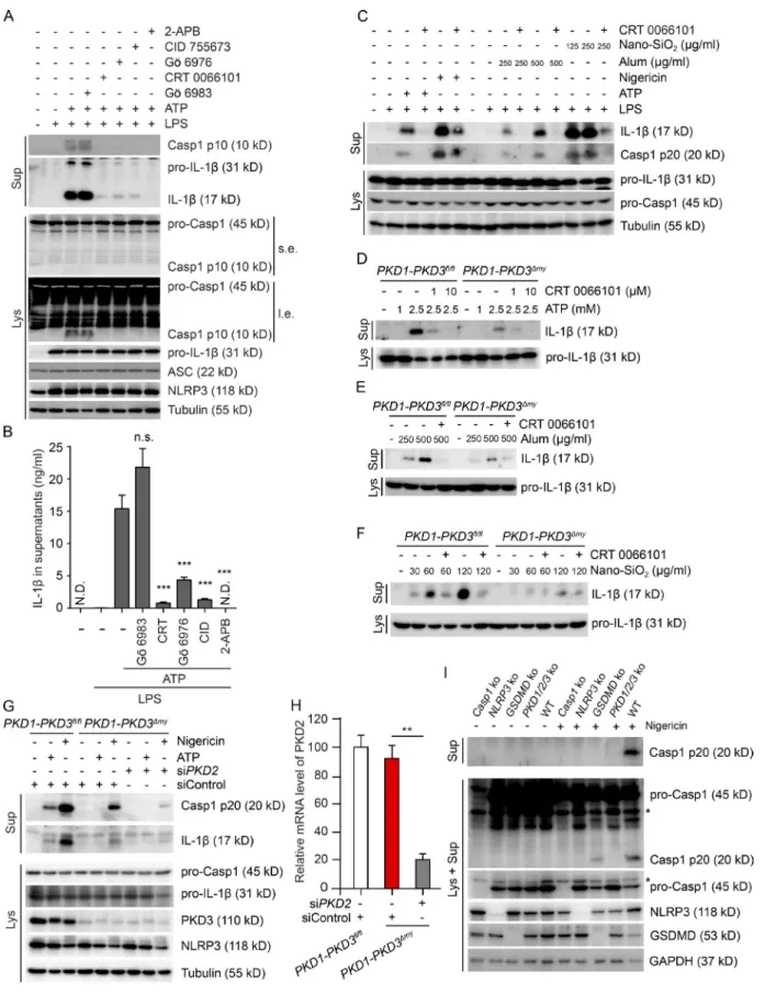
inflammasome activation, which was not affected by PKD inhibition (Fig. S1 A). We next tested whether PKD activity is required for activation of the NLRP3 inflammasome. Strik-ingly, four different PKD inhibitors (CRT 0066101, Gö 6976, CID 755673, and kb NB 142–70) almost completely abol-ished inflammasome activity in stimulated peritoneal mac-rophages and/or BMDMs without affecting expression of
NLRP3, pro–caspase-1, pro–IL-1β, and ASC (Fig. 4, A and B;
and Fig. S1, B and C). As previously reported (Lee et al., 2012),
the InsP3R antagonist 2-APB almost completely blocked
in-flammasome activation (Fig. 4, A and B; and Fig. S1 C). The PKC inhibitor Gö 6983, which does not inhibit PKD activity (Uesugi et al., 2012), had no such effect (Fig. 4, A and B; and Fig. S1 C). Inflammasome activation was also dramatically re-duced upon PKD inhibition in human PBMCs (Fig. 4 C).
The PKD family consists of three members: PKD1, PKD2, and PKD3 (Rykx et al., 2003). To further corroborate the requirement of PKD activity in NLRP3 inflammasome activation, we generated myeloid-specific PKD1-PKD3
dou-ble-KO (PKD1-PKD3Δmy) mice (Fig. S1 D). NLRP3
inflam-masome activation in BMDMs isolated from PKD1-PKD3Δmy
mice was markedly reduced as compared with cells isolated
from floxed control mice (PKD1-PKD3fl/fl; Fig. 4, D–F).
Inhi-bition was not as prominent as in PKD inhibitor–treated cells, most likely because of the remaining PKD2 activity. Indeed, additional pharmacologic inhibition of PKD (Fig. 4, D–F) or siRNA-mediated knockdown of PKD2 (Fig. 4 G) abol-ished inflammasome activity in KO cells. The development
of the myeloid lineage was not affected in PKD1-PKD3Δmy
mice (Fig. S1, E and F). Priming and release of other NF-κB–
dependent cytokines were not impaired in BMDMs from
PKD1-PKD3Δmy mice (Fig. S1, D, G, and H). To further
cor-roborate that remaining PKD2 activity in KO cells was indeed responsible for residual inflammasome activity, we generated Raw-ASC macrophages lacking PKD1, PKD2, and PKD3 (Fig. S1 I). Strikingly, caspase-1 cleavage and secretion in re-sponse to nigericin was completely abolished in these cells as compared with WT cells (Fig. 4 I). Inhibition was comparable to the one seen in NLRP3-KO cells. As expected, caspase-1 was undetectable in Caspase-1–KO cells. In line with previ-ous studies (He et al., 2015; Kayagaki et al., 2015; Shi et al., 2015), caspase-1 cleavage was partially maintained, whereas its secretion was abolished in GSD MD-KO cells (Fig. 4 I).
Flagellin-induced NLRC4-, dsDNA-induced AIM2-, and cytotoxin TcdB-induced PYR IN inflammasome activa-tion was not affected in BMDMs upon PKD inhibiactiva-tion. In-hibition of PKD upon NLRC4 inflammasome activation did not change autocleavage of caspase-1 (Fig. S2 A). The same was true for AIM2 and PYR IN inflammasome activation, of which formation of ASC specks was also unchanged (Fig. S2 B).
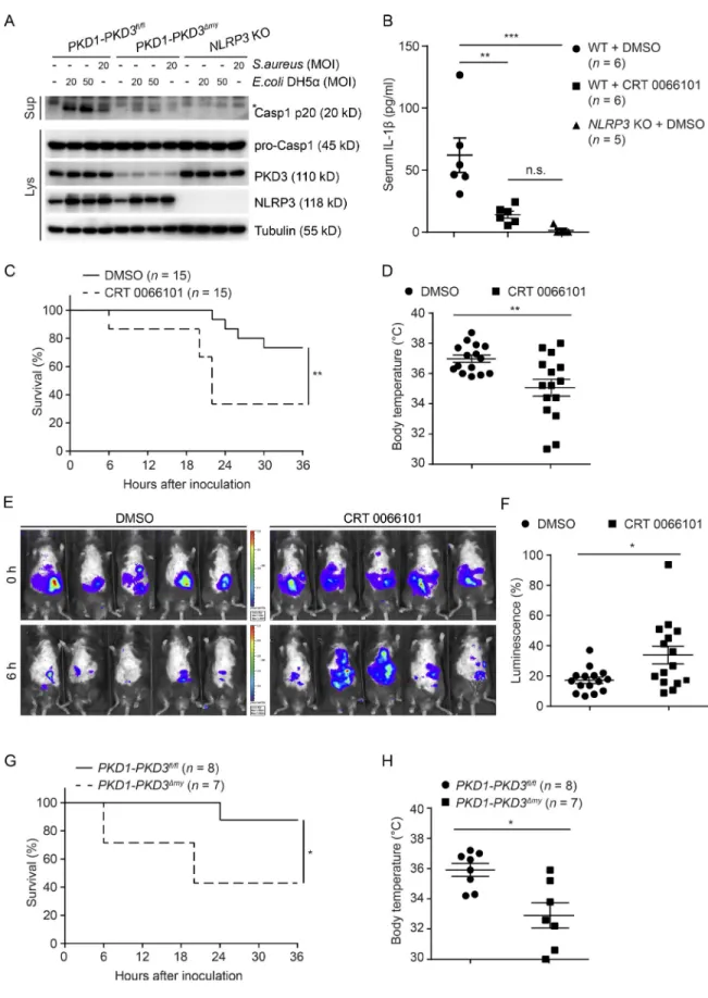
Several studies have shown that gram-negative bacteria (Escherichia coli) and gram-positive bacteria (Staphylococcus aureus) activated the NRLP3 inflammasome (Mariathasan et al., 2006; Sander et al., 2011). In line with these studies, activation of NLRP3 inflammasome-dependent cleavage of
caspase-1 in response to both E. coli DH5α and S. aureus was
abolished in NLRP3-KO cells (Fig. 5 A). In comparison with WT cells, the cleavage of caspase-1 was markedly decreased in
PKD1-PKD3Δmy cells (Fig. 5 A).
To corroborate the role of PKD in NLRP3 inflam-masome activation in vivo, we first treated mice with LPS by i.p. injection in the absence or presence of PKD inhi-Figure 1. Activation of nLrP3 inflammasome induces dAG
enrich-ment in the Golgi. (A) Confocal fluorescence imaging of LPS-primed WT BMDMs ectopically expressing a GFP-tagged DAG probe pretreated with DMSO or 10 µM U71322 for 1 h, followed by 7.5 µM nigericin stimula-tion for 20 min in the presence of DMSO or 10 µM U71322. Cells were immunostained using an antibody against giantin (a marker for Golgi). Nuclei were stained with DAPI. Regions of interest (ROI) are indicated by boxes. Bars: 10 µm; [region of interest (ROI)] 2 µm. (B) Confocal fluores-cence imaging of LPS-primed NLRP3-KO BMDMs ectopically expressing a GFP-tagged DAG probe (GFP) pretreated with DMSO or 10 µM U71322 for 1 h, followed by 7.5 µM nigericin stimulation for 20 min in presence of DMSO or 10 µM U71322. Cells were immunostained with an antibody against giantin. Nuclei were stained with DAPI. Bar, 10 µm. Data shown are representative of three independent experiments.
on October 18, 2017
jem.rupress.org
Figure 2. Activation of nLrP3 inflammasome induces mitochondrial clustering around the Golgi. (A) Confocal fluorescence imaging of LPS-primed WT and NLRP3-KO BMDMs treated or not with 5 mM ATP or 15 µM nigericin for 20 min or with 500 µg/ml alum or 125 µg/ml nano-SiO2 for 6 h. Cells
were coimmunostained using antibodies against Tom20 (a marker for mitochondria) and giantin. Nuclei were stained with DAPI. Bars, 10 µm. (B) Confocal fluorescence imaging of PMA-differentiated THP-1 cells treated or not with 15 µM nigericin for 20 min, 500 µg/ml alum, or 125 µg/ml nano-SiO2 for 6 h.
on October 18, 2017
jem.rupress.org
bition. LPS injection increased the serum IL-1β level in
DMSO- treated control mice (Fig. 5 B), whereas serum IL-1β
was almost not detectable in NLRP3-KO mice (Fig. 5 B). In comparison with DMSO-treated control mice, CRT 0066101–treated mice showed dramatically reduced serum
IL-1β levels (Fig. 5 B). We next addressed PKD-dependent
phenotypic outcomes in mice during a bacterial challenge. Inhibition of PKD in mice subjected to i.p. injections of S. aureus led to a significantly enhanced mortality as compared with infected DMSO-treated control mice (Fig. 5 C). Ac-cordingly, higher mortality was accompanied with a reduced body temperature (Fig. 5 D) as well as an increased bacterial
load (Fig. 5, E and F). Similarly, PKD1-PKD3Δmy mice showed
accelerated mortality (Fig. 5 G) and lower body temperature
(Fig. 5 H) as compared with PKD1-PKD3fl/fl control mice.
Our data are thus in line with previous findings in ASC- and IL-1β–deficient mice (Miller et al., 2007), supporting that
PKD-mediated NLRP3-dependent IL-1 β release is an
im-portant response for efficient clearance of S. aureus.
Taking all together, these data thus corrobo-rate an important role of PKD signaling in mediating NLRP3 inflammasome activity.
PKd acts downstream of mitochondrial damage and is required for the recruitment of Asc to nLrP3
Numerous studies suggest that mitochondrial damage is important for the activation of the NLRP3 inflammasome (Nakahira et al., 2011; Zhou et al., 2011; Iyer et al., 2013; Subramanian et al., 2013; Gurung et al., 2015). We thus tested whether PKD activity controls mitochondrial func-tion in response to NLRP3 inflammasome activafunc-tion. Ox-ygen consumption rates (OCRs) were dramatically lower in nigericin-stimulated cells (Fig. S3 B) than in control-treated cells. In line with a previous study (Shimada et al., 2012), nigericin-treated cells did not respond to oligomycin, FCCP, and rotenone/antimycin, whereas control-treated cells re-sponded as expected. Importantly, PKD inhibition did not prevent nigericin-induced mitochondrial damage (Fig. S3 B), indicating that improved mitochondrial function did not account for NLRP3 inflammasome inactivity upon PKD in-hibition. This was confirmed in NLRP3-null cells (Fig. S3, A and B), indicating that mitochondrial injury occurs upstream of PKD-mediated NLRP3 inflammasome activation. More-over, PKD inhibition did not affect mitochondrial clustering close to the Golgi in response to nigericin stimulation (Fig. S3 C). Interestingly, 2-APB prevented mitochondrial
cluster-ing close to Golgi (Fig. S3 C), suggestcluster-ing that InsP3-mediated
signaling is required for mitochondrial clustering.
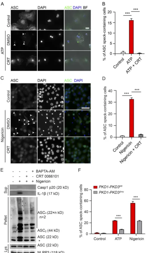
We next asked whether PKD activity was important for recruitment of ASC to NLRP3. Strikingly, almost no ASC
specks were found in stimulated BMDMs and THP-1 cells
upon PKD inhibition, whereas they were present in ∼17%
and ∼31% of respective control cells (Fig. 6, A–D).
Immuno-blotting with cross-linked pelleted protein extracts corrobo-rated reduced oligomerization of ASC upon PKD inhibition
(Fig. 6 E). Stimulation of BMDMs from PKD1-PKD3fl/fl
mice with ATP and nigericin resulted in ASC speck
forma-tion in ∼23% and ∼55% of cells, respectively. ASC speck
for-mation was reduced in cells derived from PKD1-PKD3Δmy
mice to ∼8% and ∼25%, respectively (Fig. 6 F). Overall,
these data indicate that PKD activity downstream of mi-tochondrial clustering and injury is required for the re-cruitment of ASC to NLRP3.
PKd inactivation results in retention of nLrP3 at MAMs adjacent to Golgi
NLRP3 was shown to directly bind to MAMs (Zhou et al., 2011; Yang et al., 2015). We thus further tested whether PKD deficiency affected subcellular localization of NLRP3. NLRP3 was found in small foci that predominantly
colo-calized with ASC specks in ∼35% of nigericin-stimulated
THP-1 cells. In contrast, NLRP3 staining was more diffuse
forming larger disc-like structures in ∼70% of THP-1 cells
upon PKD inhibition (Fig. 7, A and B). NLRP3-KO cells did not show any visible signal of NLRP3 immunostaining (Fig. 7 A). Conventional confocal microscopy revealed par-tial colocalization of NLRP3 with the Golgi marker giantin in PKD-inhibited cells (Fig. 7 C). 3D-SIM superresolution microscopy revealed that there was very little colocalization with giantin, indicating that NLRP3 was very close to but did not directly bind to Golgi membranes (Fig. 7 D). We thus asked whether NLRP3 was retained at MAMs close to Golgi membranes. Indeed, biochemical fractionation demonstrated increased enrichment of NLRP3 at MAMs upon PKD inhi-bition (Fig. 7 E). Consistent with findings in THP-1 cells, in stimulated PKD-inhibited BMDMs, NLRP3 was found in bright foci that were slightly bigger as compared with those in control-treated cells. Most NLRP3 localized to the Golgi region upon PKD inhibition, whereas it predominantly local-ized to the cytoplasm in control-treated cells (Fig. S2, C and D). Consistent results were observed in stimulated BMDMs
isolated from control and PKD1-PKD3Δmy mice (Fig. S2, E
and F). Altogether, these data suggest that PKD activity is required to release NLRP3 from MAMs, allowing for ASC recruitment and inflammasome activation.
PKd phosphorylates nLrP3 at ser293 to release it from MAMs
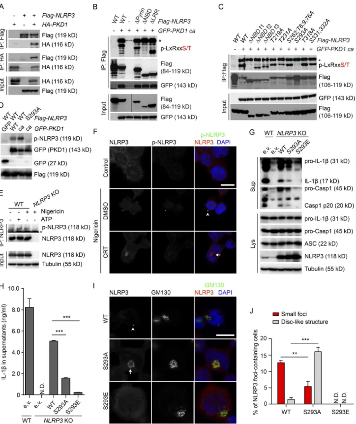
We next tested whether PKD at the Golgi interacts and phos-phorylates NLRP3 to release it from MAMs. Ectopically
Cells were coimmunostained with anti-Tom20 and anti-giantin antibodies. Nuclei were stained with DAPI. Bar, 10 µm. (C) Confocal fluorescence imaging of LPS-primed BMDMs treated or not with 1 µg/ml poly(dA:dT) for 4 h, 0.5 µg/ml flagellin for 4 h, or 0.1 nM Tcd B for 2 h. Cells were coimmunostained using antibodies against Tom20 and giantin. Nuclei were stained with DAPI. Bar, 10 µm. Data shown are representative of three independent experiments.
on October 18, 2017
jem.rupress.org
expressed PKD1 coimmunoprecipitated with ectopically expressed NLRP3 and vice versa (Fig. 8 A). Immunoblot-ting using a PKD substrate phospho-motif antibody revealed phosphorylation of ectopically expressed mouse NLRP3 in cells coexpressing constitutively active PKD1 (PKD1ca; Fig. 8 B). Expression of NLRP3 lacking the pyrin domain, the nucleotide-binding domain (NBD) or the leucine-rich repeat domain, respectively, revealed that phosphorylation occurred in the NBD (Figs. 8 B and S4 A). Expression of truncated and mutated NLRP3 identified phosphorylation of NLRP3 in the NBD at serine 293 (Ser293; Figs. 8 C and S4 A). Phosphorylation at Ser293 of ectopically expressed NLRP3 was confirmed by mass spectrometry (Fig. S4, B and C). This site is highly conserved among different species corresponding to Ser295 in human NLRP3 (Fig. S4 D). We next generated a phospho-Ser293–specific rabbit polyclonal antibody. Using this antibody, phosphorylation of ectopi-cally expressed NLRP3 was detected in cells expressing WT PKD1. Expression of PKD1ca markedly enhanced phosphor-ylation of WT, but not S293A mutant, NLRP3 (Fig. 8 D). Other kinases (Gross et al., 2009; Chuang et al., 2011; Lu et al., 2012; Martin et al., 2014; Ito et al., 2015) that have been implicated in NLRP3 inflammasome regulation did not in-duce phosphorylation of NLRP3 at Ser293 (Fig. S4 E). We next aimed at corroborating phosphorylation of endogenous NLRP3. Activation of NLRP3 inflammasome by ATP and
nigericin induced phosphorylation of NLRP3 at Ser293 (Fig. 8 E). Moreover, NLRP3 foci in nigericin-stimulated THP-1 cells and ATP-stimulated BMDMs colocalized with signals specific for phospho-NLRP3 (Ser293), whereas there were no phospho-NLRP3 foci detectable in PKD-inhibited cells (Figs. 8 F and S5 A). Loss of phosphorylation of endog-enous NLRP3 was confirmed in Raw-ASC macrophages lacking PKD1, PKD2, and PKD3 (Fig. S5 B). Hence, these data indicate that PKD phosphorylates NLRP3 at Ser293. We next asked whether PKD-mediated phosphorylation controls the activation of the NLRP3 inflammasome. To this end, we reconstituted NLRP3-deficient THP-1 cells with WT-, nonphospho (S293A)–, and phospho-mimicking
(S293E) NLRP3. Reconstitution with WT NLRP3
par-tially restored NLRP3 inflammasome activity, as indicated by
cleavage and secretion of caspase-1 and IL-1β. In comparison,
the capacity of S293A NLRP3 to restore inflammasome ac-tivity was markedly lower (Fig. 8, G and H). Reconstituted
WT NLRP3 formed foci in ∼13% of cells, whereas S293A
NLRP3 formed foci only in ∼5% of cells. Importantly,
S293A NLRP3 was retained in the Golgi region in ∼17%
cells (Fig. 8, I and J), in line with retention of endogenous WT NLRP3 at MAMs close to the Golgi in cells subjected to PKD inhibition. Together with our finding that phosphor-ylated NLRP3 can be found in the mature inflammasome, our data strongly suggest that PKD-mediated
phosphoryla-Figure 3. disruption of Golgi integrity blocks the nLrP3 inflammasome activa-tion. (A) Immunoblotting of culture superna-tants (Sup) and lysates (Lys) from LPS-primed BMDMs pretreated with ethanol (control) or 1 µg/ml, 5 µg/ml, or 25 µg/ml BFA for 1 h, fol-lowed by 7.5 µM nigericin treatment for 40 min in the presence of ethanol or BFA at the indicated concentration. Antibodies against caspase-1 (recognizing both cleaved [p20] and uncleaved protein) and IL-1β (recognizing both cleaved and uncleaved protein) were used. Tu-bulin was used as a loading control. (B) ELI SA measurements of IL-1β in culture supernatants from LPS-primed BMDMs treated as in A. The values are expressed as means ± SEM. ***, P < 0.001 (t test); n.s., not significant. (C) Confocal fluorescence imaging of LPS-primed BMDMs pretreated with ethanol or 5 µg/ml BFA for 1 h, followed by 7.5 µM nigericin treatment for 40 min in the presence of ethanol or 5 µg/ml BFA. Cells were coimmunostained with antibodies against ASC and giantin. Nuclei were stained with DAPI. Bars: 10 µm; (ROI) 2 µm. Arrow-heads indicate the ASC foci. (D) Quantification of cells containing ASC foci shown in C. ***, P < 0.001 (t test). Data shown are representative of three independent experiments.
on October 18, 2017
jem.rupress.org
Figure 4. deficiency of PKd specifically blocks the activation of the nLrP3 inflammasome. (A) Immunoblotting of culture supernatants (Sup) and lysates (Lys) from LPS-primed BMDMs pretreated with DMSO, 5 µM Gö 6983, 10 µM CRT 0066101, 5 µM Gö 6976, 30 µM CID 755673, or 50 µM 2-APB for 1 h, followed by ATP treatment for 40 min in the presence of DMSO or indicated inhibitors. l.e., long exposure; s.e., short exposure. (B) ELI SA measurements
on October 18, 2017
jem.rupress.org
tion of NLRP3 releases NLRP3 from MAMs, allowing for inflammasome maturation.
It has been demonstrated that binding of NLRP3 to MAMs is crucial for its activation (Zhou et al., 2011; Subra-manian et al., 2013). Constitutive phosphorylation of NLRP3 is thus expected to prevent its binding to MAMs and thus in-flammasome activation. Indeed, expression of S293E NLRP3 did not restore inflammasome activity in NLRP3-deficient THP-1 cells (Fig. 8, G and H). Moreover, S293E NLRP3 did not form any foci localizing diffusely throughout the cytoplasm, which is in line with its inability to bind to MAMs (Fig. 8, I and J).
Our data are thus consistent with a model in which local and timely phosphorylation of membrane-bound NLRP3 by PKD results in its release and cytoplasmic assembly of the fully active inflammasome.
PKd activity at Golgi is sufficient to activate the nLrP3 inflammasome
As shown in Fig. 1, DAG production was enhanced at the Golgi to recruit PKD to this organelle upon NLRP3 in-flammasome activation. Hence, PKD activity at the Golgi is expected to be sufficient to phosphorylate NLRP3 and to activate the NLRP3 inflammasome. Indeed, expression of WT, but not kinase-dead, PKD1 induced NLRP3 in-flammasome-dependent activation of caspase-1 and
secre-tion of IL-1β without stimulation (Fig. 9 A). Consistently,
expression of PKD1 dramatically enhanced the
activa-tion of caspase-1 and secreactiva-tion of IL-1β upon nigericin
treatment. Importantly, this effect was abolished by CRT 0066101 treatment, corroborating the importance of PKD activity for NLRP3 inflammasome activation in inhibi-tor experiments (Fig. 9 B). Strikingly, expression of GRIP tagged-PKD1, localization of which was restricted to the Golgi, but not of PKD lacking the DAG-binding domain, was sufficient to phosphorylate NLRP3 and to activate the NLRP3 inflammasome (Fig. 9, C and D). These data thus suggest that PKD activity at the Golgi is sufficient to acti-vate the NLRP3 inflammasome.
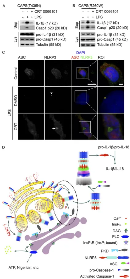
PKd inhibition blocks the activity of the nLrP3 inflammasome in cells from patients with autoactivatory mutations in nLrP3
Patients with cryopyrin-associated periodic syndrome (CAPS) suffer from autoinflammatory events caused by mutations in NLRP3 resulting in its auto-oligomerization and uncon-trolled NLRP3 inflammasome activation (Aksentijevich et al., 2007; Brydges et al., 2009; Nakamura et al., 2012). We next determined whether autoactivation of mutated NLRP3 inflammasome depends on PKD activity. PKD inhibition of LPS-stimulated PBMCs isolated from patients carrying the mutations T436N or R260W in the NLRP3 gene resulted
in a strong reduction of caspase-1 cleavage and IL-1β
secre-tion as compared with control-treated cells (Fig. 10, A and B). T436N NLRP3 was stuck in disc-like structures at the Golgi upon PKD inhibition (Fig. 10 C). These mutations result in spontaneous self-oligomerization of the NLRP3 protein (Baroja-Mazo et al., 2014). These data thus cor-roborate that PKD inhibition is sufficient to block inflam-masome activity in cells of these patients. They also indicate that PKD acts downstream of NLRP3 self-oligomerization. Indeed, a native page as well as a gel filtration assay revealed that PKD inhibition did not affect self-oligomerization of NLRP3 (Fig. S5, C and D). Given the fact that NLRP3 was retained at MAMs upon PKD inhibition, it is expected that NLRP3 at MAMs is self-oligomerized before its phosphor-ylation. Accordingly, oligomerization of reconstituted S293E NLRP3 was abolished, whereas oligomerization of reconsti-tuted S293A and WT NLRP3 were unaffected in stimulated THP-1 cells (Fig. S5 E).
Altogether, we propose a model in which MAMs local-ize close to the Golgi in response to NLRP3 inflammasome activation. This allows for PKD-induced phosphorylation of self-oligomerized NLRP3, its release from MAMs, and as-sembly of the cytosolic mature inflammasome (Fig. 10 D).
dIscussIon
In this study, we unveiled the spatial and temporal organi-zation of NRLP3 inflammasome activation. PKD-mediated
of IL-1β in culture supernatants from LPS-primed BMDMs treated as in A. The values are expressed as means ± SEM. p-values were calculated between ATP alone–treated group and ATP plus inhibitor–treated group. ***, P < 0.001 (t test); N.D., not detected; n.s., not significant. (C) Immunoblotting of culture su-pernatants (Sup) and lysates (Lys) from LPS-primed PBMCs pretreated with DMSO or 10 µM CRT 0066101 for 1 h, followed by treatment with 5 mM ATP for 40 min, 15 µM nigericin for 40 min, 250 or 500 µg/ml alum for 6 h, or 125 or 250 µg/ml nano-SiO2 for 6 h in the presence of DMSO or 10 µM CRT 0066101.
(D–F) Immunoblotting of culture supernatants (Sup) and lysates (Lys) from BMDMs isolated from LysM-Cre–negative floxed PKD1-PKD3 (PKD1-PKD3fl/fl)
control mice and LysM-Cre–positive myeloid-specific PKD1-PKD3 double-KO (PKD1-PKD3Δmy) mice. Cells were primed with LPS for 4 h. After pretreatment
with DMSO or CRT 0066101 for 1 h, cells were stimulated with ATP for 40 min (D), alum (E), or nano-SiO2 (F) as indicated for 6 h in the presence of DMSO
or 10 µM CRT 0066101. (G) Immunoblotting of culture supernatants (Sup) and lysates (Lys) from BMDMs isolated from PKD1-PKD3fl/fl control mice and
PKD1-PKD3Δmy mice. Cells were transfected with control siRNA (siControl) or siRNA against PKD2 (siPKD2) as indicated for 36 h. After LPS priming for 4 h,
cells were treated or not with 5 mM ATP or 7.5 µM nigericin for 40 min. (H) Quantitative PCR analysis of BMDMs isolated from PKD1-PKD3fl/fl control mice
and PKD1-PKD3Δmy mice were transfected with control siRNA (siControl) or siRNA against PKD2 (siPKD2) as indicated for 36 h. The level of PKD2 mRNA
relative to Hprt mRNA was analyzed by quantitative PCR. The values are expressed as means ± SEM. **, P < 0.01 (t test). (I) Immunoblotting of culture supernatants (Sup) and lysates together with culture supernatants (Lys + Sup) from Raw-ASC WT, Caspase-1–KO (Casp1 ko), NLRP3-KO (NLRP3 ko), GSD MD-KO (GSD MD ko), and PKD1/PKD2/PKD3 triple-KO (PKD1/2/3 ko) cells. LPS-primed cells were treated with or without 10 µM nigericin for 1 h. Asterisk (*) represents unspecific bands. Data shown are representative of at least three independent experiments.
on October 18, 2017
jem.rupress.org
Figure 5. PKd activity is required for nLrP3 inflammasome activation in vivo. (A) Immunoblotting of culture supernatants (Sup) and lysates (Lys) from BMDMs isolated from PKD1-PKD3fl/fl control mice, PKD1-PKD3Δmy mice, and NLRP3-KO mice. LPS-primed cells were infected with E. coli DH5α or S.
au-reus at indicated multiplicity of infection (MOI) for 3 h. (B) ELI SA analysis of serum IL-1β from LPS-injected mice. Mice were intraperitoneally pretreated with
on October 18, 2017
jem.rupress.org
signaling emanating from the Golgi close to MAMs leads to phosphorylation of NLRP3 releasing it from MAMs, a neces-sary step to allow for assembly of the mature NLRP3 inflam-masome in the cytoplasm (Fig. 10 D).
We identified the Golgi as an important element controlling the activation of the NLRP3 inflammasome in macrophages. The importance of Golgi function is reflected by our observation that mitochondria cluster close to Golgi membranes upon inflammasome activation (Fig. 2), and that disruption of Golgi integrity blocks the activation of NLRP3 inflammasome (Fig. 3). In partic-ular, disruption of Golgi by BFA blocked the activation
of NLRP3 inflammasome and secretion of IL-1β.
Rubar-telli et al. (1990) showed that IL-1β was secreted via a
BFA-insensitive pathway in LPS-activated monocytes.
In this study, the release of IL-1β was tested in response
to LPS stimulation only. Importantly, other studies have clearly demonstrated that ultrapure LPS could not trigger
IL-1β release (Martinon et al., 2004, 2006). Thus, a
plau-sible explanation is that the release of IL-1β triggered by
LPS in this study was induced through activation of other inflammasomes by contaminants. Given the fact that PKD activity is not important in the activation of other inflam-masomes, their readouts were most likely independent of Golgi-derived signaling. A more recent study by Menu et al. (2012) showed that ER stress was sufficient to activate the NLRP3 inflammasome in LPS-primed macrophages without inflammasome stimulators. Different compounds, including BFA, were used to promote ER stress through different mechanisms. ER stress induced by tunicamycin and thapsigargin, but not BFA, was sufficient to trigger NLRP3 inflammasome activity in LPS-primed BMDMs. This is in line with our study demonstrating that BFA, despite its effects on ER stress, blocks the activation of NLRP3 inflammasome by disrupting Golgi integrity.
The importance of Golgi function is further substan-tiated in our study at the molecular level. In fact, the sec-ond messenger, DAG, increased in Golgi membranes (Fig. 1), triggering local activity of the effector kinase PKD that is necessary to activate the NLRP3 inflammasome (Figs. 4 and 5). Importantly, forced targeting of PKD activity to the Golgi is sufficient to activate the NLRP3 inflammasome (Fig. 9). Finally, PKD inhibition resulted in retention of NLRP3 at
MAMs close to Golgi membranes (Fig. 7), corroborating propagation of signals from the Golgi to MAMs.
Of note, PKD is a stress kinase that senses effectors of injured mitochondria (Storz et al., 2005). Hence, in addition to DAG enrichment in the Golgi, clustering of injured mi-tochondria close to the Golgi may boost PKD activation, leading to phosphorylation of NLRP3 and its release from MAMs. Conversely, local exposure of NLRP3 with mito-chondrial effectors may also contribute to inflammasome activation. It is likely that other molecular events occur at the interface of Golgi and MAMs that contribute to NLRP3 inflammasome activation. In particular, the identified or-ganelle interplay might also be crucial to couple activation
of NLRP3 inflammasome to IL-1β secretion. In fact, signal
propagation from the Golgi to the ER mediates formation of secretory autophagosomes that have been implicated in
nonconventional secretion of IL-1β (Ponpuak et al., 2015).
Thus, our discovery will open a whole new avenue of in-teresting future research.
The importance of binding of NLRP3 to MAMs, even though it has been evidenced in the literature (Zhou et al., 2011; Yang et al., 2015), has been challenged, because the ma-ture NLRP3 inflammasome resides in the cytosol. In fact, our study integrates both observations into one coherent model highlighting the importance of the highly dynamic spatial arrangement of intracellular organelles and localization of NLRP3. Membrane binding of NLRP3 is dependent on the N-terminal sequence of its pyrin domain (Subramanian et al., 2013). The pyrin domain is essential for NLRP3 to bind ASC (Dowds et al., 2003; Agostini et al., 2004). Thus, exposure of the pyrin domain might be essential for further maturation of the NLRP3 inflammasome. Importantly, the study by Zhou et al. (2011) demonstrated that both NLRP3 and ASC were found in MAM fractions, potentially indicating that interac-tion already occurred in the MAM compartment. However, this study also showed that most ASC was cytoplasmic under both nonstimulating and stimulating conditions. We thus pro-pose that NLRP3, without or with ASC, might be released from MAMs to recruit ASC or more ASC in the cytoplasm, forming the mature inflammasome. The detailed mechanisms of phosphorylation-mediated release of NLRP3 from MAMs will be an important subject of future research. Even though speculative, phosphorylation in the NBD may change
con-DMSO or 10 mg/kg CRT 0066101 as indicated for 1 h, followed by intraperitoneal injection of 20 mg/kg LPS. Blood were collected at 2 h after LPS injection. The values are expressed as means ± SEM. **, P < 0.01; ***, P < 0.001; n.s., not significant (Mann–Whitney test). (C) The survival curve of S. aureus–infected mice. WT mice were intraperitoneally pretreated with DMSO (n = 15) or 10 mg/kg CRT 0066101 (n = 15) for 1 h, followed by intraperitoneal infection of S. aureus (7 × 10⁸ per mouse). **, P < 0.01 (Gehan–Breslow–Wilcoxon test). (D–F) Analysis of S. aureus–infected mice shown in C. Body temperature (D)
and bacterial load (E and F) of each mouse was measured at 6 h after infection. The bacterial loads were shown in percentage of luminescence at 0 h. The values are expressed as means ± SEM. *, P < 0.05; **, P < 0.01 (Mann–Whitney test). (G) The survival curve of S. aureus–infected PKD1-PKD3fl/fl mice (n = 8)
and PKD1-PKD3Δmy (n = 7) mice. Mice were treated by i.p. infection of S. aureus (7 × 10⁸ per mouse). *, P < 0.05 (Gehan–Breslow–Wilcoxon test). (H) Body
temperature of mice shown in G at 6 h after infection. The values are expressed as means ± SEM. *, P < 0.05 (Mann–Whitney test). Data shown in A are the representative of three independent experiments, whereas images in D are representative of 15 mice in each group.
on October 18, 2017
jem.rupress.org
formation of NLRP3 in a way it prevents membrane binding. Alternatively, chaperone-mediated release of phosphorylated NLRP3 might be important.
Recently, two studies showed that PKA negatively reg-ulates NLRP3 inflammasome activation by phosphorylation of NLRP3 at the very same serine residue (Guo et al., 2016; Mortimer et al., 2016). In fact, inhibitory effects of this
phos-phorylation event is fully in line with our observation that S293E NLRP3 was unable to restore inflammasome activity in NLRP3-deficient THP-1 cells. However, our data also pro-vide strong epro-vidence for PKD-mediated phosphorylation of NLRP3 to promote inflammasome activity. Taking all these findings together, this strongly suggests that the consequences of NLRP3 phosphorylation very likely depend on where Figure 6. deficiency of PKd blocks recruitment of Asc to the nLrP3 inflammasome. (A) Fluo-rescence imaging of LPS-primed BMDMs pretreated with DMSO or 10 µM CRT 0066101 for 1 h, followed by stimulation with 5 mM ATP in presence of DMSO or 10 µM CRT for 20 min. Cells were immunostained with an anti-ASC antibody. Nuclei were stained with DAPI. Bar, 10 µm. Merged pictures with bright-field (BF) microscopy signals are shown. Arrowheads indi-cate the ASC specks. (B) Quantification of ASC speck– containing BMDMs of experiments represented in A. The values are expressed as means ± SEM. ***, P < 0.001 (t test). (C) Fluorescence imaging of differen-tiated THP-1 cells pretreated with DMSO or 10 µM CRT 0066101 for 1 h, followed by stimulation with 15 µM nigericin in presence of DMSO or 10 µM CRT for 30 min. Cells were immunostained with an an-ti-ASC antibody. Nuclei were stained with DAPI. Bar, 50 µm. Arrowheads indicate ASC specks. (D) Quanti-fication of ASC speck–containing THP-1 cells of ex-periments represented in C. The values are expressed as means ± SEM. ***, P < 0.001 (t test). (E) Immuno-blotting of culture supernatants (Sup), lysates (Lys), and cross-linked pellets (Pellet) from differentiated THP-1 cells pretreated with DMSO, 10 µM CRT, or 25 µM BAP TA-AM for 1 h, followed by treatment with 15 µM nigericin in presence of DMSO, 10 µM CRT, or 25 µM BAP TA-AM for 40 min. (F) Quantification of ASC speck–containing LPS-primed BMDMs isolated from PKD1-PKD3fl/fl and PKD1-PKD3Δmy mice. Cells
were treated with 2.5 mM ATP or 7.5 µM nigericin for 20 min. The values are expressed as means ± SEM. ***, P < 0.001 (t test). Data shown are representative of at least three independent experiments.
on October 18, 2017
jem.rupress.org
Figure 7. PKd inhibition results in nLrP3 retention at MAMs close to Golgi. (A) Confocal fluorescence imaging of PMA-differentiated THP-1 cells pretreated with DMSO or 10 µM CRT for 1 h, followed by stimulation with 15 µM nigericin in the presence of DMSO or CRT for 30 min. Cells were coim-munostained with anti-NLRP3 and anti-ASC antibodies. PMA-differentiated NLRP3-KO THP-1 cells treated with nigericin and CRT was used as a negative control for anti-NLRP3 antibody immunostaining. Nuclei were stained with DAPI. Regions of interest (ROIs) are indicated by boxes. Bars: 10 µm; (ROI) 2 µm. Arrowheads indicate small NLRP3 foci; arrows indicate NLRP3 disc-like structures. (B) Quantification of cells containing small foci or disc-like structures in experiments represented in A. The values are expressed as means ± SEM. ***, P < 0.001 (t test); N.D., not detected. (C) Confocal fluorescence imaging of THP-1 cells pretreated with DMSO or 10 µM CRT for 1 h, followed by stimulation with 15 µM nigericin in the presence of DMSO or CRT for 30 min. Cells were coimmunostained with antibodies against NLRP3 and GM130. Nuclei were stained with DAPI. ROIs are indicated by boxes. Bars: 10 µm; (ROI) 2 µm. The arrowhead indicates a NLRP3 small focus, whereas the arrow indicates NLRP3 distributed in a disc-like structure. (D) 3D-SIM superresolution microscopy of differentiated THP-1 cells pretreated with 10 µM CRT for 1 h, followed by stimulation with 15 µM nigericin in the presence of 10 µM CRT for 30 min. Cells were coimmunostained with antibodies against NLRP3 and GM130. Nuclei were stained with DAPI. ROIs are indicated by boxes. Bars: 10 µm; (ROI) 2 µm. (E) Immunoblotting of lysates from indicated fractionations (Mc, crude mitochondria; Mp, pure mitochondria) isolated from THP-1 cells pretreated with DMSO or 10 µM CRT for 1 h, followed by stimulation with 15 µM nigericin (Ni) in the presence of DMSO or CRT (Ni + CRT) for 30 min. Data shown are representative of at least three independent experiments.
on October 18, 2017
jem.rupress.org
Figure 8. PKd phosphorylates nLrP3 at ser293 (in mouse, ser295 in human) to release it from MAMs. (A) Coimmunoprecipitation of exoge-nous FLAG-tagged NLRP3 with HA-tagged PKD1 and vice versa in HEK293t cells. (B) Coimmunoprecipitation of exogeexoge-nous FLAG-tagged WT (WT) NLRP3, NLRP3 lacking the pyrin domain (ΔPyrin), NLRP3 lacking the nucleotide-binding domain (ΔNBD), or NLRP3 lacking the leucine-rich repeats (ΔLRR) with GFP-tagged constitutively active mutant (ca) PKD1 in HEK293t cells. Asterisk (*) represents a band corresponding to autophosphorylation of GFP-tagged PKD1. (C) Coimmunoprecipitation of exogenous FLAG-tagged WT NLRP3, ΔNBD f1, ΔNBD f2, ΔNBD f3, S219A, T231A, S263;T6;9;76A (S263A; T266A; T269A; T276A), S293A, T318A, or S331;332A (S331A; S333A) NLRP3 with GFP-tagged constitutively active mutant (ca) PKD1 in HEK293t cells. Asterisk (*) represents
on October 18, 2017
jem.rupress.org
and when this modification occurs. In fact, phosphorylation of NLRP3 monomers by PKA may prevent its binding to MAMs, inhibiting inflammasome assembly. Local phosphory-lation of self-oligomerized NLRP3 by PKD, however, releases it from MAMs, allowing for assembly of the mature inflam-masome. It is possible, however, that other phosphorylation events mediated by other kinases or other posttranslational modifications downstream of PKD-mediated phosphory-lation control inflammasome activation. Importantly, a very recent study showed that both non and phospho-mimetic mutants of NRLP3 at serine 5 (Ser5) are inhibitory (Stutz et al., 2017), very similar to the observations we made with NLRP3 mutants at Ser293. Interestingly, it has been previously demonstrated that N-terminal amino acid residues from 2 to 9 are required for binding to membranes (Subrama-nian et al., 2013). We thus speculate that phosphorylation of Ser5, which may occur downstream of PKD-mediated phos-phorylation, prevents membrane binding of NLRP3.
Other kinases, including Syk, JNK, PKR, DAPK, BTK,
and IKKα, have been implicated in the activation of the
NRLP3 inflammasome (Jo et al., 2016). We demonstrated that none of these kinases was capable to phosphory-late NLRP3 at Ser293 (Fig. S4 E). These data are in line with the fact that none of these kinases have been shown to phosphorylate NLRP3, nor did their inactivation result in specific phenotypes we describe in this study, in partic-ular retention of NLRP3 in the Golgi–MAM compart-ment. Most recently, the kinase NEK7 has been discovered to mediate oligomerization of NLRP3, whereas its cata-lytic activity was shown to be redundant in this context (He et al., 2016; Shi et al., 2016). Our data are in line with these findings as PKD most likely phosphorylates NLRP3 downstream of its self-oligomerization and releases it from MAMs, allowing the assembly of mature inflammasome in cytosol (Fig. 10 D).
We also demonstrated that enrichment of DAG in Golgi membranes was PLC dependent (Fig. 1). Accord-ingly, PKD activity is dependent on PLC-mediated DAG production (Rozengurt et al., 2005). Our findings are fully in line with studies revealing that pharmacological inhibi-tion of PLC blocked the activainhibi-tion of the NLPR3
inflam-masome, whereas a PLC agonist was sufficient to activate the latter (Lee et al., 2012; Murakami et al., 2012). Acti-vation of PLC by G protein–coupled receptor signaling at the plasma membrane was shown to be insufficient to ac-tivate the NLRP3 inflammasome (Katsnelson et al., 2015). However, the involvement of other PLC isoforms, which are activated through G protein–independent pathways (Rhee, 2001), has not been investigated. Alternatively, PLC directly at the Golgi could be critical for PKD-mediated NLRP3 inflammasome activation (Fig. 10 D). In fact, PLC-mediated generation of DAG from phosphoinosit-ides in the Golgi complex has been reported. Both the substrates and PLC of this pathway are present in this organelle (Barker et al., 1998; Jin et al., 2001). Of note,
gain-of-function mutations in phospholipase C γ2 lead to
dominantly inherited autoinflammatory diseases (Yu et al., 2005; Everett et al., 2009; Abe et al., 2011; Ombrello et al., 2012; Koss et al., 2014). Patients carrying a gain of function
in phospholipase C γ2 showed enhanced IL-1β
produc-tion due to hyperactivaproduc-tion of the NLRP3 inflammasome, highlighting the importance of PLC-mediated signaling in the context of NLRP3 inflammasome activation (Zhou et al., 2012; Chae et al., 2015).
Several research teams have reported that Ca2+
mobi-lization plays an important role in NLRP3 inflammasome activation (Horng, 2014). Recently however, it has been
sug-gested that intracellular Ca2+ signaling is neither necessary
nor sufficient to activate the NLRP3 inflammasome (Kats-nelson et al., 2015). Our model implements PLC-dependent
local exchange of Ca2+ at MAMs that has not been
moni-tored in the latter study. Furthermore, we found that another effect of PLC activation, DAG-mediated PKD signaling from the Golgi, was critical to release NLRP3 from MAMs allow-ing for full inflammasome maturation. This may explain why
mobilization of Ca2+ by some stimuli is insufficient to activate
the NLRP3 inflammasome.
Altogether, our work thus uncovered a fundamentally new organelle interplay to be at the basis of cellular innate immune responses. Finally, we propose that interference with this signaling mechanism might be a promising avenue to treat NLRP3-related inflammatory disorders, including CAPS.
a band corresponding to autophosphorylation of GFP-tagged PKD1. (D) Immunoblotting of lysates from HEK293t cells ectopically expressing FLAG-tagged WT or S293A mutant NLRP3 together with GFP-tagged WT or ca PKD1. (E) Immunoprecipitation of endogenous NLRP3 in BMDMs isolated from WT and NL-RP3-KO mice. LPS-primed BMDMs were treated with or without 5 mM ATP or 7.5 µM nigericin as indicated for 30 min. Asterisk (*) represents an unspecific band. (F) Confocal fluorescence imaging of differentiated THP-1 cells pretreated with DMSO or 10 µM CRT 0066101 for 1 h, followed by 15 µM nigericin treatment in the presence of DMSO or 10 µM CRT 0066101 for 30 min. Cells were coimmunostained with antibodies against NLRP3 and p-NLRP3 (Ser293). Nuclei were stained with DAPI. Bar, 10 µm. The arrowhead indicates a small NLRP3 focus colocalizing with p-NLRP3 signal, whereas the arrow indicates NLRP3 distributed in a disc-like structure lacking the p-NLRP3 signal. (G) Immunoblotting of culture supernatants (Sup) and lysates (Lys) from WT and NL-RP3-KO THP-1 cells reconstituted with empty vector (e.v.), WT, S293A, or S293E mutant NLRP3. PMA-differentiated cells were treated with 15 µM nigericin for 30 min. (H) ELI SA measurements of IL-1β in culture supernatants from cells treated as in G. The values are expressed as means ± SEM. ***, P < 0.001 (t test). (I) Confocal fluorescence imaging of NLRP3-KO THP-1 cells reconstituted with WT, S293A, or S293E mutant NLRP3. Cells were coimmunostained with antibodies against NLRP3 and GM130. Nuclei were stained with DAPI. Bar, 10 µm. The arrowhead indicates a small NLRP3 focus, whereas the arrow indicates NLRP3 distributed in a disc-like structure. (J) Quantification of cells containing small foci or disc-like structures in experiments represented in I. The values are expressed as means ± SEM. **, P < 0.01; ***, P < 0.001 (t test). N.D., not detected. Data shown are representative of three independent experiments.
on October 18, 2017
jem.rupress.org
MAterIALs And MetHods Mice
NLRP3−/− mice on C57BL/6J background were obtained
from The Jackson Laboratory. Mice with targeted alleles for
PKD1 (PKD1fl/fl) were described previously (Fielitz et al.,
2008). PKD1 floxed (PKD1fl/fl) mice were provided by R.
Bassel-Duby and E.N. Olson (University of Texas Southwest-ern, Dallas, TX). Generation of PKD3 floxed mice have been
previously described (Zhang et al., 2016). We crossed PKD1fl/fl
mice and PKD3fl/fl mice on C57BL/6J background with
Lys M-Cre mice to obtain myeloid-specific PKD1-PKD3 double-KO mice. C57BL/6J WT mice were ordered from Charles River Laboratories. Mice were housed under spe-cific pathogen–free conditions. All animal experimentation was approved by the Direction des Services Vétérinaires du Bas-Rhin, France, except that bacteria infection experiments were approved by Comité Régional d’Ethique en Matière
d’Expérimentation Animale de Strasbourg (CRE MEAS).
Figure 9. PKd activity at the Golgi is required and sufficient to activate the nLrP3 inflammasome. (A) Immunoblotting of culture supernatants (Sup) and lysates (Lys) from THP-1 WT and NLRP3-KO cells infected with lentiviruses as indicated. Cells were treated with 100 nM PMA for 3 h, followed by replacement of fresh medium for 12 h. e.v., empty vector. (B) Immunoblotting of culture supernatants (Sup) and lysates (Lys) from THP-1 NLRP3-KO cells infected with lentiviruses as indicated. PMA-differentiated cells were pretreated with DMSO or 10 µM CRT 0066101 for 1 h, followed by 15 µM nigericin treatment in the presence of DMSO or 10 µM CRT 0066101 for 30 min. e.v., empty vector. (C and D) Immunoblotting of culture supernatants (Sup) and lysates (Lys) from stable THP-1 cell lines ectopically expressing GFP, GFP-tagged PKD1 WT, or a PKD1 mutant without the cysteine-rich domain (ΔCRD; C), GFP-GRIP-tagged WT, constitutively active (ca), or KD PKD1 (D). Cells were treated with 100 nM PMA for 3 h, followed by replacement of fresh medium for 12 h. Asterisk (*) represents an unspecific band. Data shown are representative of three independent experiments.
on October 18, 2017
jem.rupress.org
Figure 10. PKd activity is required for activation of the nLrP3 inflammasome in cells from cAPs patients. (A and B) Immunoblotting of culture supernatants (Sup) and lysates (Lys) from PBMCs isolated from CAPS patients carrying the NLRP3 T436N mutation (A) or the R260W mutation (B). Cells were left untreated or treated with 1 µg/ml LPS in the presence of DMSO or CRT 0066101 10 µM for 4 h. (C) Confocal fluorescence imaging of PBMCs isolated from CAPS patients carrying the NLRP3 T436N mutation were left untreated or treated with 1 µg/ml LPS in the presence of DMSO or 10 µM CRT 0066101 for 4 h. Cells were co-immunostained with antibodies against NLRP3 and ASC. Nuclei were stained with DAPI. Regions of interest (ROIs) are indicated by boxes. Bars: 10 µm; (ROI) 2 µm. (D) Model implementing iden-tified mechanisms in the activation of the NLRP3 inflammasome. The dashed black ellipse highlights the interaction of the Golgi with the MAM. Data shown in A to C are representative of ex-periments using PBMCs isolated from two CAPS patients car-rying the same mutation.
on October 18, 2017
jem.rupress.org
No exclusion of animals used for experiments was per-formed. Healthy mouse littermates were chosen randomly according to their genotypes.
reagents
Nigericin sodium salt (catalog no. N7143), BFA (B7651), ATP (A2383), and LPS from E. coli 055:B5 (L2880) were purchased from Sigma-Aldrich. CRT 0066101 (4975), kb
ND 142–70 (3962), 2-APB (1124), BAP TA-AM (2787),
U73122 (1268), CID 755673 (3327), Gö 6976 (2253), and Gö 6983 (2285) were purchased from Tocris Bioscience.
Na-no-SiO2 (tlrl-sio) and flagellin from Salmonella typhimurium
(tlrl-stfla) were obtained from InvivoGen. Alum (77161) was purchased from Thermo Fisher Scientific.
Plasmids
Mouse NLRP3 was amplified from cDNA by PCR. NLRP3 wt and NLRP3 mutants were cloned into pBOB, pLV, or pEGFP-N1 empty vectors using ligation-independent
clon-ing (LIC). Rat PKCδ C2 (DAG-binding domain), which was
amplified from GFP-C1(2) delta (plasmid #21216; Addgene),
human PKD1 wt, and PKD1 ΔCRD were cloned to pBOB
empty vector expressing C-terminal–fused EGFP tag. Golgi- localized PKD1 WT, constitutively active, and kinase-dead were cloned by a C-terminal fusion of GRIP domain of p230 as described previously (Kjer-Nielsen et al., 1999). The pX330-P2A-EGFP plasmid was generated by insert-ing P2A-EGFP sequence into EcoRI-digested pX330-U6- Chimeric_BB-CBh-hSpCas9 (plasmid #42230, Addgene) before stop codon using LIC. Guide RNA (gRNA) se-quences were inserted into BbsI-digested pX330-P2A-EGFP plasmid through ligation by T4 DNA ligase.
cell culture
All mammalian cells were cultured at 37°C, 5% CO2. Cell
lines used in this study are not listed in these databases of com-monly misidentified cell lines maintained by ICL AC. THP-1 cells (ATCC) were grown in RPMI 1640 containing 10% fetal bovine serum, 10 mM Hepes, 2.5 g/l glucose, 1 mM sodium pyruvate and gentamycin. HEK293t cells (DKFZ Heidelberg) were grown in DMEM containing 1 g/ml glucose, 10% fetal bovine serum, penicillin and streptomycin. THP-1 cells and HEK293t cells have been authenticated using short tandem repeat performed by LGC Standards. BMDMs were obtained by differentiating bone marrow progenitors from the tibia and femur in RPMI 1640 containing 30% L929-conditional medium or 50 ng/ml recombinant hM-CSF (11343113; Im-munotools), 20% heat-inactivated fetal bovine serum, peni-cillin and streptomycin for 7 d. Peritoneal macrophages were isolated from mice by peritoneal lavage on day 4 after the injection of 3.7% thioglycollate medium (1 ml/mouse). Both BMDMs and peritoneal macrophages were seeded 1 d before experiments using RPMI 1640 containing 10% heat-inacti-vated fetal bovine serum, penicillin and streptomycin. Human bloods from healthy control and CAPS patients (approved by
the Institut National de la Santé et de la Recherche Médi-cale ethics committee) were collected after the patients gave their signed informed consent. PBMCs were isolated using Ficoll-Paque PLUS (GE Healthcare) and cultured in RPMI 1640 containing 10% heat-inactivated fetal bovine serum and penicillin and streptomycin. Cell cultures were negative for mycoplasma contamination.
To activate the NLRP3 inflammasome, BMDMs, peri-toneal macrophages, or PBMCs were primed with 1 µg/ml LPS for 4 h, followed by the treatment of 2.5 mM or 5 mM ATP for 20 to 40 min, 7.5 µM or 15 µM nigericin for 20 to 40 min, 250 µg/ml or 500 µg/ml alum for 6 h, or 30
to 120 µg/ml nano-SiO2 for 6 h, whereas THP-1 cells were
differentiated by 100 nM PMA treatment for 3 h, followed by overnight incubation with fresh medium before the treat-ment of NLRP3 inflammasome activators. For the
infec-tion of BMDMs by E. coli DH5α and S. aureus (strain Xen
8.1; Xenogen), overnight cultures of bacteria were seeded in LB medium by 1/100 dilution and grew until OD600 reached 0.6 at 37°C; bacteria were collected by centrifuga-tion and washed three times with sterile 1×PBS. LPS-primed BMDMs were infected at multiplicity of infection of 20 or 50 for 3 h. For activation of AIM2- or NLRC4 inflam-masome, LPS-primed BMDMs were transfected with 1 µg/ ml poly(dA:dT) or 0.5 µg/ml flagellin, respectively, for 4 h using Lipofectamine 2000 (Life Technology) according to the manufacturer’s protocol, whereas for activation of PYR IN inflammasome, LPS-primed BMDMs were treated with 0.1 nM recombinant cytotoxin TcdB for 2 h. For inhibitors treat-ment, the cells were pretreated with inhibitors for 1 h before the treatment of cognate stimuli in present of inhibitors.
For the packaging of lentivirus, 12 µg Lenti-mix (3 µg pVSVG, 3 µg pMDL, and 3 µg pREV) plus 12 µg of gene of interest expressing plasmid were transfected into HEK293t cells (10-cm plate) using Lipofectamine 2000. After 48 h, the supernatants were collected and filtered using 0.45-µm
Millex-HV syringe filters and kept at −80°C. BMDMs and
THP-1 cells were infected in fresh medium containing 1 µg/ml polybrene (sc-134220; Santa Cruz) and 25% lentivi-rus-contained supernatant.
For the knockdown of PKD2, 1.0 × 10⁶ BMDMs were
seed in each well of a six-well plate on the day before trans-fection. Cells were transfected with 100 pmol ON-TAR GET plus Non-targeting Pool (D-001810-10-05; Dharmacon) or ON-TAR GET plus Mouse PKD2 (101540) siRNA-SMA RT pool (L-040693-00-0005; Dharmacon) using DharmaFECT 4 Transfection Reagent (T-2004-01; Dharmacon). 36 h after transfection, cells were collected for RNA isolation or treated with LPS plus ATP or nigericin for 40 min to activate the NLRP3 inflammasome.
Gene disruption using crI sPr/cas9 genome editing system
For generation of NLRP3 KO THP-1 cell lines, guide RNA
(gRNA) sequence (5′-GTA CCT GGC CAG CTT GCA GC3′)
was cloned into pX330-P2A-EGFP through ligation using
on October 18, 2017
jem.rupress.org
T4 ligase. THP-1 cells were transfected using Cell Line Nu-cleofector kit V (VCA-1003; Lonza). 24 h after transfection, GFP-positive cells were enriched by FACS (BD FACS Aria II) and seeded into 96-well plates. Obtained NLRP3-KO single-cell clones were validated by immunoblotting and sequencing of PCR-amplified targeted fragment. The
fol-lowing primers were used for PCR amplification: 5′-AAA
GAC TCA TCC GTG TGC CGT GTTC-3′ and 5′-TCC CCA
TTG AAG TCG ATC ATT AGCG-3′. Raw264.7-ASC
(Raw-ASC) WT, NLRP3-KO, Caspase-1–KO, and GSD MD-KO cell lines were generated as described previously (He et al., 2015). For the generation of PKD1/2/3 triple KO Raw-ASC cell line, three gRNAs respectively targeting PKD1 (gRNA
sequence: 5′-CTC ATG GAT GAC ATG GACG-3′), PKD2
(gRNA sequence: 5′-TAC ATC CCC CTG ATG CGCG-3′)
and PKD3 (gRNA sequence: 5′-GAC CCG ACT GAT CTC
GACG-3′) were used. Obtained PKD1/PKD2/PKD3
tri-ple-KO single-cell clones were validated by sequencing of PCR-amplified targeted fragments. The following primers
were used for PCR amplification: 5′-AGA GAA TTC GGA
TCC CTG GTT CTA AGA GTC CGG GCT-3′ and 5′-CTT
CCA TGG CTC GAG CTC TGG GGC AAC CAA GCT CC-3′
for PKD1; 5′-AGA GAA TTC GGA TCC GTG CGT GTG
CTC CAT ATT CACA-3′ and 5′-CTT CCA TGG CTC GAG
CTT ATG GGT CCA TGC CAA CTCA-3′ for PKD2; 5′-AG
A GAA TTC GGA TCC GAC TGG TCT GCT TAG GTG CA
TT-3′ and 5′-CTT CCA TGG CTC GAG CCT GAA TGG AG
C TCC TGA CCTG-3′ for PKD3.
Measurement of mitochondrial oxygen consumption
OCRs were measured using an XF96 Extracellular Flux An-alyzer (Seahorse Bioscience). Measurements were performed using assay medium (Seahorse XF base medium supple-mented with 1 mM sodium pyruvate, 2 mM l-glutamine, and 10 mM glucose) according to the Seahorse XF Cell Mito Stress Test kit (part 103015–100; Seahorse Bioscience) user
guide. 1.0 × 10⁵ THP-1 WT or NLRP3-defiecnt cells were
seed in each well in presence of 100 nM PMA. 3 h later, medium was replaced by fresh medium. The next day, cells were washed once with assay medium and equilibrated with assay medium containing DMSO or 10 µM CRT 0066101 at
37°C for 1 h in an incubator without CO2. 15 µM nigericin,
2.0 µM oligomycin, 1.0 µM FCCP, and 0.5 µM rotenone/an-timycin were sequentially injected at 9 min, 42 min, 60 min, and 78 min. Mixing, waiting, and measurement times were 3, 0, and 3 min, respectively.
Immunoprecipitation and immunoblotting
After treatments, cell supernatants and cell lysates were collected for immunoblotting analysis. Cell lysates were collected in 1× SDS sample buffer or in 1× RIPA buffer (50 mM Tris-HCl, pH 7.5, 150 mM NaCl, 1% Triton X-100, 1 mM EDTA, 1 mM EGTA, 2 mM sodium pyrophosphate,
1 mM NaVO4, and 1 mM NaF) supplemented with
prote-ase inhibitor cocktail. The immunoblot were prepared using
Tris-glycine SDS-PAGE. For the supernatants, the proteins were extracted using methanol-chloroform precipitation, separated by Tricine SDS-PAGE, and analyzed by immuno-blotting. For immunoprecipitation, FLAG-tagged NLRP3 was immunoprecipitated using anti-FLAG M2 Affinity Gel (A2220; Sigma-Aldrich), whereas HA-tagged PKD1 was done using EZview Red Anti-HA Affinity Gel (E6779; Sigma- Aldrich). After washing, bound proteins were eluted by 2× SDS sample buffer and analyzed by immunoblots for NLRP3 or PKD1 with anti-FLAG or anti-HA antibodies. For analysis of the ASC pyroptosome, pellets from whole-cell lysates were cross-linked with disuccinimidyl suberate and analyzed by immunoblotting. The immunoblots were probed overnight at 4°C or 1 h at room temperature with
anti–human IL-1β antibody (AF-201-NA; R&D Systems),
anti–human caspase-1 antibody (06-503; Merck Millipore),
anti–mouse IL-1β antibody (5129-100; BioVision), anti–
mouse caspase-1 p10 antibody (sc-514; Santa Cruz Biotech-nology), anti–mouse caspase-1 p20 (AG-20B-0042-C100; AdipoGen), anti-ASC antibody (sc-22514; Santa Cruz Bio-technology), anti-NLRP3 antibody (G-20B-0014-C100;
AdipoGen); anti-GAP DH (G9545; Sigma-Aldrich), anti-
tubulin (T9026; Sigma-Aldrich), anti–phospho-PKD/PKCμ
(Ser916) antibody (2051; Cell Signaling Technology), anti– phospho-(Ser/Thr) PKD substrate antibody (4381; Cell Sig-naling Technology), rabbit anti–phospho-NLRP3 (Ser295) antibody (homemade: serine-phosphorylated “RKPSRIL FLC” peptide was used for immunization of rabbits; after the purification using serine-phosphorylated peptide, phos-phorylation-specific antibody was obtained through deple-tion using nonphosphorylated peptide), anti-PKD3 antibody (5655; Cell Signaling Technology), anti-PKD antibody (C-20; sc-639; Santa Cruz Biotechnology), anti-GSD MD (20770–1-AP; Proteintech), anti-GM130 antibody (11308-(20770–1-AP; Pro-teintech), anti-calnexin antibody (10427-2-AP; ProPro-teintech), anti-Tom20 antibody (sc-11415; Santa Cruz Biotechnology), anti-FLAG antibody (F1804; Sigma-Aldrich); and anti-HA antibody (sc-805; Santa Cruz Biotechnology).
Immunofluorescence microscopy
After treatments, cells plated on coverslips (9–15 mm) were fixed in 4% paraformaldehyde for 15 min at room tempera-ture. Cells were permeabilized for 10 min using 0.25% Triton X-100 in PBS. After blocking with 10% normal goat serum for 1 h, cells were incubated with anti-ASC antibody (1:100 dilution; sc-22514; Santa Cruz Biotechnology), anti-NLRP3 antibody (1:100 dilution; G-20B-0014-C100; AdipoGen; previously approved for immunostaining; Man et al., 2014), anti–phospho-NLRP3 (Ser295) antibody (generated as de-scribed in the methodology section Immunoprecipitaion and immunoblotting above; 1:50), anti-giantin antibody (1:300 di-lution; ALX-804-600-C100; Enzo Life Science), anti- giantin antibody (1:300 dilution; AB24586; Abcam), anti-GM130 antibody (1:300 dilution; 11308-AP; Proteintech), and an-ti-Tom20 antibody (1:500 dilution; sc-11415; Santa Cruz
on October 18, 2017
jem.rupress.org