HAL Id: inserm-00593989
https://www.hal.inserm.fr/inserm-00593989
Submitted on 28 Sep 2011
HAL is a multi-disciplinary open access
archive for the deposit and dissemination of
sci-entific research documents, whether they are
pub-lished or not. The documents may come from
teaching and research institutions in France or
abroad, or from public or private research centers.
L’archive ouverte pluridisciplinaire HAL, est
destinée au dépôt et à la diffusion de documents
scientifiques de niveau recherche, publiés ou non,
émanant des établissements d’enseignement et de
recherche français ou étrangers, des laboratoires
publics ou privés.
mesial temporal lobe epilepsy.
Mélanie Langlois, Pierre-Olivier Polack, Hélène Bernard, Olivier David,
Stéphane Charpier, Antoine Depaulis, Colin Deransart
To cite this version:
Mélanie Langlois, Pierre-Olivier Polack, Hélène Bernard, Olivier David, Stéphane Charpier, et al..
Involvement of the thalamic parafascicular nucleus in mesial temporal lobe epilepsy.. Journal of
Neu-roscience, Society for NeuNeu-roscience, 2010, 30 (49), pp.16523-35. �10.1523/JNEUROSCI.1109-10.2010�.
�inserm-00593989�
Neurobiology of Disease
Involvement of the Thalamic Parafascicular Nucleus in
Mesial Temporal Lobe Epilepsy
Me´lanie Langlois,
1Pierre-Olivier Polack,
3He´le`ne Bernard,
1Olivier David,
2Ste´phane Charpier,
3Antoine Depaulis,
1and Colin Deransart
11Equipe 9, Dynamique des Re´seaux Synchrones Epileptiques, and2Equipe 5, Neuroimagerie Fonctionnelle et Me´tabolique, Grenoble Institut des Neurosciences, Institut National de la Sante´ et de la Recherche Me´dicale U 836-Université Joseph Fourier-Commisariat à l’Energie Atomique Centre Hospitalier Universitaire, 38700 La Tronche, France, and3Centre de Recherche de l’Institut du Cerveau et de la Moelle Épinie`re, Universite´ Pierre et Marie Curie/Institut National de la Sante´ et de la Recherche Me´dicale, Unite´ Mixte de Recherche-S 975, Centre National de la Recherche Scientifique Unite´ Mixte de Recherche 7225, Hoˆpital Pitie´-Salpeˆtrie`re, 75013 Paris, France
Mesial temporal lobe epilepsy (MTLE) is characterized by focal seizures, associated with hippocampal sclerosis, and often resistance to
antiepileptic drugs. The parafascicular nucleus (PF) of the thalamus is involved in the generation of physiological oscillatory rhythms. It
receives excitatory inputs from the cortex and inhibitory inputs from the basal ganglia, a system implicated in the control of epileptic
seizures. The aim of this study was to examine the involvement of the PF in the occurrence of hippocampal paroxysmal discharges (HPDs)
in a chronic animal model of MTLE in male mice. We recorded the local field potential (LFP) and the extracellular and intracellular
activity of hippocampal and PF neurons during spontaneous HPDs
in vivo. The end of the HPDs was concomitant with a slow
repolar-ization in hippocampal neurons leading to an electrical silence. In contrast, it was associated in the PF with a transient increase in the
power of the 10 –20 Hz band in LFPs and a depolarization of PF neurons resulting in a sustained firing. We tested the role of the PF in the
control of HPDs by single 130 Hz electrical stimulation of this nucleus and bilateral intra-PF injection of NMDA and GABA
Aantagonist
and agonist. High-frequency PF stimulation interrupted ongoing HPDs at an intensity devoid of behavioral effects. NMDA antagonist and
GABA
Aagonist suppressed hippocampal discharges in a dose-dependent way, whereas NMDA agonist and GABA
Aantagonist increased
HPDs. Altogether, these data suggest that the PF nucleus plays a role in the modulation of MTLE seizures.
Introduction
The parafascicular nucleus (PF) of rodents, also called
centrome-dian/parafascicular complex (CM/PF) in primates, belongs to the
intralaminar system of the thalamus. Its input– output
connec-tivity confers to this structure a key position to interfere with both
cortical and basal ganglia functions. Indeed, the PF receives
affer-ent fibers from the somatosensory and motor cortices, reticular
thalamic nucleus (nRT), zona incerta, entopeduncular nucleus,
substantia nigra pars reticulata (SNr), mesencephalic reticular
formation, and pedunculopontine nucleus and sends
glutama-tergic projections to motor, somatosensory, and entorhinal
cor-tices and to subcortical targets, such as the striatum, subthalamic
nucleus, and SNr (Berendse and Groenewegen, 1991; Groenewegen
and Berendse, 1994; Mouroux et al., 1995; Descheˆnes et al., 1996;
Van der Werf et al., 2002; Smith et al., 2004). The PF participates
in cognitive (sleep wake cycle, reward mechanisms), sensory, and
motor processes (Pavlides et al., 1987; Van der Werf et al., 2002;
Saade´ et al., 2007) as well as sensorimotor coordination (Liu et al.,
2008). Recent clinical studies showed that deep brain stimulation
(DBS) of the CM/PF improves behavioral and motor processes in
patients with traumatic brain injuries (Schiff et al., 2007) and has
therapeutic effects in severe generalized epileptic syndromes
(Velasco et al., 2000, 2001, 2002, 2006). However, its effect on
epilepsies such as the mesial temporal lobe epilepsy (MTLE) has
never been tested (Cendes et al., 2002). MTLE is a focal epilepsy
syndrome, with recurrent seizures mostly confined to the
hip-pocampus and ipsilateral temporal lobe. Its is associated with a
unilateral hippocampal sclerosis, characterized by an extensive
neuronal loss in the CA1 and CA3 regions and the hilus of the
dentate gyrus, a reactive gliosis, and strong neuroplasticity
(Engel, 1996; Wieser, 2004). Most patients with MTLE become
resistant to antiepileptic drugs (Engel, 1996; Cendes et al., 2002),
and surgical resection of the sclerotic temporal lobe has become
an alternative therapy (Ojemann, 1987; Chabarde`s et al., 2005).
Here, we test the hypothesis that the PF could control focal
epileptic seizures, using a recently characterized mouse model of
MTLE. In this model, unilateral intrahippocampal injection of
kainic acid (KA) initially induces a nonconvulsive focal status
epilepticus followed by a latent period of approximately 2 weeks,
during which recurrent spontaneous hippocampal paroxysmal
discharges (HPDs) and hippocampal sclerosis progressively
de-Received March 4, 2010; revised Sept. 24, 2010; accepted Sept. 30, 2010.
This work was supported by the Institut National de la Sante´ et de la Recherche Me´dicale, the Agence Nationale de la Recherche (ANR-06-NEURO-005-01), and the Re´gion Rhone-Alpes (Cluster Handicap, Vieillissement, et Neu-rosciences). We thank Dr. Isabelle Guillemain for critical reading of this manuscript and Anne-Marie Godeheu for histological processing. M.L. received a PhD fellowship from the Ministe`re Franc¸ais de la Recherche.
Correspondence should be addressed to Colin Deransart, Equipe 9, Dynamique des Re´seaux Synchrones Epilep-tiques, Centre de recherche Inserm U 836-UJF-CEA-CHU, Grenoble Institut des Neurosciences, Universite´ Joseph Fourier, Baˆtiment Edmond J. Safra, Domaine de la Merci, 38700 La Tronche Cedex, France. E-mail: colin.deransart@ ujf-grenoble.fr.
DOI:10.1523/JNEUROSCI.1109-10.2010
velop (Suzuki et al., 1995; Riban et al., 2002). Hippocampal
sei-zures sporadically spread to the cerebral cortex and are resistant
to most antiepileptic drugs (Riban et al., 2002). Altogether, this
murine model reproduces most of electroclinical, histological,
and pharmacological features of MTLE. To examine the
corre-lated changes occurring in the networks and neurons of the PF
and hippocampus during MLTE, we performed in this model in
vivo local field potential (LFP) recordings as well as extracellular
and intracellular recordings from the hippocampus and PF
nu-cleus. The putative remote control of hippocampal seizures by
the PF nucleus was further investigated by testing the effects of
130 Hz stimulations and pharmacological manipulations of this
nucleus on the occurrence of hippocampal paroxysms.
Materials and Methods
Animals. Experiments were performed on 63 8-week-old C57BL/6 male
mice (Janvier) weighing 26 –35 g, housed in individual cages with food and water ad libitum, and kept in a 12 h light/dark cycle. All animal experimentations were performed in accordance with the rules of the European Committee Council Directive of November 24, 1986 (86/609/ EEC), and all procedures were approved by the local department of the veterinarian services for the use and care of animals (agreement #380612) as well as by the ethical committees of our institutes. All efforts were made to minimize animal suffering and reduce the number of animals used in each series of experiments.
Freely moving experiments. Mice were stereotaxically injected under
general anesthesia (4% chloral hydrate in 0.9% NaCl; 10 ml/kg, i.p.) with 50 nl of a 20 mMsolution of KA (Sigma) in 0.9% NaCl (i.e., 1 nmol) into the right dorsal hippocampus [anteroposterior (AP),⫺1.9; mediolateral (ML),⫺1.5; dorsoventral (DV), ⫺2 mm] with bregma as the reference (Paxinos and Franklin, 2001), using a stainless-steel cannula [outer di-ameter (o.d.), 0.28 mm; Cortat SA] connected to a 0.5l microsyringe (Hamilton) via PE20 tubing filled with distilled water. Each injection was performed over 1 min using a micropump (CMA/100; Carnegie Medi-cin) as in previous studies (Riban et al., 2002; Heinrich et al., 2006). At the end of the injection, the cannula was left in place for an additional 1 min period to limit reflux along the cannula track.
After intrahippocampal injection, all mice were implanted with (1) two monopolar surface electrodes placed over the left and right fron-toparietal cortex for pharmacological experiments or a bipolar electrode within the right frontal sensorimotor cortex (AP,⫹1.4; ML, ⫺1.6; DV, ⫺2 mm from bregma) for signal analysis; (2) a monopolar electrode placed over the cerebellum (reference electrode); and (3) a bipolar elec-trode inserted into the injected hippocampus. The elecelec-trodes were made of stainless-steel wire isolated by polyester (diameter, 0.125 mm; FE245840; Goodfellow). They were inserted in the skull above the cortex and the cerebellum. The bipolar electrode was formed of two twisted polyester insulated stainless-steel wires. It was aimed at the right hip-pocampus with the same coordinates as for the injection site.
For recordings or stimulations within the PF, mice were implanted bilaterally with bipolar electrodes (AP,⫺2.3; ML, ⫾ 0.6; DV, ⫺3.7 mm from bregma). For local drug applications, they were implanted with stainless-steel injection guide cannulae (o.d., 0.40 mm; inner diameter, 0.3 mm; Phymep; AP,⫺2.3; ML, ⫾0.6; DV, ⫺2.6 mm from bregma). These guide cannulae were positioned 1.1 mm above the target areas. Stainless-steel stylets (o.d., 0.27 mm; Cortat) were inserted into the guide cannulae to keep their patency. All of the electrodes were soldered to a female microconnector (BLR150Z; Fischer Elektronik) fixed to the skull by cyanolytic glue and dental acrylic cement. Animals were allowed a week for recovery after surgery.
In vivo extracellular and intracellular recording experiments. Extracel-lular (single-unit) and intracelExtracel-lular recordings were performed in vivo from 10 adult (12–16 weeks old) C57BL/6 male mice, 4 weeks after they received an intrahippocampal injection of KA. Animals were initially anesthetized with chloral hydrate (4% chloral hydrate in 9% NaCl; 10 ml/kg, i.p.). A cannula was then inserted into the trachea, and the animal was placed in a stereotaxic frame. Wounds and pressure points were
repeatedly (every 2 h) infiltrated with lidocaine (2%). Once the surgical procedures had been completed (see below), mice were maintained in a narcotized and sedated state by injections of fentanyl (3g 䡠 kg⫺1, i.p.; Janssen-Cilag) and haloperidol (600g 䡠 kg⫺1, i.p.; Janssen-Cilag) re-peated every 20 –30 min (Charpier et al. 1999; Slaght et al., 2002). To obtain long-lasting stable intracellular recordings, mice were immobi-lized withD-tubocurarine (0.2 mg, i.p., every hour; Sigma) and artificially
ventilated. The degree of anesthesia was assessed by continuously mon-itoring the LFPs and heart rate, and additional doses of fentanyl, halo-peridol, andD-tubocurarine were administrated at the slightest change
toward an awaked pattern. Body temperature was maintained (36.5– 37.5°C) with a homeothermic blanket. At the end of the experiments, animals received an overdose of sodium pentobarbital (60 mg/kg, i.p.; Dolethal; Centravet).
Electrophysiological recordings and analysis. For standard LFP
record-ings, animals were placed in Plexiglas boxes (14⫻ 14 ⫻ 24.5 cm) for habituation at least 1 h before recordings. LFPs were recorded in awake freely moving animals using a digital acquisition system (Coherence 3NT; Deltamed) with a sampling rate of 256 Hz and analog bandpass filtering between 1 and 90 Hz. Each animal was recorded at regular in-tervals (three times/week) for 1 month after KA injection to monitor the progressive development of HPDs (i.e., epileptogenesis) and their stabi-lization and recurrence (i.e., chronic phase) as described previously (Riban et al., 2002). Only animals showing reproducible and recurrent HPDs were used in this study.
For the quantification of seizure reproducibility, the number, mean, and cumulative duration of hippocampal discharges were measured ev-ery week during three 20 min periods and then averaged. For local drug applications, the number, mean, and cumulative duration of hippocam-pal discharges were measured during three and six 20 min periods before and after local drug application, respectively. For PF electrical stimula-tion experiments, animals were recorded for 1 h before the stimulastimula-tion sessions. During the recording and stimulation sessions, the mice were continuously watched to detect changes in their behavior.
Mice were directly connected to a miniature headstage preamplifier (MPA8I; Multi Channel Systems), and signals were amplified, filtered (2000⫻, bandpass 1 Hz to 5 kHz; FA32I; Multi Channel Systems), and stored to hard disk (16 bit analog-to-digital converter; 2 kHz sampling frequency; CED Power1401; Cambridge Electronic Design) using the Spike2 software.
Time–frequency analysis of 36 HPDs and concomitant right PF and sensorimotor cortex activity collected in six freely moving mice was per-formed using an in-house developed toolbox for dynamical analysis of intracerebral LFP. The amplitude (square root of power) of oscillatory activity between 1 and 60 Hz was obtained using standard time–fre-quency analysis based on the Morlet wavelet transform (Le Van Quyen et al., 2001). The first and last spikes of each discharge were used to define its onset and termination, respectively. The time window of analysis of each discharge was defined to contain 6 s before HPDs onset and after HPDs termination. For each frequency, the amplitude was computed on seven periods length sliding time window, providing an effective frequency-specific time resolution. Time–frequency sampling of the time–frequency plane was 50 ms/1 Hz. Time–frequency data were nor-malized using the standard procedure: for each frequency, the mean of the baseline was subtracted to the data and then demeaned data were divided by the standard deviation of the baseline. Baseline was defined as the 4 s preceding each HPD. Finally, the typical time–frequency pattern of recorded HPDs was defined as the median value over HPDs of the normalized time–frequency charts computed as such. Before median averaging, time–frequency charts were resampled linearly on an arbitrary time scale to normalize HPD duration.
Neurons from the hippocampus, presumably located within the CA1 region, were recorded within 1 mm of the LFP electrode at the following coordinates: 2.3 mm posterior to the bregma, 0.6 mm lateral to the mid-line, and 1.5–2 mm under the cortical surface. Intracellular and extracel-lular recordings of thalamic neurons were made from the same region of the PF nucleus ipsilateral to the injected hippocampus at the following stereotaxic coordinates: 2.3 mm posterior to the bregma, 0.6 mm lateral to the midline, and 2.75–3.75 mm ventral to the brain surface. The
re-corded thalamic cells were subsequently confirmed as PF neurons after examination of their morphology and their localization (see below). In all experiments, the intracellular or single-unit extracellular recordings were simultaneously performed with the corresponding ipsilateral hip-pocampal LFP.
Intracellular recordings were obtained with glass micropipettes filled with 2Mpotassium acetate (50 –70 M⍀), and the reference electrode was
placed in a muscle at the opposite side of the head. For single-unit extra-cellular recordings and juxtaextra-cellular labeling (see below), glass electrodes were filled with 0.5MNaCl and 1.7% neurobiotin (10 –20 M⍀; Vector
Laboratories).
Intracellular recordings were obtained under current-clamp condi-tions using the active bridge mode of an Axoclamp-2B amplifier (Molec-ular Devices). Data were digitized with a sampling rate of 10 kHz (intracellular and extracellular signals) or 1 kHz (LFP) for off-line anal-ysis. Measurements of apparent membrane input resistance and time constant were based on the linear electrical cable theory applied to an idealized isopotential neuron (Rall, 1969). Membrane input resistance was assessed by measurement of the mean (nⱖ 10) membrane potential change at the end of hyperpolarizing current pulses of⫺0.4 nA (200 ms duration, applied every 1.25 s), and the membrane time constant was the time taken for the membrane potential to reach 63% of its final value. Average membrane potential of hippocampal and thalamic neurons was determined from the average membrane potential of all interictal periods. When a tip potential was recorded after termination of the intracellular recording, the membrane potential values were corrected accordingly. The amplitude of action potentials was calculated as the potential differ-ence between their voltage threshold, measured as the membrane poten-tial at which the dV/dt exceeded 10 V s⫺1(Mahon et al., 2003), and the peak of the spike waveform. Numerical values are given as means⫾ SD unless stated otherwise.
Cumulative histograms of the binned action potential discharge of extracellularly recorded neurons were performed by first encoding in time the position of the peak of LFP spikes and action potentials in two separate channels using the memory buffer function and then using the “stimulus histogram” function of the Spike 2 software (CED Software; Cambridge Electronic Design).
Morphological identification of recorded PF neurons. Extracellularly
re-corded neurons were labeled by juxtacellular injection of neurobiotin (Pinault, 1996). Briefly, positive current pulses (1– 8 nA; 200 ms) were applied at a frequency of 2.5 Hz through the bridge circuit of the ampli-fier. The current was slowly increased while the electrode was advanced toward the neuron in 1m steps (LSS-1000 Inchworm motor position-ing system; Burleigh Instruments) until the cell discharge was driven by the injected current. Current pulses were applied for a 10 –30 min period to obtain a reliable labeling of neuronal processes. At 1–2 h after the injection, the animal received a lethal dose of pentobarbital and was perfused via the ascending aorta with 200 ml of 0.3% glutaraldehyde and 4% paraformaldehyde in phosphate buffer (PB), 0.1M, pH 7.4. Brains were postfixed for 2 h in the same fixative solution without glutaralde-hyde and then immersed in 30% sucrose PB at 4°C until sectioning. Frozen sections of fixed brains were cut at 50 –70m in the frontal plane and serially collected in PB. After several rinses in PB, neurobiotin was revealed by incubation of the sections in the avidin– biotin peroxidase complex (1:100; Vector Laboratories) in PB containing 0.3% Triton X-100 for at least 12 h at 4°C. Incubated sections where washed in PB (two times for 10 min) before immersion in a solution containing 0.005% 3,3⬘-diaminobenzidine tetrahydrochloride (Sigma), 0.4% nickel-ammo-nium sulfate, and 0.0006% H2O2. After several washes in PB, sections were mounted on gelatin-coated slides, counterstained with safarin, and dehy-drated through alcohol to xylene for light microscopic examination. The location of labeled neurons within the PF thalamic nucleus was confirmed using the atlas of Paxinos and Franklin (2001).
Intracerebral electrical stimulations. All stimulations were performed
with a Grass S88H stimulator (Grass Technologies) delivering square current pulses of constant current. Before starting each stimulation ses-sion, a period of 1 h was allowed for habituation of the animals to the test cage. Thereafter, the animals, equipped with bipolar intra-PF electrodes, received a 5 s electrical stimulation applied within the first 2 s of an HPD
onset. The parameters used (frequency, 130 Hz; monophasic mode, pulse width, 60s) were determined according to previous stimulation studies of other structures (e.g., SNr, subthalamic nucleus) showing sup-pressive effects on epileptic seizures (Vercueil et al., 1998; Velísek et al., 2002; Deransart and Depaulis, 2004; Feddersen et al., 2007). Additional trials were made with 20 Hz stimulations. The stimulation intensity was progressively increased from 5 to 150A by 5 A steps, with at least a 1 min interval between two stimulations, until the observation of (1) HPD interruption, defined as the return to baseline of hippocampus activity within 2 s after stimulation onset for at least 15 s (antiepileptic thresh-old), (2) antiepileptic threshold associated with a slight head and/or trunk straightening without paw repositioning (behavioral threshold), or (3) behavioral threshold associated with paw repositioning and/or clonus of the limbs or contralateral whole body turning around the axis (motor threshold) (Deransart and Depaulis, 2004). These changes in behavior were measured by continuous observation of the animal by the experimenter. Such thresholds were determined for different modes of stimulation (referential vs bipolar; unilateral vs bilateral).
Intraparafascicular drug applications.D-(
E)-2-amino-4-methyl-5-phos-phono-3-pentanoic acid (CGP40116; Ciba Geigy), NMDA (Sigma), musci-mol (5-aminomethyl-3-hydroxyisoxazole; Sigma), and picrotoxin (Sigma) were dissolved in NaCl 0.9%, aliquoted, and kept frozen at⫺80°C.
After a 60 min reference period of LFPs recording, animals were gently handled and received within 1 min a bilateral injection of (1) 2 or 4 pmol (10 or 20M) of CGP40116, a competitive inhibitor of NMDA glutama-tergic receptors; (2) 17.5 or 35 pmol of muscimol, a GABAAreceptors agonist; (3) 5 or 10 pmol of NMDA, the glutamatergic NMDA receptors agonist; or (4) 2.5 or 5 pmol of picrotoxin, a GABAAreceptors antago-nist, in a volume of 200 nl per side. These compounds and the doses used were determined according to previous studies in other animal models of epilepsy (Deransart et al., 1996, 1998, 1999; Nail-Boucherie et al., 2005). Since HPD generally occurs when animals are in a state of quiet wakeful-ness, we used only low doses of drugs to avoid behavioral side effects, like awakening, that may have artificially decreased the occurrence of HPDs. Injection cannulae (o.d., 0.28 mm) were connected to a 10l Hamilton microsyringe moved by a micropump (CMA/100; Carnegie Medicin). Each animal received the drug and the vehicle (NaCl 0.9%) according to a Latin-square design with at least 48 h between two injections. Each mouse was thus used as its own control and was injected a maximum of four times. LFP activity was then recorded for 120 min. The animals were continuously watched to detect any behavioral effects induced by the drug injection.
Effects of pharmacological injections on spontaneous LFPs recorded in the cortex and hippocampus were evaluated with the amplitude spec-trum of the fast Fourier transform computed on 6 s time windows of interictal activity taken every 20 min before and after injection.
Histology. After completion of each freely moving experiment, the
animals were killed with an overdose of pentobarbital (60 mg/kg, i.p.; Dolethal; Centravet) and their brains were removed, frozen, and cut in 20 m coronal sections. These sections were stained with cresyl violet and each recording, stimulation, or injection site was localized with reference to the atlas of Paxinos and Franklin (2001). Only data from animals with (1) correct location of the hippocampal electrode, (2) correct histological feature in the injected hippocampus (Riban et al., 2002), and (3) injec-tion or stimulainjec-tion sites located within the boundaries of the PF were kept in statistical analyses.
Statistics. For the follow-up of HPD stability, data were expressed as
mean⫾ SEM of cumulative duration, mean duration, and number of HPDs per 20 min period. The data collected at different weeks after KA injection were compared using a nonparametric ANOVA for related samples (Friedman test). Paired comparisons between two time points were done using a nonparametric test for related samples (Wilcoxon test) (Siegel, 1956).
For deep brain stimulation experiments, thresholds were expressed as mean ⫾ SEM and were compared using the nonparametric tests of Kruskal–Wallis and Mann–Whitney when independent groups were concerned (localization study, comparisons between modes of stimula-tion) or the nonparametric tests of Friedman and Wilcoxon for related
samples (comparisons between parameters of stimulations, comparison between periods).
For local drug application experiments, data were expressed as mean⫾ SEM of percentage of cumulative duration, mean duration, and number of HPDs per 20 min period, compared with vehicle condition (NaCl, 100%). Within each period, the means between test condi-tions were compared using a nonparametric ANOVA (Friedman test). Then, paired com-parisons versus control conditions (vehicle only) were performed using a nonparametric test for related samples (Wilcoxon test).
Statistical analysis was performed with Sig-maStat 3.0 (SPSS). The significance level for all statistical analysis was set at p⬍ 0.05.
Results
Correlated changes in hippocampus, PF
nucleus, and sensorimotor cortex local
field potentials during hippocampal
paroxysmal discharges
Recurrent HPDs (Fig. 1 A) were observed
in the ipsilateral hippocampus 2 weeks
af-ter KA injection and were recorded
there-after during the 6 week duration of the
study. These discharges generally
oc-curred at rest and were associated with
behavioral arrest and/or mild facial
au-tomatisms as described previously (Riban
et al., 2002; Heinrich et al., 2006; Meier et
al., 2007). In between HPDs, isolated
spikes or bursts of three to four spikes
were observed in the hippocampus. Only
occasionally, a propagation of the
parox-ystic activity to the cerebral cortex,
lead-ing to a generalized clonic seizure, was
recorded. The stability of HPD was first
assessed in nine mice from 2 to 7 weeks
after KA injection using the mean and
cu-mulated duration as well as the number of
HPDs by 20 min periods. These variables
were found very stable over the 6 weeks,
with no significant statistical differences
(Table 1).
We performed time–frequency power
analysis (Fig. 1) of LFPs recorded in the
hippocampus, PF nucleus, and
sensori-motor cortex of freely moving mice
dis-playing established recurrent HPD (n
⫽
36 HPDs from 6 mice; 28
⫾ 5 d after
KA injection) (Fig. 1 A). Oscillatory hippocampal activities were
mostly below 40 Hz (Fig. 1 B) with two distinct time-dependent
behaviors: (1) hippocampal LFP power drastically increased in
the 2–10 Hz frequency band at the beginning of the HPD and
then progressively decreased, and (2) a weaker increase of power
was also observed between 20 and 40 Hz, which was maintained
throughout the HPD (Fig. 1 B top left map, C). In the PF (n
⫽ 36
HPDs from 6 mice) and the sensorimotor cortex (n
⫽ 28 HPDs
from 3 mice), a progressive increase of synchronous oscillatory
activity was detected between 5 and 20 Hz (Fig. 1 B, C). The power
averaged in this frequency band reached its maximum value in
the PF just after termination of the HPD (Fig. 1C). This increase
of synchronous oscillatory activity was then further analyzed
(Fig. 1 B, right maps), and a transient increase in the 10 –20 Hz
band was found to occur during the last second of the HPD. Such
dynamic changes in LFP activity were not observed in the
hippocampus.
These data show that although no paroxysmal-like activity
was found in the PF during HPD, the specific electrical changes
observed in this structure suggests a functional link between PF
and hippocampus during HPD.
Intracellular activities of hippocampal neurons during
spontaneous HPD in vivo
To further understand the relationships between hippocampal
and PF neuronal activity, we determined the neuronal substrate
of HPD by simultaneously recording hippocampal LFPs and
in-Figure 1. Parafascicular and sensorimotor cortex local field potential activities during HPDs. A, Simultaneous LFP recording of the activity of the hippocampus (top), PF (middle), and sensimotor cortex (bottom) during a spontaneous HPD occurring in a freely moving mouse. B, Left, Median average over seizures of LFP power in the time–frequency plane (n⫽36intra-PFandhippocampal LFPs from 6 mice; n⫽ 28 cortical LFPs from 3 mice). It was possible to derive a standard pattern of power during a HPDs by normalizing the time axis of the time–frequency representation before median averaging (correspondence of start and end of HPDs was assumed; white lines). Note in the PF the increase of the power in the 5 and 20 Hz bands during HPD. Right, Analysis of LFPs 3 s before and after HPD termination (vertical line corresponding to the last spike of the HPD) revealed an increase in 10 –20 Hz synchronous oscillatory activity of the PF nucleus and sensorimotor cortex within the second before HPD termination. The color scale indicates the increase (shades of red) or decrease (shades of blue) of power expressed in standard deviations of baseline activity taken during the 5 s before HPD onset. C, Time series of LFP power averaged between 5 and 20 Hz for hippocampal (Hipp; blue), parafascicular (green), and cortical (Cx; red) LFPs. Note the progressive increase in time of the PF LFP power leading to a maximum at the HPD termination.
tracellular activities. Recorded hippocampal neurons were
lo-cated in the CA1 region of the epileptic hippocampus in the
vicinity (1 mm) of the KA injection site. These neurons had
elec-trical membrane properties and intrinsic firing patterns similar
to those described previously from CA1 pyramidal neurons
re-corded from normal rats in vivo (Henze and Buzsa´ki, 2001) and
KA-treated mice in vitro (Le Duigou et al. 2008). Their action
potential had an amplitude of 63.5
⫾ 4.5 mV (n ⫽ 8 cells), a
duration of 1.3
⫾ 0.5 ms (n ⫽ 8 cells), and a voltage threshold of
⫺51.6 ⫾ 7.1 mV (n ⫽ 8 cells), and was followed by a
high-amplitude afterhyperpolarization (10.6
⫾ 7.4 mV; n ⫽ 8 cells)
(Fig. 2 A). Membrane input resistance and membrane time
con-stant were 31.1
⫾ 14.8 M⍀ and 3.9 ⫾ 2.4 ms, respectively (n ⫽ 8
cells) (Fig. 2 A). In response to injection of suprathreshold
cur-rent pulses, hippocampal cells discharged repetitively with a tonic
and regular firing pattern (Fig. 2 A).
In absence of paroxysmal activity in the corresponding LFP,
hippocampal neurons were highly polarized, with a mean
mem-brane potential of
⫺78.1 ⫾ 9.6 mV (n ⫽ 8 cells), and displayed
small-amplitude bursts of depolarizing postsynaptic potentials
(dPSPs) that remained, in the vast majority of the cells (seven of
eight), subthreshold for action potential generation (Fig. 2 B
1,
arrow). The remaining neuron showed a moderate background
spontaneous firing of 1.76 Hz.
The occurrence of interictal-like activities, which consisted of
single or short clusters (two to five paroxysmal events) of
high-voltage sharp waves as described previously (Riban et al., 2002),
was coincident in hippocampal cells with an increase in both
duration and amplitude of the grouped dPSPs (Fig. 2 B
1, crossed
arrow). A progressive temporal summation (lasting 0.5–1 s) of
these interictal excitatory synaptic events (Fig. 2 B
2, arrow) could
eventually lead to a sustained depolarization (up to 33.7
⫾ 9.2
mV; n
⫽ 126 interictal events from 8 cells) generating bursts of
action potentials (13.4
⫾ 5.3 action potentials; n ⫽ 126 interictal
events from 8 cells) (Fig. 2B
2). These paroxysmal depolarizing shifts
slowly decayed within 3.3
⫾ 1.5 s (n ⫽ 126 interictal events, 8 cells).
Table 1. Stability of HPDs
Time after KA injection (weeks)
2 3 4 5 6 7
Number of HPDs 24⫾ 2 21⫾ 2 22⫾ 2 19⫾ 1 21⫾ 1 19⫾ 1
Cumulated duration of HPDs (s) 407⫾ 43 347⫾ 40 325⫾ 27 349⫾ 28 369⫾ 26 380⫾ 34 Mean duration of HPDs (s) 17⫾ 1 16⫾ 2 17⫾ 1 19⫾ 1 18⫾ 1 21⫾ 2
Averaged number of, cumulated, and mean durations by 20 min periods of HPD recorded in nine mice from 2 to 7 weeks after KA injection.
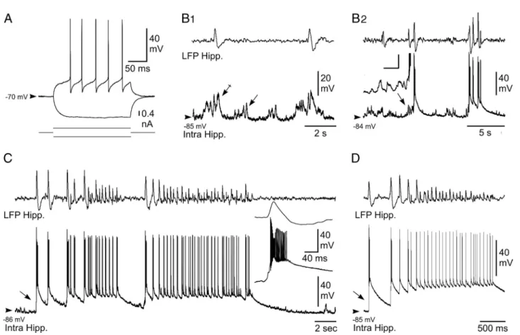
Figure 2. Intracellular activity of hippocampal neurons from the KA-injected mouse in vivo. A, Voltage responses (top traces) of a neuron located in the CA1 region of the epileptic hippocampus to hyperpolarizing (average of 5 successive trials) and depolarizing (single-response) current pulses (bottom traces). B, Intracellular correlate (bottom traces) of LFP (top traces) interictal-like activities. B1, Subthreshold events. In between the interictal spikes, the hippocampal (Hipp.) neuron displayed bursts of dPSPs (arrow), which were amplified (crossed arrow) when coincident with
paroxysmal LFP. B2, Suprathreshold events. Brief clusters of LFP spikes could be correlated with neuronal bursting, which were generated by large-amplitude depolarization gradually constructed
by temporal summation of excitatory synaptic events (inset; calibration: 30 mV, 20 ms). C, D, Intracellular activity (bottom trace) of pyramidal neurons associated with LFP seizure activity (top trace). Note the abrupt depolarization at the onset of seizure (arrows) and the sustained neuronal depolarization throughout the local paroxysm. The inset in C, is the enlargement of the initial neuronal paroxysmal shift indicated by the arrow. Arrowheads indicate the mean interictal membrane potential. Records shown in A–C and D are from two separate experiments.
HPDs were characterized in the LFP by
the repetition of high-voltage biphasic
sharp waves (44.0
⫾ 14.4 waves; n ⫽ 58
HPDs from 8 cells) (Fig. 2C,D) lasting
16.9
⫾ 7.6 s and having an interval
fre-quency (2.9
⫾ 1.3 Hz) similar to that
cal-culated from behaving KA-treated mice
(see Table 1). Contrasting with the
inter-ictal discharges, the start of HPDs was
concomitant with an abrupt (Fig. 2C,
in-set) and large (33.6
⫾ 8.2 mV; n ⫽ 58
HPDs from 8 cells) depolarization, arising
from a relatively quiescent membrane
tential and causing a burst of action
po-tentials (Fig. 2C,D). This initial ictal
neuronal event was followed by a
sus-tained
membrane
depolarization
of
19.2
⫾ 6.9 mV (n ⫽ 58 HPDs from 8
cells), which was maintained throughout
the HPDs (Fig. 2C,D). This prolonged
de-polarization was mostly crowned by
re-peated sharp depolarizing potentials,
which occurred from the decay phase of
the preceding one and generated a cluster
of action potentials (4.0
⫾ 1.8 action
po-tentials; n
⫽ 58 epileptic HPDs from 8
cells). At the end of the HPDs, hippocampal
neurons slowly repolarized, within 6.3
⫾
3.6 s (n
⫽ 58 HPDs from 8 cells), toward
their interictal membrane potential.
These findings provide, to our
knowl-edge, the first in vivo intracellular
descrip-tion of the interictal and ictal events
occurring in the hippocampus of a mouse
model of MTLE.
Intracellular activity of PF neurons
during HPD
To investigate the functional coupling between hippocampus
and thalamic PF nucleus during HPD, we performed in vivo
in-tracellular recordings of PF neurons simultaneously with LFP
activities in the ipsilateral CA1 region. The five recorded neurons,
from five different mice, had an average membrane resistance of
29.7
⫾ 9.3 M⍀ and a membrane time constant of 10.5 ⫾ 3.7 ms
(Fig. 3A). Their action potentials had an amplitude of 58.1
⫾ 6.8
mV and a duration of 0.9
⫾ 0.3 ms, and were followed by an
afterhyperpolarization of large amplitude (9.2
⫾ 4.4 mV) (Fig.
3A). When applied from the resting potential, suprathreshold
current pulses elicited sustained tonic firing (Fig. 3A
1).
More-over, the injection of negative current pulses was immediately
followed by a postanodal excitation, likely caused by a
low-threshold calcium potential (Fig. 3A
2, top) generating a burst of
action potentials (Fig. 3A
1,A
2), and/or induced a sag potential (Fig.
3A
2, bottom), likely caused by a hyperpolarization-activated inward
cationic current. Altogether, these electrical membrane properties
were consistent with those classically described in thalamocortical
cells (Jahnsen and Llina´s, 1984).
During interictal periods, the intracellular activity of PF
neu-rons was characterized by a sustained barrage of
high-frequency, small-amplitude, depolarizing synaptic potentials
corresponding to a mean membrane potential of
⫺58.5 ⫾ 3.8
mV (n
⫽ 161 interictal periods from 5 neurons). This
back-ground synaptic activity caused an irregular discharge of
ac-tion potentials, with a mean frequency of 7.9
⫾ 7.0 Hz (n ⫽ 5
neurons) (Fig. 3 B, C).
The intracellular activity of PF neurons (n
⫽ 3) was clearly
affected by the occurrence of HPDs. The onset of hippocampal
paroxysms was correlated with a slight hyperpolarization of PF
neurons (
⫺1.1 ⫾ 0.5 mV; n ⫽ 27 from three cells) and a transient
decrease in the mean firing rate by 30.5
⫾ 10.5% (n ⫽ 27 from
three cells) (Fig. 3 B, C), which could lead to a complete
interrup-tion in cell discharge. A striking finding was the robust increase in
PF neurons firing that coincided with the termination of HPDs
(Fig. 3 B, C). This sustained excitation had a duration of 3.0
⫾
1.8 s (n
⫽ 27 from three cells) and resulted in a mean firing rate of
17.7
⫾ 9.5 Hz (Fig. 3B,C). Similar exacerbated activities in PF
neurons could be also observed immediately after interictal
dis-charges in the hippocampal LFPs (result not shown). Continuous
injection of negative DC currents (n
⫽ 3 cells, three animals)
revealed that these discharges in PF neurons at the end of the
seizure were attributable to a sustained membrane depolarization
sculpted by the temporal summation of high-frequency dPSPs
(Fig. 3D).
These data confirm and extend the correlated changes found
in the thalamic LFPs as a function of the different phases of the
HPDs, and further indicate a mirror-like firing activity in
hip-pocampal and thalamic cells.
Figure 3. Intracellular activity of PF thalamic neurons is disrupted by hippocampal seizures in vivo. A, Electrophysiological properties of recorded PF thalamic neurons. A1, Voltage responses (top traces) of a PF thalamic neuron to hyperpolarizing (average
potential from 10 successive trials) and depolarizing (single-response) current pulses (bottom traces). Note the tonic firing induced by the positive current and the postinhibitory excitatory rebound evidenced by the averaging of action potentials (arrow). A2, In
these two other PF cells, the current-induced hyperpolarization was clearly followed by a robust postanodal excitation, reminiscent of a low-threshold calcium potential, crowned by a burst of sodium spikes. A sag potential, likely caused by the hyperpolarization-activated inward cationic current, could also detected in some cells (bottom, arrow). B, C, Disruption of the spontaneous intracel-lular activity of PF neurons (bottom trace) during paroxysmal activity in the ipsilateral hippocampal (Hipp.) LFP (top trace). B, The thalamic cell was slightly hyperpolarized during the epileptic episode, and its firing was transiently interrupted. A prolonged train of action potentials promptly followed the HPD. C, In this other neuron, the firing rate was decreased during the HPD and then dramatically augmented at the termination of hippocampal paroxysms (dashed boxes). D, The excitatory postictal rebound in PF neurons resulted from summed dPSPs. At the end of the HPD (top), the thalamic neuron, which was hyperpolarized by DC current injection (⫺1.0 nA; middle trace), displayed a sustained membrane depolarization. As shown by the expansion of the record segment (bottom) indicated by the asterisk, the excitatory rebound resulted from the temporal summation of individual dPSPs (oblique lines). Arrowheads indicate the mean interictal membrane potential.
Extracellular activity of PF neurons during HPDs
To further extend the statistical analysis of the functional
rela-tionships between hippocampus and PF nucleus during HPDs,
we then recorded the spontaneous activity of PF neurons in
KA-treated mice by extracellular recordings of 30 single units from
seven mice. Recorded cells, which were morphologically
identi-fied by juxtacellular injection of neurobiotin (see Materials and
Methods), were located within the PF nucleus (Fig. 4 A
1). We
could discriminate two populations of PF
cells according to their distinctive
pat-terns of electrical activity which were
af-fected, or not, by the occurrence of HPDs.
However, all PF neurons showed an
ar-rhythmic spontaneous spike activity
(2.9
⫾ 0.53 Hz) during the interictal
peri-ods (Fig. 4 A
2,A
3).
The first subpopulation of PF cells
(Fig. 4 A
2) (n
⫽ 19), the activity of which
was changed during HPDs, had a mean
firing frequency during interictal periods
of 3.04
⫾ 0.77 Hz (from 0.1 to 9.48 Hz).
During interictal clusters of isolated
spikes (interictal discharges) (Fig. 4 B
1,B
2)
and HPDs (Fig. 4 B
3,B
4), the firing rate of
this set of thalamic neurons decreased,
reaching the mean values of 2.4
⫾ 0.6 Hz
(range, 0 – 4.89 Hz; n
⫽ 375 interictal
clus-ters of isolated spikes) and 1.44
⫾ 0.28 Hz
(range, 0 – 4.2 Hz; n
⫽ 131 HPDs),
respec-tively. The transition between interictal
clusters of isolated spikes or HPDs and
postictal periods was characterized in
these PF cells by a drastic increase in their
firing rate (Fig. 4 B
2,B
4), the end of
hip-pocampal paroxysms being concomitant
with an abrupt and transient excitation of
these neurons. Before this increase of
fir-ing rate, the mean discharge frequency of
PF thalamic cells during hippocampal
par-oxysmal events was 2.5
⫾ 0.26 Hz, whereas
the rebound of excitation had a mean
fre-quency of 11
⫾ 0.68 Hz and lasted 2.02 ⫾
0.12 s (n
⫽ 102 interictal clusters of isolated
spikes and HPDs, 19 cells). This increase in
the firing rate of PF cells concomitant with
the end of hippocampal events was
statisti-cally significant ( p
⬍ 0.001;
Mann–Whit-ney rank sum test).
The second category of PF cells (n
⫽ 11),
which were also located within the
bound-aries of the PF nucleus, did not show any
significant change in their firing rate
be-tween ictal and interictal periods (interictal
mean frequency, 2
⫾ 0.63 Hz; range, 0.084–
6.58 Hz; mean frequency during clusters of
isolated spikes, 2.53
⫾ 0.8 Hz; range, 0.054–
8.88 Hz; mean frequency during HPDs, 3⫾
0.85 Hz; range, 0.86 –7.34 Hz; n
⫽ 11 cells).
We found no correlation between the
firing pattern and the morphology of the
PF neurons.
Finally, 13 neurons located outside the
PF (e.g., ventral posteromedial,
centrolat-eral, or posterior thalamic nuclei) were recorded in the same
conditions, and their firing patterns did not show any detectable
correlation with hippocampal LFP and were not modified by
the occurrence of HPDs (interictal mean frequency, 3.25
⫾
1.15 Hz; range, 0.031–13.6 Hz; mean frequency during
clus-ters of isolated spikes, 3.83
⫾ 1.3 Hz; range, 0.67–15.1 Hz;
mean frequency during HPDs, 2.75
⫾ 0.85 Hz; range, 0.62–9.4
Hz; n
⫽ 13 cells).
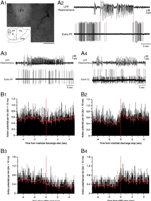
Figure 4. Extracellular recordings from PF neurons during HPDs. A1, Microphotograph of a PF neuron labeled by juxtacellular injection
of neurobiotin. The cell body (filled circle in the inset, schematic coronal plane drawing) was located in the central region of the lateral part of the PF nucleus. Anteriority from the interaural line was 1.62 mm. Fr, Fasciculus retroflexus; Po, posterior thalamic nucleus; VPM, ventral posteromedial thalamic nucleus. Scale bar, 100m.A2–A4, Single-spike activity of thalamic neurons (bottom traces) during paroxysmal
activity in the ipsilateral hippocampus (top traces). A2, Disruption of the spontaneous extracellular activity of the PF neuron shown in A1
during the HPD (between the dashed vertical lines) and representative of the first subpopulation of PF cells. A3, Extracellular activity of
another PF neuron affected by the HPD occurrence and representative of the second subpopulation of PF cells. No change in their firing rate was observed. A4, Simultaneous recording of two centrolateral thalamic nucleus (CL) neurons during HPD. Note that the activity of these
two cells is not affected by the HPD. B1–B4, Mean cumulative histograms of binned action potential discharge of 19 PF neurons aligned to
hippocampal epileptic events (5 s before and after the start of short clusters of isolated spikes (B1, B3) and HPDs (B2, B4). Note slight
decrease of the activity of PF neurons after the beginning of hippocampal clusters of isolated spikes (B1) or HPDs (B3). Contrarily, the mean
firingrateofPFneuronsincreased500msbeforetheendofclusterofisolatedspikes(B2)andHPDs(B4)andlastedapproximately1.5safter
Altogether, our intracellular and
extra-cellular recordings from the PF
demon-strate that the firing pattern of most of PF
neurons is drastically modified by the
oc-currence of HPDs.
Suppressive effect of high-frequency
stimulation of the PF
nucleus
To test the hypothesis of a role of the PF in
the control of HPDs, we then examined
the effects of high-frequency stimulation
(HFS) of this structure. Such stimulations
are now commonly used for DBS in
hu-man patients and are suggested to result in
an overall suppression of the local
neuro-nal activity (McIntyre et al., 2004),
al-though this effect is very much debated
(Gradinaru et al., 2009). In this
experi-ment, we tested single ipsilateral,
con-tralateral, or bilateral 5 s stimulations of
the PF using either monophasic and
bi-polar modes (frequency, 130 Hz; pulse
width, 60
s) in 18 mice. In a few mice,
the effects of lower frequency (20 Hz)
were also tested to mimic the behavior
of PF neurons observed at termination
of HPDs.
Ipsilateral stimulations
In nine animals with both electrode tips
located within the ipsilateral PF nucleus,
bipolar stimulations interrupted the
on-going HPDs at a mean threshold of 25
⫾
1.9
A. The antiepileptic and behavioral
thresholds were significantly lower than
the motor threshold (73 and 65%
de-creases, respectively; p
⫽ 0.004; Wilcoxon
test) (Fig. 5A). A slight but not significant
difference was observed between
antiepi-leptic and behavioral thresholds ( p
⫽
0.25; Wilcoxon test). Furthermore, time–
frequency analysis revealed that ipsilateral
130 Hz stimulations of the PF at the
anti-epileptic threshold suppressed all
oscilla-tory activities between 0 and 20 Hz at the hippocampal and
cortical levels (Fig. 5C).
When a referential mode was used (monopolar stimulation
with one PF electrode as the cathode and the reference
elec-trode over the cerebellum as the anode), the thresholds were slightly
increased (⬃6%), although no significant differences were observed
(results not shown).
To investigate the specificity of the PF as a target for deep brain
stimulation, we compared the thresholds for ipsilateral bipolar
stimulations between sites located within the ipsilateral PF (in
sites, n
⫽ 9) and adjacent regions (out sites, n ⫽ 6; namely, the
posterior thalamic nuclei group, the ventral anterior pretectal
nucleus, the dorsal anterior pretectal nucleus, and the
periven-tricular fiber system) (Fig. 5D). The thresholds for antiepileptic,
behavioral, and motor effects were significantly lower when
stim-ulations were applied within the PF region ( p
⫽ 0.013, 0.016, and
0.013, respectively; Mann–Whitney test) (Fig. 5 B, D). The
possi-bility to interrupt HPDs by stimulations of adjacent regions,
although with higher current intensities, could account for
spreading phenomenons as well as the involvement of other
pu-tative neuronal networks. Similarly to intracerebral
pharmaco-logical manipulations, such a gradient effect could also be
observed when stimulations were applied to other structures
(Feddersen et al., 2007).
Then we tested the effect of 20 Hz stimulations, which
corre-sponds to the mean firing rate of PF neurons during the rebound
of excitation observed at the end of HPDs. In two mice equipped
with bipolar electrodes in the ipsilateral PF, 20 Hz stimulation did
not interrupt ongoing HPDs, whatever the amount of current
applied (up to 150
A). However, transient interruptions of
HPDs were observed, which lasted the duration of the
stimula-tion (results not shown). This suggests that 20 Hz stimulastimula-tion of
the PF interferes with HPDs without terminating them.
Contralateral and bilateral stimulations
Contralateral stimulations of the PF also interrupted ongoing
HPDs (n
⫽ 9 mice) with a mean antiepileptic threshold of 35 ⫾
Figure 5. Effects of 130 Hz stimulation of the PF nucleus on HPDs. A, Intensity thresholds according to stimulation mode: ipsilateral, contralateral, or bilateral stimulations (n⫽ 9, 9, and 4 mice respectively; *p ⬍ 0.05, Wilcoxon test, compared with ipsilateral stimulation values;#p⬍ 0.05, Wilcoxon test, compared with antiepileptic threshold values). B, Site specificity of ipsilateral 130 Hz stimulation. Intensity thresholds according to electrode tip localization, inside versus outside the boundaries of the PF nucleus (n⫽ 9 mice inside; n ⫽ 6 mice outside; *p ⬍ 0.05, Mann–Whitney test, compared with thresholds obtained with stimulations applied within the PF nucleus). C, Time–frequency chart of power averaged over hippocampal and cortical derivations and over 24 hippocampal HPDs interrupted by 130 Hz stimulation of the PF nucleus. Before averaging, the power was frequency normalized according to interictal activity. The color scale indicates the increase (shades of red) or decrease (shades of blue) of power expressed in standard deviations of the interictal activity. D, Mouse’s brain coronal sections showing histological recon-struction of the electrode tip localization inside the PF nucleus (black dots) and outside the boundaries of the PF nucleus (open triangles). IA, Interaural; ml, medial lemniscus; fr, fasciculus retroflexus.
3.2
A and higher behavioral (⫹10%; p ⫽ 0.25) and motor
thresholds (
⫹95%; p ⫽ 0.004; Wilcoxon test). All of these
thresh-olds were significantly higher than for ipsilateral stimulations
(antiepileptic, p
⫽ 0.03; behavioral, p ⫽ 0.006; motor, p ⫽ 0.001;
Mann–Whitney test) (Fig. 5A). When bilateral stimulations were
applied (n
⫽ 4 mice), bipolar 130 Hz stimulation interrupted
HPDs with threshold values that were similar to ipsilateral
stim-ulations ( p
⫽ 0.12) and significantly lower than contralateral
stimulations ( p
⫽ 0.017) (Fig. 5A).
Effects of PF glutamatergic neurotransmission modulation
on HPDs
The PF receives strong glutamatergic inputs mainly arising from
the sensorimotor cortex. To determine the potential role of this
glutamatergic neurotransmission on the PF, the effects of (1)
blockade and (2) potentiation of NMDA-mediated
glutamater-gic neurotransmission on the occurrence of HPDs were
exam-ined by local application of CGP40116 (n
⫽ 8 mice) or NMDA
(n
⫽ 9 mice), respectively.
Bilateral intra-PF injection of CGP40116 (2 and 4 pmol per
side) significantly suppressed the number of HPDs (data not
shown) and their cumulated duration for up to 100 min at the
highest dose (Fig. 6 A), compared with the vehicle condition.
During the first 40 min after injection, the number and
cumu-lated duration of HPDs were decreased by
⬎95%, compared with
vehicle (NaCl injection, 100%) (Fig. 6 A). Ictal activity then
re-turned to baseline (Fig. 6 A, reference periods Ref1, Ref2, and
Ref3) within 2 h. Moreover, the latency for the reoccurrence of
HPDs after drug injection was significantly increased at 2 or 4
pmol per side in a dose-dependent way (Fig. 6 B). At either dose,
the interictal activity in the hippocampus or in the cortex during the
0 –20 and 20 – 40 min periods after injection was not altered as
indi-cated by the stability of the shape of the LFP amplitude spectrum
between before and after injection periods (supplemental Fig. S1A,
available at www.jneurosci.org as supplemental material).
In addition, the suppressive effects on HPDs were observed
only when injections were performed inside the PF (Fig. 6 D,
black dots, tips of injection cannulae). In three additional animals
injected outside the PF (namely, the dorsal anterior pretectal
nucleus, deep mesencephalic nucleus, and ventral tegmental
area), no significant effects on HPD suppression or reoccurrence
latency (16
⫾ 4 min outside the PF vs 48 ⫾ 6 min inside the PF at
4 pmol/side) were observed (Fig. 6 B, D, open triangles).
Finally, at the doses used, no behavioral effects were observed:
mice remained quiet in their test cage and explored their
envi-ronment or groomed. In two mice injected with a higher dose (8
pmol/side), suppression of HPDs was observed, along with
pros-trations beginning within the first minutes after the injection and
lasting for up to 60 min (results not shown).
By contrast, bilateral intra-PF injection of NMDA
signifi-cantly increased by 100 and 202%, respectively, the number (data
not shown) and cumulative duration of HPDs during the first 20
min after injection of the lowest dose (5 pmol/side) compared
with vehicle injection (NaCl, 100%) (Fig. 6 A). This worsening
effect was mostly over (19 and 36%, respectively) 40 min after
injection and back to the baseline level, at 60 min after injection
(Fig. 6 A). No effects on the mean duration of HPDs were
ob-served (data not shown). At this dose, no behavioral or motor
side effects were observed at any time. At the highest dose of
NMDA (10 pmol/side), behavioral side effects such as sniffing or
exploring behaviors that clearly interfered with the occurrence of
HPDs were observed. Nonetheless, the latency of reoccurrence of
the first HPDs after drug injection significantly decreased after
injection of both 5 and 10 pmol per side, compared with NaCl
(Fig. 6C). However, no generalizations of the HPDs by spreading
to the cortex were ever observed.
In five additional animals injected outside the boundaries of the
PF (namely, the ventromedial thalamic nucleus, mediodorsal
tha-lamic nucleus, dorsal anterior pretectal nucleus, superior cerebellar
peduncle, and posterior thalamic nuclei group), no significant
ef-Figure 6. Effects of bilateral intra-PF injections of CGP40116 and NMDA on HPDs. A, Percentage (mean and SEM) of cumulated duration of HPDs per 20 min period before [reference (Ref)] and after intra-PF microinjections of CGP40116 (4 pmol/side; n⫽8;*p⬍0.05;Wilcoxontest)orNMDA(5pmol/side;n⫽9;*p⬍0.05;Wilcoxontest)comparedtovehicle(NaCl).B,ReoccurrencelatencyofHPDsaftervehicle (NaCl), CGP40116 (2 and 4 pmol/side) intra-PF microinjections, and CGP40116 (4 pmol/side) injected outside the boundaries of the PF nucleus (OUT; n⫽8;*p⬍0.05;Wilcoxontest).C,Reoccurrencelatency of HPDs after vehicle (NaCl), NMDA (5 and 10 pmol/side) intra-PF microinjections, and NMDA (5 pmol/side) injected outside the boundaries of the PF nucleus (OUT; n⫽9;*p⬍0.05;Wilcoxontest).D,Example ofmouse’sbraincoronalsectionsshowinghistologicalreconstructionofthemicroinjectionsitesatwhichbilateralintra-PFinjectionsofCGP40116(2and4pmol/side)resultedinasuppressionofHPDs(blackdots, tips of the injection cannulae) and sites at which the same doses of CGP40116 were without effects on HPDs occurrence (open triangles). IA, Interaural; ml, medial lemniscus; fr, fasciculus retroflexus.
fects on HPD suppression or reoccurrence latency (14
⫾ 3 min
outside vs 4
⫾ 1 min inside at 5 pmol/side) were observed (Fig. 6C).
Effects of PF GABAergic neurotransmission modulation
on HPDs
To determine the potential role of GABAergic
neurotransmis-sion from the basal ganglia structures (mainly the SNr) on the
PF neurons, the effects of (1) potentiation and (2) blockade of
GABA
A-mediated neurotransmission were examined by local
applications of muscimol (n
⫽ 8) or picrotoxin (n ⫽ 8),
respectively.
Bilateral intra-PF injection of muscimol (17.5 and 35 pmol per
side) significantly suppressed the number of HPDs (data not shown)
and their cumulated duration for up to 80 min at the highest dose,
compared with vehicle injection (Fig. 7A). At the lowest dose,
bilat-eral injection of muscimol induced a significant suppression in the
number of HPDs for 40 min and a significant decrease in cumulated
duration for 20 min after injection, compared with vehicle injection
(data not shown). At the highest dose, the number and cumulated
duration of HPDs decreased by
⬎90%duringthefirst20minperiod
after injection, compared with the vehicle condition (NaCl, 100%)
(Fig. 7A). It then progressively returned to baseline within 2 h.
More-over, the latency for the reoccurrence of HPDs after drug injection
increased significantly at 17.5 or 35 pmol per side in a
dose-dependent way (Fig. 7B). At either dose, the interictal activity in the
hippocampus or the cortex during the 0 –20 and 20 – 40 min periods
after injection was not altered, as indicated by the stability of the
shape of the LFP amplitude spectrum between preinjection and
postinjection periods (supplemental Fig. S1B, available at www.
jneurosci.org as supplemental material).
In addition, the suppressive effects were observed only when
injections were performed within the PF (Fig. 7 B, D, black dots,
tips of injection cannulae). In seven additional animals injected
outside the PF (namely, in the superior cerebellar peduncle,
ven-tromedial thalamic nucleus, posterior hypothalamic area,
periac-queductal gray, lateral hypothalamic area, deep mesencephalic
nucleus, and ventral tegmental area), no significant effects on
HPD suppression or reoccurrence latency (15
⫾ 2 min outside
the PF vs 50
⫾ 6 inside the PF at 35 pmol/side) were observed
(Fig. 7 B, D, open triangles).
Finally, at the doses used, no behavioral effects were observed.
In two mice injected with a higher dose of muscimol (70 pmol/
side), behavioral and motor side effects such as forelimb
stereo-typies in upright position and rotations were observed, beginning
within the first minutes after the injection and lasting for up to 60
min. In this case, LFP recordings were contaminated by
move-ment artifacts.
Bilateral intra-PF injection of picrotoxin induced a significant
increase in the number and cumulative duration of HPDs during
the first hour after injection at the lowest dose (2.5 pmol/side)
(Fig. 7A). The cumulated durations of HPDs were worsened by
107, 195, and 52% at 20, 40, and 60 min, respectively, compared
with vehicle (NaCl, 100%) (Fig. 7A). The latency for the
reoccur-rence of HPDs after drug injection significantly increased at 2.5
pmol/side (Fig. 7C). At this dose, neither behavioral nor motor
side effects were observed. At the dose of 5 pmol/side, picrotoxin
also induced an aggravation in both the number and cumulated
duration of HPDs (35 and 42%, respectively, between 40 and 60
min after injection) lasting up to 60 min. However, at this dose,
motor side effects that interfered with the occurrence of HPDs
were noticed, but no generalizations of the HPDs by spreading to
the cortex were ever observed. In four additional animals injected
outside the boundaries of the PF (namely, in the lateral habenular
nucleus, ethmoid nucleus, dorsal anterior pretectal nucleus,
sub-commissural nucleus), no significant effects on HPD suppression
Figure 7. Effects of bilateral intra-PF injections of muscimol and picrotoxin on HPDs. A, Percentage (mean and SEM) of cumulated duration of HPDs per 20 min period, before [reference (Ref)] and after intra-PF microinjections of muscimol (35 pmol/side; n⫽ 8; *p ⬍ 0.05; Wilcoxon test) or picrotoxin (2.5 pmol/side; n ⫽ 8; *p ⬍ 0.05; Wilcoxon test) compared to vehicle (NaCl). B, Reoccurrence latency of HPDs after vehicle (NaCl), muscimol (17.5 and 35 pmol/side) intra-PF microinjections, and muscimol (35 pmol/side) injected outside the boundaries of the PF nucleus (OUT;
n⫽8;*p⬍0.05;Wilcoxontest).C,ReoccurrencelatencyofHPDsaftervehicle(NaCl),picrotoxin(2.5and5pmol/side)intra-PFmicroinjections,andpicrotoxin(2.5pmol/side)injectedoutsidethe
boundaries of the PF nucleus (OUT; n⫽ 8; *p ⬍ 0.05; Wilcoxon test). D, Example of mouse’s brain coronal sections showing histological reconstruction of the microinjection sites at which bilateral intra-PF injections of muscimol (17.5 and 35 pmol/side) resulted in a suppression of HPDs (black dots, tips of the injection cannulae) and sites at which the same doses of muscimol were without effects on HPDs occurrence (open triangles). IA, Interaural; ml, medial lemniscus; fr, fasciculus retroflexus.