HAL Id: inserm-00517199
https://www.hal.inserm.fr/inserm-00517199
Submitted on 13 Sep 2010
HAL is a multi-disciplinary open access
archive for the deposit and dissemination of
sci-entific research documents, whether they are
pub-lished or not. The documents may come from
teaching and research institutions in France or
abroad, or from public or private research centers.
L’archive ouverte pluridisciplinaire HAL, est
destinée au dépôt et à la diffusion de documents
scientifiques de niveau recherche, publiés ou non,
émanant des établissements d’enseignement et de
recherche français ou étrangers, des laboratoires
publics ou privés.
Intravenous administration of 99mTc-HMPAO-labeled
human mesenchymal stem cells after stroke: in vivo
imaging and biodistribution.
Olivier Detante, Anaïck Moisan, Julien Dimastromatteo, Marie-Jeanne
Richard, Laurent Riou, Emmanuelle Grillon, Emmanuel Barbier,
Marie-Dominique Desruet, Florence de Fraipont, Christoph Segebarth, et al.
To cite this version:
Olivier Detante, Anaïck Moisan, Julien Dimastromatteo, Marie-Jeanne Richard, Laurent Riou, et al..
Intravenous administration of 99mTc-HMPAO-labeled human mesenchymal stem cells after stroke:
in vivo imaging and biodistribution.. Cell Transplantation, Cognizant Communication Corporation,
2009, 18 (12), pp.1369-79. �10.3727/096368909X474230�. �inserm-00517199�
Copyright 2009 Cognizant Comm. Corp. E-ISSN 1555-3892 www.cognizantcommunication.com
Intravenous Administration of
99mTc-HMPAO-Labeled Human Mesenchymal
Stem Cells After Stroke: In Vivo Imaging and Biodistribution
Olivier Detante,*†‡ Anaı¨ck Moisan,†§¶ Julien Dimastromatteo,†¶# Marie-Jeanne Richard,†**††‡‡
Laurent Riou,†¶ Emmanuelle Grillon,*† Emmanuel Barbier,*† Marie-Dominique Desruet,*§¶
Florence De Fraipont,†,††‡‡ Christoph Segebarth,*† Assia Jaillard,*†§§ Marc Hommel,†‡¶¶
Catherine Ghezzi,†¶ and Chantal Remy*†
*INSERM, U836, Grenoble Cedex 9, France †Universite´ Joseph Fourier, Grenoble Cedex 9, France
‡Unite´ Neuro-Vasculaire, Neurologie, Centre Hospitalier Universitaire de Grenoble, Grenoble Cedex 9, France §Service de Biophysique et Me´decine Nucle´aire, Centre Hospitalier Universitaire de Grenoble, Grenoble Cedex 9, France
¶Radiopharmaceutiques Biocliniques, INSERM, U877, Faculte´ de Me´decine, La Tronche, France #ERAS Labo, Saint Nazaire-les-Eymes, France
**Unite´ Mixte de The´rapie Cellulaire et Tissulaire, Centre Hospitalier Universitaire de Grenoble, Grenoble Cedex 9, France ††Unite´ de Cance´rologie biologique et biothe´rapie, Centre Hospitalier Universitaire de Grenoble, Grenoble Cedex 9, France
‡‡INSERM, U823, Institut Albert Bonniot, La Tronche Cedex, France
§§Neuroradiologie/IRM, Centre Hospitalier Universitaire de Grenoble, Grenoble Cedex 9, France ¶¶INSERM U003, Centre d’Investigation Clinique, Centre Hospitalier Universitaire, Grenoble Cedex 9, France
Human mesenchymal stem cells (hMSC) are a promising source for cell therapy after stroke. To deliver these cells, an IV injection appears safer than a local graft. We aimed to assess the whole-body biodistribution of IV-injected99mTc-HMPAO-labeled hMSC in normal rats (n= 9) and following a right middle cerebral artery
occlusion (MCAo, n= 9). Whole-body nuclear imaging, isolated organ counting (at 2 and 20 h after injec-tion) and histology were performed. A higher activity was observed in the right damaged hemisphere of the MCAo group [6.5± 0.9 × 10⁻³% of injected dose (ID)/g] than in the control group (3.6± 1.2 × 10⁻³%ID/
g), 20 h after injection. In MCAo rats, right hemisphere activity was higher than that observed in the contralateral hemisphere at 2 h after injection (11.6± 2.8 vs. 9.8 ± 1.7 × 10⁻³%ID/g). Following an initial
hMSC lung accumulation, there was a decrease in pulmonary activity from 2 to 20 h after injection in both groups. The spleen was the only organ in which activity increased between 2 and 20 h. The presence of hMSC was documented in the spleen, liver, lung, and brain following histology. IV-injected hMSC are transiently trapped in the lungs, can be sequestered in the spleen, and are predominantly eliminated by kidneys. After 20 h, more hMSC are found in the ischemic lesion than into the undamaged cerebral tissue. IV delivery of hMSC could be the initial route for a clinical trial of tolerance.
Key words: Human mesenchymal stem cell (hMSC); Stroke; Brain ischemia; Cell transplantation; Biodistribution; Nuclear imaging
INTRODUCTION plied later on after the stroke onset with the aim of en-hancing brain plasticity (29,41,45,53). It has recently been shown that this could be achieved by cell trans-Stroke is the leading cause of acquired disability in
adults in industrialized countries. Admission to special- plantation (8,36–38).
Among various cell types, human mesenchymal stem ized stroke units (25,52) and thrombolysis (43,54) are
the only effective strategies for the early treatment of cells (hMSC), derived from bone marrow, offer the ad-vantage of not originating from a tumoral or modified acute ischemic stroke. Unfortunately, the brief time
win-dow limits thrombolysis to a small fraction of patients. source (28). Moreover, they are poorly immunogenic (1,34,48) and do not lead to the ethical problems associ-Thus, an alternative strategy is needed that could be
ap-Received June 4, 2008; final acceptance September 1, 2009. Online prepub date: October 1, 2009.
Address correspondence to Olivier Detante, Grenoble Institut des Neurosciences, BP 170, 38042 Grenoble Cedex 9, France. Tel: 00.33.(0)4.56.52.05.88; Fax: 00.33.(0)4.56.52.05.98; E-mail: ODetante@chu-grenoble.fr
ated with embryonic stem cells. The use of hMSC for been performed in various pathological conditions such as chronic paraplegia (18) and myocardial infarction cell therapy relies on their capacity to graft and survive
for a long time in the tissue of interest (12). Bone mar- (5,32). Briefly, radiolabeled MSC were mainly detected in the lungs, liver, kidney, infarcted myocardium, and row-derived hMSC are identified as multipotent
progen-itor cells that differentiate both into mesenchymal and spleen following IV injection. A recent human case re-port used bone marrow mononuclear cells radiolabeled nonmesenchymal lineages, including neurons and
endo-thelia (56). As such, MSC promote structural and func- with99mTc (17). Nine days after ischemic stroke, these
cells were injected intra-arterially. Twenty-four hours tional repair in several organs, notably in the brain after
stroke in rodent models using either the intracerebral later, the labeled cells were observed, using in vivo nu-clear imaging, in the damaged brain, liver, and spleen (10,14,35), intra-arterial (50), or intravenous (IV) (13,
15,34,39,46,51,57) administration. Despite a potential (17). To our knowledge, no study assess the biodistribu-tion of IV-injected radiolabeled hMSC after stroke. better efficiency of local delivery (intracerebral route),
systemic IV administration appears to be safer and eas- These data are however necessary to evaluate the risk of a systemic cell therapy after stroke and/or its therapeutic ier than local brain grafting following stroke in the
clini-cal setting as it is less invasive. Intracerebral delivery is potential, quantifying the hMSC homing to the ischemic lesion.
limited to small graft sites into large ischemic lesions
whereas IV injection allows cell distribution into vascu- Thus, the aim of our study was to evaluate the whole-body biodistribution of 99mTc-HMPAO-labeled hMSC
larized and viable areas of the lesion. In humans, the
first clinical trial demonstrated that IV delivery of following IV injection in normal rats and in rats sub-jected to focal cerebral ischemia.
hMSC is feasible and safe after stroke (4).
In experimental studies showing a benefit of hMSC
MATERIALS AND METHODS
administration following stroke, there are little data
re-ported about the quantity of hMSC localizing in the All animal procedures conformed strictly to French government guidelines for the use and care of animals brain lesion and about cell redistribution and/or
elimina-tion from other organs. MSC imaging can provide such (license #380806 for O. Detante). Surgical procedures were conducted under aseptic conditions and every ef-data. MSCs have previously been labeled with
paramag-netic particles for magparamag-netic resonance imaging (MRI) fort was made to reduce animal suffering. Anesthesia was induced by inhalation of 5% isoflurane (Fore`ne, Ab-(23) and organs of interest such as the kidneys, liver
(21,26), heart (22,32), or brain (27,30) have been studied bott Laboratory) in 30% O2 in air and maintained
throughout all surgical and imaging procedures with 2– in vivo. Unfortunately, MRI has a poor sensitivity and
whole-body cell MRI is challenging in experimental 2.5% isoflurane through a facial mask. conditions.
hMSC Labeling hMSC radioactive labeling (7) and nuclear imaging
are better suited for whole-body biodistribution studies. The hMSC were isolated from bone marrow aspirate from a healthy donor who gave informed consent. Isola-Because of its short decay time (6 h) and its emission
energy well adapted to gamma-cameras, thus ensuring tion and culture procedures were conducted in the Cell Therapy Unit (biotherapy team of general clinical re-high image quality, 99mTc is better suited than 111In for
short tracking of hMSC in vivo (2,6).99mTc is a radioac- search centre, University Hospital of Grenoble), in
ac-cordance with good laboratory practices. Cell cultures tive agent widely used in clinical practice for
scintigra-phy in nuclear medicine. Technetium 99m-hexamethyl- were performed according to previously described meth-ods (42,48).
propylene amine oxime (99mTc-HMPAO) is a lipophilic
complex that is reduced into a hydrophilic complex by For each experiment, approximately 7× 10⁶ hMSC
were incubated for 15 min in 370 MBq of 99m
Tc-a glutTc-athione-dependent mechTc-anism Tc-after trTc-apping into
the cell (44). It provides a stable labeling for in vivo cell HMPAO prepared according to the instructions of the manufacturer (Ceretec, GE Healthcare). The prepara-tracking, and many cell types have already been labeled
with 99mTc-HMPAO to study cell biodistribution over tion was used within 30 min after reconstitution, as
rec-ommended (47). Cells were rinsed twice and resus-periods of up to 24 h. In unconditioned mice, Allers et
al. (3) mainly observed lung, liver, and spleen activity pended in 1 ml of phosphate-buffered saline (PBS) prior to IV injection. These labeling conditions were chosen 24 h after IV injection of hMSC labeled with 99m
Tc-HMPAO. Gao et al. (19) confirmed these results in according to preliminary in vitro assays (data not shown). Flow cytometry was used to check radiolabeled healthy rats after IV injection of111In-labeled MSC. The
authors observed an early lung activity, which redistrib- cell viability using 7-amino-actinomycin D (7-AAD, BD PharMingen) at 24 and 48 h after labeling. Colony form-uted in the liver, kidneys, spleen, and long bones 48 h
evaluate the in vitro proliferation capacity of the labeled Whole-Body Nuclear Imaging
hMSC. Immediately after radiolabeled hMSC injection, the rats were installed in a cradle. The cradle was placed Animal Model and Experimental Groups vertically in front of a dual-head small-animal gamma-camera (gamma-imager, Biospace Lab, Paris, France). Eighteen adult Sprague-Dawley rats (Janvier, France)
weighing between 250 and 300 g were randomly allo- The two detectors were asymmetrically positioned so that scintigraphic image acquisition of the upper and cated into two groups: 1) a group that underwent a
tran-sient right middle cerebral artery occlusion (MCAo) lower halves of the animal body could be simultane-ously performed by each detector, respectively. Gamma-(n= 9); 2) an age-matched control group without
cere-bral ischemia (n= 9). acquisition software (Biospace Lab) was used for image acquisition. The energy window for99mTc was 122–170
To perform the MCAo, rectal temperature was
main-tained at 37.0± 0.5°C with a feedback-controlled heat- keV, and decay correction was automatically applied to image counts recorded in the list mode. Image acquisi-ing electrical blanket connected to a rectal probe. Focal
brain ischemia was induced by intraluminal occlusion of tion lasted for 120 min and began immediately follow-ing 99mTc- hMSC injection in all nine control and nine
the right MCA (40). Briefly, the right carotid arterial
tree was isolated. A cylinder of melted adhesive (length MCAo animals (initial imaging). A second, 120-min ac-quisition was initiated 18 h following the IV injection in 2 mm, diameter 0.38 mm) attached to a nylon thread
(diameter 0.22 mm) was advanced from the lumen of four control and four MCAo rats (late imaging). These animals were awakened between imaging sessions. the external carotid artery into the internal carotid artery
up to 5 mm after the external skull base. The rats were
awakened and tested for spontaneous circling and fore- Biodistribution by Isolated Organ Counts
limb flexion during the occlusion period. After 90 min, Immediately following completion of the initial scin-rats were reanesthetized and the thread was removed. tigraphic image acquisition (i.e., 120 min after 99m
Tc-hMSC injection), five control and five MCAo rats were Brain MRI euthanized using a lethal dose of pentobarbital by intra-cardiac injection under isoflurane anesthesia. The eutha-MRI (7 T horizontal magnet) was performed to
mea-sure the ischemic lesion volume with a T2 spin echo nasia of the remaining four control and four MCAo rats
was performed immediately following the late image ac-weighted imaging (T2WI) (TR/TE= 2500/60 ms, field
of view= 30 mm, thickness = 1 mm, matrix = 128 × 128, quisition (20 h after injection). Following euthanasia, samples from the left and right cerebral hemisphere, two averages) in five rats, 1–3 days after MCAo. Rectal
temperature was maintained at 37.0± 0.5°C with a feed- heart, thyroid, salivary gland, stomach, liver, spleen, kid-ney, lung, and skeletal (hind limb) muscle were quickly back-controlled heating water blanket. The whole
ische-mic lesion was manually delineated on each slice. Le- obtained, rinsed in physiological saline, and weighed. Samples of blood and urine were also collected. 99m
Tc-sion volumes were computed by multiplying the number
of pixels by pixel surface area and slice thickness.To hMSC activity was assessed using a gamma-well counter (Cobra II, Packard Instruments) with a 120–160 assess blood–brain barrier permeability 1 week after
MCAo, T2WI and T1WI (same spatial resolution) before keV 99mTc energy window. All tissue counts were
cor-rected for background and decay during the time of and after an IV injection of gadolinium were performed
in four additional rats. counting. Histology Intravenous Injection of hMSC
99mTc-hMSC (in 1 ml of PBS) were injected as a slow Samples of lung, spleen, liver, and brain were
ob-tained prior to gamma-well counting from both groups. bolus into the saphenous vein. One week after cerebral
ischemia, the MCAo group received an injection of They were stored at−80°C and cut using a cryostat into 20-µm sections. Transplanted hMSC were identified 3.4± 1.2 × 10⁶ cells (corresponding to an injected
ac-tivity of 78.8± 35.1 MBq). The control group received with a human-specific monoclonal antibody to nuclear antigen (MAB1281; 1/2000; Chemicon, CA, USA). This an injection of 3.2± 1.1 × 10⁶cells (76.4± 24.7 MBq).
The injected dose was within the effective therapeutic primary antibody was incubated overnight at 4°C. A flu-orescent anti-mouse secondary antibody (1/500; Jackson dose range for IV grafts (15,34,51).To assess the
biodis-tribution of99mTc-HMPAO alone,99mTc-HMPAO (81.1± laboratory, MA, USA) was then applied for 1 h. All cell
nuclei were counterstained blue with Hoechst prior to 3.6 MBq) alone was injected in four additional rats, 1
week after a right MCAo.No immunosuppressive agents examination under epifluorescent microscopy (Nikon Eclipse E600, Japan). Prior to this study, the histological were used in agreement with previous studies (16,34).
methodology was set up on brains that had received an Cerebral Lesion Size by MRI
intracerebral hMSC grafts after stroke. All rats in MCAo group, awakened over the course of the artery occlusion period (90 min), exhibited neuro-Data Analysis
logical deficits as a consequence of the ongoing cerebral ischemia. The control rats had no deficit. In addition, Image Analysis. Gamma-vision+ software (Biospace
the cerebral lesion (infarcted tissue and edema) was au-Lab, Paris, France) was used for examiner-blind analysis
thenticated by MRI, 1–3 days following MCAo. Mean of planar images. Regions of interest (ROI) were drawn
volume was 262.5± 193.7 mm3.The four additional rats
on the whole brain, lung, and both left and right kidneys.
that were submitted to MCAo and received gadolinium No ROI was drawn on the liver because of possible lung
1 week later exhibited lesion with a markedly increased superimposition. ROI activities were expressed as counts
blood–brain barrier permeability. per minute per mm (cpm/mm2). Eighteen-hour decay
correction was applied to the ROI quantifications that
Whole-Body Nuclear Imaging were performed on late images. Initial and late ROI
ac-Immediately after labeling, 99mTc-HMPAO-labeled
tivities were then normalized towards the injected dose
hMSC were injected in rats. Each group received the expressed as GBq and therefore expressed as cpm/mm2/
same number of cells (3.4± 1.2 × 10⁶ cells for the
GBq.
MCAo group; 3.2± 1.1 × 10⁶ cells for control group)
Biodistribution Analysis. Organ, blood, and urine
ac-and the same injected radioactivity (78.8± 35.1 MBq tivities of 99mTc-hMSC were expressed as cpm,
decay-for MCAo group; 76.4± 24.7 MBq for control group). corrected to the time of injection, and normalized to the
Images in Figure 1 were obtained from most representa-tissue weight (g) and to the injected dose also expressed
tive rats in each group and are given as an example. as cpm. For the purpose of clarity, these results, initially
Table 1 shows quantitative imaging data (corrected for expressed as percent of the injected dose per gram of
decay) useful for result comparison. sample weight (%ID/g), were finally expressed as 10−3
A trend towards higher activity in the whole brain %ID/g.
following ischemia was observed in the MCAo group (23.3± 3.5 cpm/mm2/GBq at 2 h and 8.7± 1.8 cpm/
Statistical Analysis
mm2/GBq at 20 h) when compared with control rats
All results are expressed as mean± SD.
Between-(20.3± 3.5 cpm/mm2/GBq at 2 h and 7.9± 2.1 cpm/
group comparison of mean values was performed using
mm2/GBq at 20 h).
the unpaired Student t-test after checking the
homogene-An early and significant lung trapping was observed in ity of variance (Levene’s test). Paired t-test was used for
all rats as indicated by the initially high pulmonary activ-within-group comparison (left vs. right hemisphere). A
ity. This phenomenon was transient as the lung activity value of p< 0.05 was considered as significant.
decreased dramatically between the initial and late imaging sessions (control group, from 2209.9± 487.2 to 153.5 ±
RESULTS
75.8 cpm/mm2/GBq; MCAo group, from 2749.2± 544.6
hMSC Labeling to 231.3± 39.6 cpm/mm2/GBq).
Cultured hMSC expressed CD73, CD90, and CD105 Finally, the route of 99mTc-hMSC elimination was
but were CD45− and CD34− (data not shown). These mostly renal as shown from in vivo planar images.
Kid-cells differentiate into osteoblasts, chondrocytes, and ad- ney radioactivity was stable over time. No significant ipocytes as previously described (42). difference was observed between the MCAo and control
The efficiency of99mTc-HMPAO hMSC labeling (i.e.,
groups.
percentage of radiotracer activity incorporated into cells Concerning whole-body quantification, we observed over the total activity of radiotracer mixed with cells) that, at 20 h, 27.6± 6.6% and 29.5 ± 7% of the 2-h ac-was 26.7± 11.6%. In vitro assays showed that more tivities are still visualized for control and MCAo groups, than 96% of the initial99mTc activity remained into
via-respectively. ble hMSC 4 h after labeling.
Biodistribution by Isolated Organ Counts Using flow cytometry with 7-AAD, we observed that
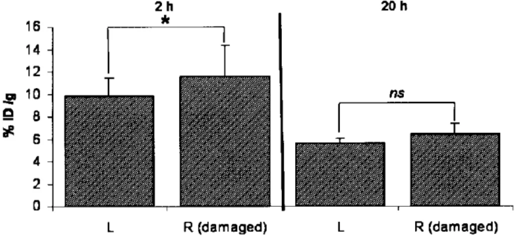
the 99mTc-HMPAO-labeled hMSC viability was 97.9% At 2 h following 99mTc-hMSC injection, the right
damaged hemisphere activity in MCAo rats tended to be and 96.1%, respectively, at 24 and 48 h after labeling
(compared to⬃98% viability of unlabeled cells). Cells higher than in controls (11.6± 2.8 × 10⁻³%ID/g vs. 7.9± 2.5× 10⁻³ %ID/g; p= 0.059). A marked difference ap-were also cultured to evaluate their capacity to form
col-onies. After 10 days of cell culture, no colony was de- peared 20 h after injection (6.5± 0.9 × 10⁻³ %ID/g vs.
3.6± 1.2 × 10⁻³ %ID/g; p= 0.010). One out of 10,000
tected for 99mTc-HMPAO-labeled hMSC whereas 29%
Figure 1. Whole-body nuclear imaging. The two detectors of a small-animal gamma-camera were vertically and asymmetrically
positioned. Image acquisition was started immediately following the IV injection of99mTc-hMSC for 2 hours (A, B). A second, late
planar image was also acquired from 18 to 20 h following the injection (C, D). Regions of interest were drawn on the whole brain, lung, and kidneys. Displayed images were obtained from the most representative rats of each group at each time. Note that the activity scales are different between 2- and 20-h images because of the large difference of activities due to the radioactive decay. L: left; R: right; MCAo: middle cerebral artery occlusion.
hemisphere of MCAo rats. This proportion was half of One week after MCAo, IV injection of 99m
Tc-HMPAO alone led to higher activity into the left normal this in the control rats. Moreover, the late contralateral
hemisphere activity was significantly higher in the hemisphere than in the right damaged one, 2 and 20 h after injection (mean activity right/left hemisphere: MCAo group when compared with the control group
(Table 2). Within the MCAo group, the initial ipsilateral 0.93± 0.02 and 0.89 ± 0.10, respectively), despite an al-tered blood–brain barrier in the lesion.
hemisphere activity was significantly higher than the
contralateral one (11.6± 2.8 × 10⁻³ %ID/g vs. 9.8± 1.7 × The organ counts indicated that hMSC could colonize an ischemic lesion in the brain after a systemic injection 10−3%ID/g, p= 0.024) (Fig. 2).
Table 1. Whole-Body Nuclear Imaging
0–2 Hours 18–20 Hours
Control MCAo Control MCAo
(n= 9) (n= 9) p (n= 4) (n= 4) p
Brain 20.3± 3.5 23.3± 3.5 ns 7.9± 2.1 (−62 ± 6%) 8.7± 1.8 (−65 ± 1%) ns
Lung 2209.9± 487.2 2749.2± 544.6 0.042 153.5± 75.8 (−94 ± 3%) 231.3± 39.6 (−91 ± 3%) ns
Right kidney 382.6± 151.6 379.4± 123.6 ns 353.3± 72.2 (+13 ± 28%) 372.4± 56.6 (+6 ± 33%) ns
Left kidney 314.4± 101.3 340.4± 143.0 ns 310.0± 39.8 (+1 ± 21%) 330.3± 53.8 (−6 ± 33%) ns
Two-hour image acquisition was started immediately following the intravenous injection of99mTc-hMSC. A second, late planar image was acquired from 18 to 20 h following injection. Results, corrected for decay, are expressed as counts/minute/mm2/injected GBq (cpm/mm2/GBq): mean± SD. Middle cerebral artery occlusion (MCAo) group was compared with control group (unpaired t-test). Percent change (mean ± SD) was calculated per group from values obtained in the four animals that were imaged at 0–2 and at 18–20 h after injection. ns, no significant difference.
Table 2. Whole-Body hMSC Biodistribution by Isolated Organ Counting
2 Hours 20 Hours
Control MCAo Control MCAo
Organ (n= 5) (n= 5) p (n= 4) (n= 4) p Left brain 7.9± 2.5 9.8± 1.7 ns 3.6± 1.2 (−54%) 5.6± 0.5 (−43%) 0.022 Right brain 7.9± 2.5 11.6± 2.8 ns (0.059) 3.6± 1.2 (−54%) 6.5± 0.9 (−44%) 0.010 Blood 169.9± 69.8 145.5± 41.4 ns 22.8± 4.0 (−87%) 32.0± 11.5 (−78%) ns Heart 76.3± 17.4 103.4± 30.5 ns 26.3± 3.2 (−66%) 29.1± 9.8 (−72%) ns Thyroid 34.5± 9.9 82.9± 52.5 ns 19.4± 4.0 (−44%) 24.5± 7.5 (−70%) ns Salivary gland 43.0± 7.7 49.7± 24.8 ns 14.2± 1.7 (−67%) 20.6± 2.6 (−59%) 0.006 Stomach 66.4± 21.2 120.4± 59.7 ns 25.2± 3.0 (−62%) 35.5± 20.3 (−71%) ns Liver 1363.9± 320.0 1508.1± 265.4 ns 1011.5± 182.2 (−26%) 1311.3 ± 380.6 (−13%) ns Spleen 851.9± 266.7 796.7± 274.1 ns 1434.0± 468.3 (+68%) 1825.6 ± 522.8 (+129%) ns Kidney 2607.3± 661.7 3094.8± 657.7 ns 2261.0± 438.7 (−13%) 3356.4 ± 165.1 (+8%) 0.003 Lung 18696.0± 14675.6 11578.6 ± 11552.6 ns 1181.7± 862.7 (−94%) 485.5± 241.6 (−96%) ns Urine 22479.6± 4112.5 27325.4± 15781.0 ns 1105.7± 732.3 (−95%) 1555.1 ± 1433.3 (−94%) ns Muscle 32.8± 15.8 27.0± 20.4 ns 5.4± 1.5 (−84%) 10.2± 3.0 (−62%) 0.028
A comparison between control and middle cerebral artery occlusion (MCAo) groups was performed (unpaired t-test). Tracer activity was assessed 2 or 20 h following99mTc-hMSC systemic injection and is expressed as percentage of the injected dose per gram of organ (10−3%ID/ g): mean± SD. All tissue counts were corrected for background and decay. Both right (damaged) and left hemisphere were counted for the MCAo group whereas whole brain activity was assessed for the control group. Percent change was calculated per group from mean values obtained across the five animals that were sacrificed at 2 h and across the four that were sacrificed at 20 h after injection. ns, no significant difference.
despite initial cell entrapment in the lungs. Lung trap- 11,578.6± 11,552.6 × 10−3 %ID/g to 485.5± 241.6 × 10⁻³ %ID/g). The IV injection of 99mTc-HMPAO alone
ping was observed on in vivo images and was confirmed
by the isolated organ count. Delayed hMSC redistribu- after MCAo led to a much smaller lung activity at both observation time points (2,340.7± 180.7 × 10⁻³ %ID/g tion then occurred as observed following isolated lung
counts in both groups (control group, from 18,696.0± at 2 h after injection and 1,213.9± 468.1 × 10⁻³%ID/g at 20 h).
14,675.6× 10−3 %ID/g at 2 h after injection to 1,181.7±
862.7× 10⁻³ %ID/g at 20 h; MCAo group, from Isolated organ counts also confirmed that the kidneys
Figure 2. Cerebral distribution of IV injected99mTc-hMSC after ischemic lesion shown by
gamma-well counting. A within-group comparison (paired t-test, MCAo group) showed a higher activity in the right (R) damaged hemisphere than in the left (L) one (p= 0.024 at 2 h; p = 0.070 at 20 h following hMSC injection). Tracer activity is expressed as percent of the injected dose per gram of organ (10−3%ID/g): mean± SD.
were predominantly involved in the elimination of99mTc- Histology
hMSC as indicated by the high renal and urinary
ac-tivity. Epifluorescence microscopy images suggested the presence of human cells in lungs, liver, spleen, and brain Finally, activity in the spleen increased significantly
from 2 to 20 h after injection. Such an increase was in (damaged and contralateral hemispheres for MCAo group, and control brain only 20 h following hMSC in-contrast with the overall activity decrease observed in all
other evaluated organs. These data suggested that hMSC jection) (Fig. 3). No human nuclei were observed in the brain of control rats 2 h following hMSC IV injection. could be sequestered in the spleen.
We did not observe any correlation between the cere- Technical difficulties to obtain histological data from fragile samples as lungs or damaged brain did not allow bral lesion size and the cellular uptake in the different
organs. a histological cell quantification.
Figure 3. Histology. hMSC were identified as green with a human-specific monoclonal antibody (MAB1281). All cell nuclei were
counterstained blue with Hoechst medium. Epifluorescence microscopy merged images showed human cells in lungs, liver, spleen, and brain (in the damaged right hemisphere and the contralateral for MCAo group, and in control brain 20 h following systemic injection of hMSC). No human cells were observed in brains of the control group 2 h after injection. R, right; L, left; MCAo, middle cerebral artery occlusion. Scale bar: 50µm.
DISCUSSION Imaging was not able to detect this difference due to a lack of sensitivity. These data about hMSC distribution Our aim was to evaluate the whole-body
biodistribu-are in accordance with previously published experimen-tion of IV-injected hMSC after cerebral ischemia. Our
tal studies that showed the benefit of IV injected MSC results indicate that: 1) some hMSC seems to be able to
and the survival of grafted cells in the ischemic lesion migrate towards an ischemic brain lesion after a
sys-by histology (13,15,16,57). However, we presently temic injection despite the initial cell entrapment in the
showed that only few injected hMSC are observed in lungs; 2) after 20 h, more hMSC are found in the
ische-the brain (1/10,000 in ische-the ischemic hemisphere corre-mic lesion than into the undamaged cerebral tissue; 3)
sponding to 300 cells/3× 106injected hMSC). This
pro-hMSC can be sequestered in the spleen and are
predomi-portion corresponds to previous histological results that nantly eliminated by kidneys; 4) planar nuclear imaging
showed a poor graft survival in ischemic brain lesion (1 does not reach the sensitivity of gamma-well counting
to 4/10,000) 4 days after IV injection of bone marrow-to study the biodistribution of 99mTc-HMPAO-labeled
derived CD133+ cells (9). By histology, Li et al. (34) hMSC.
observed that 4% of 3× 10⁶ hMSC injected
intrave-Cell Labeling and Viability nously 24 h after stroke entered the rat brain. In the present study, hMSC were delivered 1 week after stroke
111In labeling of hMSC at a dose of 30 Bq/cell does
and a lower fraction of the injected hMSC was found not induce cell adverse effects (7), whereas
radiation-into the brain. The optimal time of transplantation de-induced cell damage was observed using 111In labeling
pends on the desired effect: acute neuroprotection or of hematopoietic progenitor cells (11). In our
experi-neuroregeneration in a stabilized lesion (20). Acute ment,99mTc labeling (5 Bq/cell) induces a loss of hMSC
hMSC injection after stroke could induce the migration ability to form colonies. In vitro, no deleterious effect
of a bigger number of cells into the damaged tissue. In-on cell proliferatiIn-on was observed with HMPAO alIn-one
deed, most of the chemoattractant agent levels (as cyto-and loss of hMSC proliferation seems to be due to the
kines, chemokines, adhesion molecules) are elevated radioactive agent (99mTc) (data not shown). However, our
during the first days poststroke and return to baseline in results of flow cytometry with 7-AAD demonstrate a
1 week (20,58). However, an early hMSC injection after good cell viability over the experimental period (2 days).
stroke is not possible in clinical trials in case of autolo-The alteration of the ability of radiolabeled hMSC to
gous transplantation (culture delay) (4). Our study sug-proliferate must be carefully considered as it may alter
gests that, 1 week after stroke, hMSC migrated towards their therapeutic benefit. For future experiments,
espe-the ischemic brain lesion. cially if related to long-term follow-up as is possible
Concerning the whole-body biodistribution, the when using 111In labeling, only a small fraction of
hMSC homing in nontarget organs such as the spleen grafted cells must therefore be radiolabeled and used to
that was observed in the present study as well as in oth-track the rest of the unlabeled cell population (17).
ers must be carefully considered following either IV IV-Injected hMSC Are Attracted to Cerebral (3,19,32) or intra-arterial injection (17).
Ischemic Lesion
Graft Route The apparent hMSC biodistribution within the brain
following injection might be due to the release and bio- After IV administration of hMSC, transient early lung trapping was observed in the present study without distribution of the99mTc-HMPAO. To control this point,
we injected 99mTc-HMPAO alone in rats submitted to any obvious respiratory problem. Lung activity then
de-creased dramatically from 2 to 20 h after injection, sug-MCAo 1 week earlier. Despite an increased blood–brain
barrier permeability (observed by MRI), we observed a gesting the migration of cells across the lung capillary network. No lung trapping was observed after IV injec-higher activity in the left normal hemisphere than in the
damaged right, 2 and 20 h after injection (mean activity tion of 99mTc-HMPAO alone. The phenomenon of lung
trapping was previously observed in normal rats follow-right/left hemisphere= 0.93 ± 0.02 and 0.89 ± 0.10,
re-spectively). The opposite ratios were observed after in- ing IV injection of 111In-labeled MSC, which were
de-tected initially in the lungs and later on in the liver and jection of labeled hMSC (MCAo group: 1.18± 0.09 and
1.16± 0.10) (Fig. 2). These results suggest that a cell other organs (19). MSC lung clearance increased follow-ing vasodilatation with sodium nitroprusside (19). MSC release of 99mTc-HMPAO would yield an
underestima-tion of the amount of hMSC into the right damaged redistribution from the lungs to nontarget organs (liver, kidney, spleen) and to infarcted myocardium was also hemisphere.
Together with isolated brain counting and histology, observed following IV injection in a canine model (32). All these results suggest transient vascular lung trapping our results suggest that brain hemispheres, especially the
tis-apy Unit (Grenoble University Hospital) for their friendly
sue. Thus, IV injection of hMSC could be efficiently
technical support.
used despite the initial lung accumulation, which can be reduced using a vasodilatator.
REFERENCES
Other graft routes, such as arterial (50) or
intra-1. Aggarwal, S.; Pittenger, M. F. Human mesenchymal stem
cerebral delivery, could avoid lung entrapment and
cells modulate allogeneic immune cell responses. Blood
thereby increase the number of grafted cells in the target
105:1815–1822; 2005.
tissue. Intracerebral transplantation of human bone mar- 2. Allan, R. A.; Sladen, G. E.; Bassingham, S.; Lazarus, C.; row-derived CD133+ cells in rats 1 h and 3 days after Clarke, S. E.; Fogelman, I. Comparison of simultaneous
99mTc-hmpao and 111In oxine labelled white cell scans in
stroke resulted in good graft survival (7%) in the
trans-the assessment of inflammatory bowel disease. Eur. J.
plant site associated with a functional benefit (9).
How-Nucl. Med. 20:195–200; 1993.
ever, IV injection of these cells resulted in poor graft
3. Allers, C.; Sierralta, W. D.; Neubauer, S.; Rivera, F.;
survival (1 to 4/10,000) (9). Lappalaien et al. (33) also Minguell, J. J.; Conget, P. A. Dynamic of distribution of showed that111In labeled neural progenitors can be
de-human bone marrow-derived mesenchymal stem cells after transplantation into adult unconditioned mice.
Trans-tected into ischemic hemisphere by nuclear imaging
plantation 78:503–508; 2004.
after intra-arterial but not after IV injection. This is
con-4. Bang, O. Y.; Lee, J. S.; Lee, P. H.; Lee, G. Autologous
sistent with in vivo MRI study showing brain
localiza-mesenchymal stem cell transplantation in stroke patients.
tion of MSC after intra-arterial but not IV injection (55). Ann. Neurol. 57:874–882; 2005.
However, in this study (55), authors reported a clear risk 5. Barbash, I. M.; Chouraqui, P.; Baron, J.; Feinberg, M. S.; Etzion, S.; Tessone, A.; Miller, L.; Guetta, E.; Zipori, D.;
of vascular occlusion after intra-arterial cell delivery
ac-Kedes, L. H.; Kloner, R. A.; Leor, J. Systemic delivery
cording to Doppler flow data. Concerning myocardial
of bone marrow-derived mesenchymal stem cells to the
infarction, intracoronary stem cell delivery was also
infarcted myocardium: Feasibility, cell migration, and
found to be more efficient than IV injection (5,24). body distribution. Circulation 108:863–868; 2003. Although intra-arterial injection via the carotid artery 6. Becker, W.; Schomann, E.; Fischbach, W.; Bo¨rner, W.;
Gruner, K. R. Comparison of 99Tcm-hmpao and 111
In-(17) or intracerebral grafts (31,49) are feasible for
treat-oxine labelled granulocytes in man: First clinical results.
ment of stroke, IV cell injection is less invasive and
Nucl. Med. Commun. 9:435–447; 1988.
technically easier than a surgical procedure in the setting
7. Bindslev, L.; Haack-Sorensen, M.; Bisgaard, K.; Kragh,
of pilot clinical trials (4). Clinical studies using intrace- L.; Mortensen, S.; Hesse, B.; Kjaer, A.; Kastrup, J. Label-rebral grafts in a few patients after stroke reported lim- ling of human mesenchymal stem cells with indium-111 for SPECT imaging: Effect on cell proliferation and
dif-ited results (31,49). Moreover, the IV injection allows
ferentiation. Eur. J. Nucl. Med. Mol. Imaging 33:1171–
cell distribution into vascularized and viable areas of the
1177; 2006.
lesion and not only into localized graft sites.In the
fu-8. Bliss, T.; Guzman, R.; Daadi, M.; Steinberg, G. K. Cell
ture, more investigations focusing on the peripheral de- transplantation therapy for stroke. Stroke 38:817–826; livery of stem cells are needed to optimize the route and 2007.
9. Borlongan, C. V.; Evans, A.; Yu, G.; Hess, D. C.
Limita-the timing of grafting, and to explain Limita-the mechanisms of
tions of intravenous human bone marrow CD133+ cell
cell migration towards damaged tissue. Currently phase
grafts in stroke rats. Brain Res. 1048:116–122; 2005.
2 clinical trials investigating cell therapy tolerance after
10. Borlongan, C. V.; Lind, J. G.; Dillon-Carter, O.; Yu, G.;
stroke need to be performed using IV cell injection and Hadman, M.; Cheng, C.; Carroll, J.; Hess, D. C. Bone additional experimental studies are needed before inva- marrow grafts restore cerebral blood flow and blood brain
barrier in stroke rats. Brain Res. 1010:108–116; 2004.
sive local delivery is advocated.
11. Brenner, W.; Aicher, A.; Eckey, T.; Massoudi, S.; Zuhayra, M.; Koehl, U.; Heeschen, C.; Kampen, W. U.;
CONCLUSION Zeiher, A. M.; Dimmeler, S.; Henze, E. 111In-labeled
CD34+ hematopoietic progenitor cells in a rat myocardial
IV-injected hMSC are transiently trapped in the
infarction model. J. Nucl. Med. 45:512–518; 2004.
lungs, can be sequestered in the spleen, and are
predomi-12. Chamberlain, G.; Fox, J.; Ashton, B.; Middleton, J.
Con-nantly eliminated by kidneys. After 20 h, more hMSC cise review: Mesenchymal stem cells: Their phenotype, are found in the ischemic lesion than into the undam- differentiation capacity, immunological features, and
po-tential for homing. Stem Cells 25:2739–2749; 2007.
aged cerebral tissue. Thus, IV delivery of hMSC could
13. Chen, J.; Li, Y.; Katakowski, M.; Chen, X.; Wang, L.; Lu,
be the initial route for a clinical trial of tolerance.
D.; Lu, M.; Gautam, S. C.; Chopp, M. Intravenous bone marrow stromal cell therapy reduces apoptosis and
pro-ACKNOWLEDGMENTS: O.D. benefited from an INSERM
grant. J.D. benefited from a grant of “Association Nationale motes endogenous cell proliferation after stroke in female rat. J. Neurosci. Res. 73:778–786; 2003.
de la Recherche Technique (ANRT).” The study was funded
by INSERM/DHOS grant and J. Fourier Grenoble University 14. Chen, J.; Li, Y.; Wang, L.; Lu, M.; Zhang, X.; Chopp, M. Therapeutic benefit of intracerebral transplantation of
“UJF-Vivier de la Recherche Me´dicale.” The authors thank
Pr Marie Favrot for scientific support, and the technicians of bone marrow stromal cells after cerebral ischemia in rats. J. Neurol. Sci. 189:49–57; 2001.
Ther-15. Chen, J.; Li, Y.; Wang, L.; Zhang, Z.; Lu, D.; Lu, M.; paramagnetic nanoparticles. Magn. Reson. Med. 50:767– 776; 2003.
Chopp, M. Therapeutic benefit of intravenous
administra-tion of bone marrow stromal cells after cerebral ischemia 28. Jiang, Y.; Jahagirdar, B. N.; Reinhardt, R. L.; Schwartz, R. E.; Keene, C. D.; Ortiz-Gonzalez, X. R.; Reyes, M.; in rats. Stroke 32:1005–1011; 2001.
16. Chen, J.; Zhang, Z. G.; Li, Y.; Wang, L.; Xu, Y. X.; Lenvik, T.; Lund, T.; Blackstad, M.; Du, J.; Aldrich, S.; Lisberg, A.; Low, W. C.; Largaespada, D. A.; Verfaillie, Gautam, S. C.; Lu, M.; Zhu, Z.; Chopp, M. Intravenous
administration of human bone marrow stromal cells in- C. M. Pluripotency of mesenchymal stem cells derived from adult marrow. Nature 447:880–881; 2007.
duces angiogenesis in the ischemic boundary zone after
stroke in rats. Circ. Res. 92:692–699; 2003. 29. Jin, K.; Wang, X.; Xie, L.; Mao, X. O.; Zhu, W.; Wang, Y.; Shen, J.; Mao, Y.; Banwait, S.; Greenberg, D. A. Evi-17. Correa, P. L.; Mesquita, C. T.; Felix, R. M.; Azevedo,
J. C.; Barbirato, G. B.; Falca˜o, C. H.; Gonzalez, C.; Men- dence for stroke-induced neurogenesis in the human brain. Proc. Natl. Acad. Sci. USA 103:13198–13202; 2006. donc¸a, M. L.; Manfrim, A.; de Freitas, G.; Oliveira, C. C.;
Silva, D.; Avila, D.; Borojevic, R.; Alves, S.; Oliveira, 30. Kim, D.; Chun, B.; Kim, Y.; Lee, Y. H.; Park, C.; Jeon, I.; Cheong, C.; Hwang, T.; Chung, H.; Gwag, B. J.; Hong, A. C. J.; Dohmann, H. F. Assessment of intra-arterial
in-jected autologous bone marrow mononuclear cell distribu- K. S.; Song, J. In vivo tracking of human mesenchymal stem cells in experimental stroke. Cell Transplant. 16: tion by radioactive labeling in acute ischemic stroke. Clin.
Nucl. Med. 32:839–841; 2007. 1007–1012; 2007.
31. Kondziolka, D.; Wechsler, L.; Goldstein, S.; Meltzer, C.; 18. De Haro, J.; Zurita, M.; Ayllon, L.; Vaquero, J. Detection
of111In-oxine-labeled bone marrow stromal cells after in- Thulborn, K. R.; Gebel, J.; Jannetta, P.; DeCesare, S.;
Elder, E. M.; McGrogan, M.; Reitman, M. A.; Bynum, travenous or intralesional administration in chronic
para-plegic rats. Neurosci. Lett. 377:7–11; 2005. L. Transplantation of cultured human neuronal cells for patients with stroke. Neurology 55:565–569; 2000. 19. Gao, J.; Dennis, J. E.; Muzic, R. F.; Lundberg, M.;
Caplan, A. I. The dynamic in vivo distribution of bone 32. Kraitchman, D. L.; Tatsumi, M.; Gilson, W. D.; Ishimori, T.; Kedziorek, D.; Walczak, P.; Segars, W. P.; Chen, marrow-derived mesenchymal stem cells after infusion.
Cells Tissues Organs 169:12–20; 2001. H. H.; Fritzges, D.; Izbudak, I.; Young, R. G.; Marcelino, M.; Pittenger, M. F.; Solaiyappan, M.; Boston, R. C.; 20. Guzman, R.; Choi, R.; Gera, A.; De Los Angeles, A.;
Andres, R. H.; Steinberg, G. K. Intravascular cell replace- Tsui, B. M. W.; Wahl, R. L.; Bulte, J. W. M. Dynamic imaging of allogeneic mesenchymal stem cells trafficking ment therapy for stroke. Neurosurg. Focus 24:E15; 2008.
21. Hauger, O.; Frost, E. E.; van Heeswijk, R.; Deminie`re, C.; to myocardial infarction. Circulation 112:1451–1461; 2005.
Xue, R.; Delmas, Y.; Combe, C.; Moonen, C. T. W.;
Grenier, N.; Bulte, J. W. M. MR evaluation of the glomer- 33. Lappalainen, R. S.; Narkilahti, S.; Huhtala, T.; Liima-tainen, T.; Suuronen, T.; Na¨rva¨nen, A.; Suuronen, R.; ular homing of magnetically labeled mesenchymal stem
cells in a rat model of nephropathy. Radiology 238:200– Hovatta, O.; Jolkkonen, J. The spect imaging shows the accumulation of neural progenitor cells into internal or-210; 2006.
22. Hill, J. M.; Dick, A. J.; Raman, V. K.; Thompson, R. B.; gans after systemic administration in middle cerebral ar-tery occlusion rats. Neurosci. Lett. 440:246–250; 2008. Yu, Z.; Hinds, K. A.; Pessanha, B. S. S.; Guttman, M. A.;
Varney, T. R.; Martin, B. J.; Dunbar, C. E.; McVeigh, 34. Li, Y.; Chen, J.; Chen, X. G.; Wang, L.; Gautam, S. C.; Xu, Y. X.; Katakowski, M.; Zhang, L. J.; Lu, M.; Janaki-E. R.; Lederman, R. J. Serial cardiac magnetic resonance
imaging of injected mesenchymal stem cells. Circulation raman, N.; Chopp, M. Human marrow stromal cell ther-apy for stroke in rat: Neurotrophins and functional recov-108:1009–1014; 2003.
23. Hinds, K. A.; Hill, J. M.; Shapiro, E. M.; Laukkanen, ery. Neurology 59:514–523; 2002.
35. Li, Y.; Chopp, M.; Chen, J.; Wang, L.; Gautam, S. C.; M. O.; Silva, A. C.; Combs, C. A.; Varney, T. R.;
Bala-ban, R. S.; Koretsky, A. P.; Dunbar, C. E. Highly efficient Xu, Y. X.; Zhang, Z. Intrastriatal transplantation of bone marrow nonhematopoietic cells improves functional re-endosomal labeling of progenitor and stem cells with large
magnetic particles allows magnetic resonance imaging of covery after stroke in adult mice. J. Cereb. Blood Flow Metab. 20:1311–1319; 2000.
single cells. Blood 102:867–872; 2003.
24. Hofmann, M.; Wollert, K. C.; Meyer, G. P.; Menke, A.; 36. Lindvall, O.; Kokaia, Z. Recovery and rehabilitation in stroke: stem cells. Stroke 35:2691–2694; 2004.
Arseniev, L.; Hertenstein, B.; Ganser, A.; Knapp, W. H.;
Drexler, H. Monitoring of bone marrow cell homing into 37. Lindvall, O.; Kokaia, Z. Stem cells for the treatment of neurological disorders. Nature 441:1094–1096; 2006. the infarcted human myocardium. Circulation 111:2198–
2202; 2005. 38. Lindvall, O.; Kokaia, Z.; Martinez-Serrano, A. Stem cell
therapy for human neurodegenerative disorders-how to 25. Indredavik, B.; Bakke, F.; Slordahl, S. A.; Rokseth, R.;
Haˆheim, L. L. Stroke unit treatment. 10-year follow-up. make it work. Nat. Med. 10(Suppl.):S42–50; 2004. 39. Liu, Z.; Li, Y.; Qu, R.; Shen, L.; Gao, Q.; Zhang, X.; Lu, Stroke 30:1524–1527; 1999.
26. Ittrich, H.; Lange, C.; Togel, F.; Zander, A. R.; Dahnke, M.; Savant-Bhonsale, S.; Borneman, J.; Chopp, M. Axo-nal sprouting into the denervated spiAxo-nal cord and synaptic H.; Westenfelder, C.; Adam, G.; Nolte-Ernsting, C. In
vivo magnetic resonance imaging of iron oxide-labeled, and postsynaptic protein expression in the spinal cord after transplantation of bone marrow stromal cell in stroke arterially-injected mesenchymal stem cells in kidneys of
rats with acute ischemic kidney injury: Detection and rats. Brain Res. 1149:172–180; 2007.
40. Longa, E. Z.; Weinstein, P. R.; Carlson, S.; Cummins, R. monitoring at 3T. J. Magn. Reson. Imaging 25:1179–
1191; 2007. Reversible middle cerebral artery occlusion without
cra-niectomy in rats. Stroke 20:84–91; 1989. 27. Jendelova, P.; Herynek, V.; DeCroos, J.; Glogarova, K.;
Andersson, B.; Hajek, M.; Sykova, E. Imaging the fate of 41. Macas, J.; Nern, C.; Plate, K. H.; Momma, S. Increased generation of neuronal progenitors after ischemic injury in implanted bone marrow stromal cells labeled with
super-the aged adult human forebrain. J. Neurosci. 26:13114– 51. Shen, L. H.; Li, Y.; Chen, J.; Zacharek, A.; Gao, Q.; Kapke, A.; Lu, M.; Raginski, K.; Vanguri, P.; Smith, A.; 13119; 2006.
42. Moriscot, C.; de Fraipont, F.; Richard, M.; Marchand, M.; Chopp, M. Therapeutic benefit of bone marrow stromal cells administered 1 month after stroke. J. Cereb. Blood Savatier, P.; Bosco, D.; Favrot, M.; Benhamou, P. Human
bone marrow mesenchymal stem cells can express insulin Flow Metab. 27:6–13; 2007.
52. Silvestrelli, G.; Parnetti, L.; Paciaroni, M.; Caso, V.; and key transcription factors of the endocrine pancreas
de-velopmental pathway upon genetic and/or microenviron- Corea, F.; Vitali, R.; Capocchi, G.; Agnelli, G. Early ad-mission to stroke unit influences clinical outcome. Eur. J. mental manipulation in vitro. Stem Cells 23:594–603;
2005. Neurol. 13:250–255; 2006.
53. Thored, P.; Wood, J.; Arvidsson, A.; Cammenga, J.; 43. National institute of neurological disorders and stroke
rt-PA stroke study group. Tissue plasminogen activator for Kokaia, Z.; Lindvall, O. Long-term neuroblast migration along blood vessels in an area with transient angiogenesis acute ischemic stroke. N. Engl. J. Med. 333:1581–1587;
1995. and increased vascularization after stroke. Stroke 38:
3032–3039; 2007. 44. Neirinckx, R. D.; Burke, J. F.; Harrison, R. C.; Forster,
A. M.; Andersen, A. R.; Lassen, N. A. The retention 54. Wahlgren, N.; Ahmed, N.; Da´valos, A.; Ford, G. A.; Grond, M.; Hacke, W.; Hennerici, M. G.; Kaste, M.; mechanism of technetium-99m-hm-pao: Intracellular
reac-tion with glutathione. J. Cereb. Blood Flow Metab. 8:S4– Kuelkens, S.; Larrue, V.; Lees, K. R.; Roine, R. O.; Soinne, L.; Toni, D.; Vanhooren, G. Thrombolysis with 12; 1988.
45. Ohab, J. J.; Fleming, S.; Blesch, A.; Carmichael, S. T. A alteplase for acute ischaemic stroke in the safe implemen-tation of thrombolysis in stroke-monitoring study (sits-neurovascular niche for neurogenesis after stroke. J.
Neu-rosci. 26:13007–13016; 2006. most): An observational study. Lancet 369:275–282; 2007.
55. Walczak, P.; Zhang, J.; Gilad, A. A.; Kedziorek, D. A.; 46. Onda, T.; Honmou, O.; Harada, K.; Houkin, K.; Hamada,
H.; Kocsis, J. D. Therapeutic benefits by human mesen- Ruiz-Cabello, J.; Young, R. G.; Pittenger, M. F.; van Zijl, P. C. M.; Huang, J.; Bulte, J. W. M. Dual-modality moni-chymal stem cells (hMSCs) and Ang-1 gene-modified
hMSCs after cerebral ischemia. J. Cereb. Blood Flow toring of targeted intraarterial delivery of mesenchymal stem cells after transient ischemia. Stroke 39:1569–1574; Metab. 28:329–340; 2008.
47. Piera, C.; Pavı´a, A.; Bassa, P.; Garcı´a, J. Preparation of 2008.
56. Woodbury, D.; Schwarz, E. J.; Prockop, D. J.; Black, [99mTc]hm-pao. J. Nucl. Med. 31:127–128; 1990.
48. Plumas, J.; Chaperot, L.; Richard, M.; Molens, J.; Bensa, I. B. Adult rat and human bone marrow stromal cells dif-ferentiate into neurons. J. Neurosci. Res. 61:364–370; J.; Favrot, M. Mesenchymal stem cells induce apoptosis
of activated t cells. Leukemia 19:1597–1604; 2005. 2000.
57. Wu, J.; Sun, Z.; Sun, H.; Wu, J.; Weisel, R. D.; Keating, 49. Savitz, S. I.; Dinsmore, J.; Wu, J.; Henderson, G. V.;
Stieg, P.; Caplan, L. R. Neurotransplantation of fetal por- A.; Li, Z.; Feng, Z.; Li, R. Intravenously administered bone marrow cells migrate to damaged brain tissue and cine cells in patients with basal ganglia infarcts: A
prelim-inary safety and feasibility study. Cerebrovasc. Dis. 20: improve neural function in ischemic rats. Cell Transplant. 16:993–1005; 2007.
101–107; 2005.
50. Shen, L. H.; Li, Y.; Chen, J.; Cui, Y.; Zhang, C.; Kapke, 58. Yan, Y.; Sailor, K. A.; Lang, B. T.; Park, S.; Vemuganti, R.; Dempsey, R. J. Monocyte chemoattractant protein-1 A.; Lu, M.; Savant-Bhonsale, S.; Chopp, M. One-year
fol-low-up after bone marrow stromal cell treatment in mid- plays a critical role in neuroblast migration after focal ce-rebral ischemia. J. Cereb. Blood Flow Metab. 27:1213– dle-aged female rats with stroke. Stroke 38:2150–2156;