HAL Id: hal-03042133
https://hal.archives-ouvertes.fr/hal-03042133
Submitted on 7 Jan 2021
HAL is a multi-disciplinary open access
archive for the deposit and dissemination of sci-entific research documents, whether they are pub-lished or not. The documents may come from teaching and research institutions in France or abroad, or from public or private research centers.
L’archive ouverte pluridisciplinaire HAL, est destinée au dépôt et à la diffusion de documents scientifiques de niveau recherche, publiés ou non, émanant des établissements d’enseignement et de recherche français ou étrangers, des laboratoires publics ou privés.
Force Measurement of Living Professional Phagocytes of
the Immune System
Anna Mularski, Florence Niedergang
To cite this version:
Anna Mularski, Florence Niedergang. Force Measurement of Living Professional Phagocytes of the Immune System. Australian Journal of Chemistry, CSIRO Publishing, 2020, 73 (3), pp.104. �10.1071/CH19409�. �hal-03042133�
Force measurement of living professional phagocytes of the immune system 1 2 Anna Mularski1,2,3 and Florence Niedergang1,2,3 3 4 1. Inserm, U1016, Institut Cochin, Paris, France. 5 2. CNRS, UMR 8104, Paris, France. 6 3. Université Paris Descartes, Sorbonne Paris Cité, Paris, France. 7 8 9 Abstract 10 11 In higher organisms, the professional phagocytes of the immune system (dendritic cells, 12 monocytes/macrophages and neutrophils), are responsible for pathogen clearance, the 13 development of immune responses via cytokine secretion and presentation of antigens 14 derived from the internalized material and the normal turnover and remodelling of 15 tissues and disposal of dead cells. These functions rely on the ability of phagocytes to 16 migrate and adhere to sites of infection, dynamically probe their environments to make 17 contact with phagocytic targets, and to perform phagocytosis, a mechanism of 18 internalization of large particles, microorganisms and cellular debris for intracellular 19 degradation. The cell generated forces that are necessary for the professional 20 phagocytes to act in their roles as ‘first responders’ of the immune system have been the 21 subject of mechanical studies in recent years. Methods of force measurement such as 22 atomic force microscopy, traction force microscopy, micropipette aspiration, magnetic 23 and optical tweezers, and exciting new variants of these, have accompanied classical 24 biological methods to perform mechanical investigations of these highly dynamic 25 immune cells. 26 27 28 Introduction 29 30 Phagocytosis is the cellular mechanism of the internalization of large particles, 31 microorganisms and cellular debris that can be up to several microns in size (1). Single 32 celled eukaryotes, such as amoebae and the slime mold, Dictyostelium discoideum, rely 33 on phagocytosis for nutrition (2). In higher organisms, the professional phagocytes of 34 the immune system (dendritic cells, monocytes/macrophages and neutrophils), are 35 responsible for pathogen clearance, the development of immune responses via cytokine 36 secretion and presentation of antigen derived from the internalized material and the 37 normal turnover and remodelling of tissues and disposal of dead cells (3). These cells 38 vary in size, morphology and the mode with which they carry out these vital immune 39 functions, however all of these cells perform phagocytosis with great efficiency. 40 41 The ability of phagocytes to migrate to sites of infection is instrumental in their role as 42 the ‘first responders’ of the immune system. Many immune cells migrate along chemical 43 gradients to encounter and engulf phagocytic targets (4-6). The signaling mechanisms 44 underlying migration have been determined in mesenchymal cells in particular (7, 8). 45 According to a classification based on morphology and molecular players, cells with 46 strong adhesion properties and protrusion activity at their leading edge are classified as 47
belonging to the mesenchymal migration mode, while cells with a less adhered 48 phenotype and a higher contractile activity have an amoeboid migration. 49 50 In mesenchymal cells, effective migration requires cells to attach to their surrounding 51 environment and generate traction against the material of which the surrounding 52 environment is composed. The cell then contracts, which exerts traction on the 53 underlying substrate generating strong cellular forces at the cell’s leading edge. The 54 contraction is followed by the release of the cell’s uropod and forward motion of the cell 55 is achieved (9-11). This mode of cellular migration is referred to as ‘the towing 56 mechanism’. Mesenchymal cells form strong focal adhesions, integrin-based structures 57 use adhere to the substrate and to serve as mechanosensors to sense local stiffness (12). 58 These adhesions with the substrate contain stress fibers that enable the large cellular 59 contractions required for their motility of mesenchymal cells (13). More recently 60 though, studies have appeared that investigated the mechanical mechanism of migration 61 and adhesion of the phagocytic cells of the immune system, which have long been 62 known to migrate at greater speeds with different (and shorter-lived) adhesive 63 architecture than the larger, highly contractile mesenchymal cells. These studies have 64 revealed significant variation from earlier mechanistic studies of mesenchymal cells 65 such as endothelial cells and fibroblasts. In addition, the physical properties of the cells’ 66 environment also play an important role in defining the migration type of immune cells. 67 This is especially true for dendritic cells, as described in a recent review (14). 68 69 Not all phagocytes migrate to the sites of infection, some reside in niches such that 70 phagocytic targets are encountered via the directed movement of lymph or blood flow, 71 this can be a passive or stochastic event. (15-17). However, both migratory and non-72 migratory phagocytes also actively survey their environment with continuous, dynamic 73 probing to maximise encounters with phagocytic targets: phagocytic cells must be in 74 physical contact with their target as phagocytosis is initiated by the triggering of surface 75 receptors. Phagocytes probe their environments with actin driven protrusions, the 76 result of the dynamic formation and disassembly of actin networks directly under the 77 membrane surface. Flannagan et al. have demonstrated that these membrane 78 protrusions greatly enhance the frequency of receptor engagement with stationary or 79 randomly moving target particles (18). Cells that were unable to form these membrane 80 protrusions failed to engage stationary particles (18, 19). Indeed, Nimmerjahn et al. (20) 81 demonstrated that membrane protrusions allow phagocytes to increase their scanning 82 radius by allowing access to spaces their cell bodies cannot access. 83 84 Once phagocyte and target are in physical contact, phagocytosis can be initiated by the 85 triggering of surface receptors for opsonins like immunoglobulins (IgG), in the case of 86 the Fc receptor (FcR) mediated phagocytosis, or complement, in the case of integrin 87 (αMβ2) mediated phagocytosis. These opsonins coat the particulate antigen, labelling it 88 for internalisation and eventual degradation by phagocytosis. This process is shown 89 schematically in Figure 1. Phagocytosis can also be triggered by receptors that bind 90 directly to surface determinants of microorganisms, such as mannose receptors, 91 scavenger receptors or Dectin-1 (21). Receptor triggering (Figure 1A) initiates a 92 transient burst of actin polymerization that forms the phagocytic cup (Figure 1B) and 93 provides the force with which membrane ruffles extend to engulf particulate matter. 94 Actin polymerisation continues at pseudopod tips, while depolymerisation of the actin at 95 the base of the phagocytic cup occurs, facilitating the membrane resupply required 96
(Figure 1C) (21). Closure of the phagosome occurs when membrane extensions meet, 97 promoted by actin and dynamin activities (Figure 1D), after which the phagosome is 98 internalised by the phagocyte (Figure 1E). After the cell achieves this significant 99 mechanical feat, the phagosome is degraded via series of fission and fusion events with 100 endocytic compartments. 101 102 103 Figure 1: Schematic representation of phagocytosis. Phagocytosis begins with physical 104 contact between phagocyte and target (A) such that ligand-receptor binding can initiate 105 phagocytosis, beginning with a transient burst of actin polymerization that forms the 106 phagocytic cup (B) and provides the force with which membrane folds extend to engulf 107 particulate matter. Actin polymerisation continues at pseudopod tips, while 108 depolymerisation of the actin at the base of the phagocytic cup occurs, facilitating the 109 membrane resupply required (C). Closure of the phagosome occurs when membrane 110 extensions meet (D), after which the phagosome is internalised by the phagocyte (E). 111 112 The efficiency of the professional phagocytes in performing these vital immune 113 functions can be significantly reduced in patients with chronic illnesses (22, 23). This 114 can be due to microbes co-opting phagocytes to better invade host cells and/or to evade 115 immune responses (24). This is the case in particular after viral infections of 116 macrophages promoting the development of bacterial super-infections. Conversely, 117 improper regulation of macrophage function in a chronic inflammatory environment has 118 been implicated in several diseases including cancer, rheumatoid arthritis and 119 atherosclerosis (25). Given this therapeutic significance, the professional phagocytes 120 have been the subject of intense study in medicine and cell biology. Significant progress 121 has been made in the elucidation of the signalling pathways governing the process, from 122 the triggering of phagocytic receptors to phagosome closure, internalisation and 123 degradation (1, 26). More recently however, physical studies investigating the forces 124 involved in the mechanical feats required for phagocytosis to occur have provided new 125 avenues for investigation. Studies featuring force measurement of living phagocytic cells 126 from traditional force measurement methods, such as atomic force microscopy, 127 micropipette aspiration, magnetic and optical tweezers, as well less established and very 128 exciting methods such as traction force microscopy and new variants of these will form 129 the basis of this review. 130 131 132 133 134
Migration studies of the professional phagocytes 135 136 Cells are mechanically attached to neighboring cells and the extracellular matrix (ECM). 137 Forces generated by a cell are transmitted through these adhesions (tractions). Cellular 138 tractions are very small (pN to nN range) and occur across small length scales (nm to 139 µm range) making direct measurement of these forces difficult. A way around this 140 difficulty is to measure the deformations to flexible materials induced by cellular 141 tractions. Cellular tractions can thus be determined provided a substrate of well-defined 142 material properties. A broad family of techniques, operating on this principle, allow for 143 measurement of cellular tractions (27). The majority of migration studies of the 144 professional phagocytes have utilized a technique referred to as traction force 145 microscopy (TFM). As this technique is less commonly used than other more established 146 methods such as atomic force microscopy, or magnetic and optical tweezers, the basic 147 principles will be outlined here. Briefly, in standard (2D) TFM, small (~200 nm) 148 fluorescent beads are mixed into silicone or polyacrylamide substrates of known 149 stiffness and coated with extracellular matrix proteins to facilitate cellular adhesion. The 150 fluorescent beads serve as fiduciary markers that can be tracked in space and time with 151 optical microscopy. Typically, a TFM experiment requires optical imaging of the 152 distribution of beads at the substrate surface in a stressed state, and the imaging the 153 beads again in an unstressed state, usually after lysing the cell under observation. 154 Computational algorithms are then used to analyse the resulting images and to 155 determine the displacement of the beads and the forces that would be required to cause 156 such displacement. The typical critique of standard TFM is that tractions are collected in 157 2D and therefore the method is ‘blind’ to forces exerted in the z direction. There are two 158 exciting variations to this approach that correct for this limitation, 2.5 and 3D TFM. In 159 2.5D TFM, the same experimental conditions apply as in standard TFM in that cells exert 160 forces on a planar, or 2D substrate, but bead displacement is observed in x, y and z. This 161 requires significantly more complex computation. The result though, is that cell 162 generated forces exerted on the substrate in x, y and z can be quantified (28-30). It is 163 important to note here that there is some confusion in the naming of the TFM variants. 164 In some studies, 2.5D TFM is referred to as ‘3D TFM’ to reflect that forces are measured 165 in x, y and z, but as the cellular forces being exerted on a planar substrate, ‘2.5D TFM’ is 166 more appropriate. ‘Truly’ 3D TFM, the measurement of cell generated forces in x, y and z 167 within a 3D substrate, is both technically and computationally demanding and 168 performed in very few laboratories in the world. The method requires the preparation 169 of porous 3D substrates of known and consistent material properties and very complex 170 computation for the tracking of fiduciary markers in 3D (31). To the best of our 171 knowledge, 2.5D and 3D TFM have not yet been applied to the study of the professional 172 phagocytes of the immune system. As it is standard 2D TFM that is overwhelmingly 173 present in the literature of the professional phagocytes of the immune system, hereafter, 174 when TFM is discussed, it is standard 2D TFM that is being referred to. The differences 175 between the techniques outlined in this section are summarised in Table 1 with 176 references for further reading and some links to open source code helpful to those who 177 wish to get started with TFM.’ 178 179 180 181 182 183
184
185
Technique Measurement Major strengths Major limitations Refs Traction force microscopy (TFM) Cellular traction forces in x, y plane • Relatively simple fabrication of substrates, using standard lab supplies • Standard fluorescence microscope for image acquisition • Generally computationally demanding, though some ImageJ plugins are available that can be used to get started: https://sites.google.com/site/q ingzongtseng/tfm (32) And some open source MATLAB code: https://github.com/dkovari/TF Matlab(33) • Requires lysis for determination of ‘null force’ state (32-37) 2.5D TFM Cellular traction forces in x, y and z • Relatively simple fabrication of substrates, using standard lab supplies • More computationally demanding than TFM • Requires lysis for determination of ‘null force’ state • Requires confocal microscope for image acquisition (28-30) 3D TFM Cellular traction forces in x, y and z within a 3D matrix • Near native measurement
of traction forces • More computationally demanding than TFM and 2.5D TFM • Requires lysis for determination of ‘null force’ state • Requires confocal microscope for image acquisition • Preparation of substrates more difficult than in TFM (31) Micropillar Cellular traction forces in x, y • Forces are independent for each pillar making computation relatively simple • Small tractions easier to measure than in TFM • Doesn’t require lysis of cells for determination of ‘null force’ state • Requires microfabrication and functionalisation of micropillar substrate (38, 39) Table 1: Cellular traction force measurement techniques. 186 187 Smith et al. (36) and Jannat et al.(37) used TFM to study the migration of neutrophils. 188 Neutrophils are key players in the inflammatory response to injury and pathogens. They 189 are activated by chemoattractants leading to firm adhesion and rapid migration (20 190 µm/min). Neutrophils have also been known to turn quickly in response to chemical 191 cues (13). The authors found that neutrophils achieve this motility not through frontal 192 towing but rather by ‘tail contraction’ or ‘rearward-squeezing’ where traction forces are 193 concentrated in the cells uropod and the cell is pushed forward through a squeezing 194 mechanism, demonstrated to be dependent on ROCK and myosin activity. The authors 195 also found that the magnitude of neutrophil tractions were very small compared to 196 those generated by mesenchymal cells (consistent with the need to move quickly 197 towards phagocytic targets). 198 199 Ricart et al. (39) performed similar experiments on dendritic cells (DCs). DCs are 200 initiators of the adaptive immune response. They are stationed throughout the 201 periphery awaiting pathogen entry after which, they mature and migrate to lymph 202
nodes where they orchestrate lymphocyte activation. Given the extensive migration they 203 are required to undertake, it is very important that they can interpret external cues to 204 migrate through different microenvironments. The authors used a micropillar array 205 detector, as they found that the traction forces exerted by DCs were too weak to reliably 206 quantify using TFM. PDMS micropillar arrays were fabricated with an effective stiffness 207 of ~1.5 kPa. Tips of the micropillar were microcontact printed with fibronectin to 208 promote cell adhesion. DCs were allowed to adhere and exposed to chemokine gradients 209 and timelapse imaging revealed the deflection of micropillars due to cell migration. 210 Forces were calculated by multiplying the deflection of each individual micropillar by 211 the spring constant of the micropillars. These experiments revealed that the maximal 212 stress was at the leading edge of the cell, an indicator of the towing model of migration, 213 with significantly less force exerted by DCs than neutrophils. 214 215 In two studies by Hind et al. (34, 35) macrophage migration was studied using TFM. 216 Macrophages play an important role in the innate immune response by clearing 217 pathogens through phagocytosis and activating the adaptive immune response through 218 cytokine production and antigen presentation. They need to be able to migrate to site of 219 infection. The TFM experiments of Hind et al. (34) revealed that the magnitude of 220 macrophage tractions was dependent on the stiffness of the substrate. This result, from 221 experiments with substrates of four different stiffnesses, is important: macrophages 222 migrate through very different microenvironments in the different parts of the 223 organism. Jannat et al. (37) observed the same trend with neutrophils though with 224 substrates of only two different stiffnesses. Like DCs, macrophages were found to 225 migrate using the towing mechanism of motility with the interesting difference that 226 prior to macrophage contraction with which the towing mode of migration begins, 227 macrophages were found to extend and attach a pseudopod to the substrate. The 228 authors also demonstrate that macrophages are mechanoresponsive cells, hypothesing 229 that this ability may offer an advantage to cells that migrate through tissues of 230 significantly different density. In a later publication from the same group, Hind et al. 231 (35) used TFM to study the motility of differently polarized macrophages. Macrophages 232 can become polarized by cues in their environments resulting in functional changes. 233 Two main polarisations were obtained after in vitro treatment: pro-inflammatory M1 234 macrophages induced with IFNg and LPS, and anti-inflammatory M2 macrophages 235 treated with IL4. M0 macrophages were differentiated with M-CSF and not further 236 activated. The TFM experiments reported by Hind et al. demonstrate that M1 237 macrophages generate significantly less force than M0 macrophages, which in turn, 238 generate significantly less force than M2 macrophages. The same trend was observed for 239 motility. Related work demonstrated that macrophages use the amoeboid or the 240 mesenchymal migration modes depending of their subsets and activation status (40). 241 More importantly, as the authors state, differences in motility could serve as therapeutic 242 targets in a number of diseases specifically associated with macrophage activity and 243 roles in the different phases of an inflammatory process. This sentiment could be 244 extended to all of the phagocytes discussed here and these studies emphasise the 245 importance of studying the force generation of individual cell types: their motilities 246 differ according to their functions. 247 248 249 Adhesion studies 250 251
Rather than focal adhesions, monocyte-derived macrophages form podosomes. These 252 are F-actin rich cone shaped structures that are ~600 nm in height, of submicron 253 diameter and form perpendicular to the substrate, surrounded by a ring of integrins and 254 other actin binding proteins (41). These adhesive structures have recently been 255 investigated using atomic force microscopy (AFM). The versatility of this technique in 256 biological applications is long established and as such the operating principles will not 257 be discussed here. A recent and comprehensive overview of AFM in molecular and cell 258 biology applications can be found in Dufrêne et al. (42). 259 260 In an AFM and correlative fluorescence microscopy study, Labernadie et al. (43) 261 determined that podosomes are very dynamic, with a typical lifespan of ~10 min with 262 oscillations in height and stiffness recorded throughout the lifetime of the structure. 263 Average podosome height and stiffness were accurately measured (578 ± 209 nm and 264 43.8 ± 9.3 kPa) and the authors found that these properties did not vary significantly 265 with changes in substrate. These AFM measurements of podosome height and stiffness 266 were performed, as is required with AFM, from the dorsal side. This is possible due to 267 the relative stiffness and significant height of the podosome structure when compared 268 with the plasticity of the overlying plasma membrane. The authors acknowledge that the 269 determination of individual podosome forces is beyond the capability of a traditional 270 AFM setup, as this would require measurement from the ventral side. It seems that this 271 limitation prompted the authors next research direction, as some years later, the 272 authors presented a novel, and complementary, AFM based method called ‘protrusion 273 force microscopy’ (PFM) to address this point (44). The differences in these two 274 approaches are shown in Table 2. 275 276
Method Atomic force microscopy (AFM) Protrusion force microscopy (PFM)
Experimental configuration
Podosome size
measurement Dimensions of intracellular podosome structures measured from dorsal side Dimensions of extracellular podosome structures measured from ventral side
Force
measurement Stiffness of podosomes determined by deflection of cantilever Protrusion force calculated from deformations in formvar sheet to which
cell is adhered Table 2: Differences between atomic force microscopy (AFM) and protrusion force 277 microscopy (PFM) for podosome studies. 278 279 280 PFM involves the use of AFM to measure local protrusions generated on a compliant 281 formvar sheet by macrophage podosomes. The forces generated by individual 282 podosomes were then estimated using a model that accounted for the mechanical 283 properties of the formvar sheet. It is interesting to note, that the name ‘protrusion force 284 microscopy’ was chosen by way of analogy with TFM. PFM is indeed complementary to 285 TFM as it addresses its major limitation: that forces in the z direction are not measured. 286 Given that the dominant adhesive structure in macrophages is the podosome (that 287 exerts force in the z direction), PFM has the potential to provide additional data on the 288
force generation of these cells, that until now have predominantly been investigated 289 using TFM. On application of their new method to living macrophages, Labernadie et al. 290 found that single podosomes generate a protrusion force that increases with the 291 stiffness of the substrate – the hallmark of mechanosensing activity. The authors also 292 observed that these protrusions oscillate with a constant period and determined that 293 combined actomyosin contraction and actin polymerization were required for their 294 continuation. 295 296 297 Probing the environment 298 299 Cell membrane protrusions can be characterized according to shape. Veil like, several 300 micron broad protrusions are termed ruffles or lamellipodia. Tube like protrusions, 301 varying from a length of a few hundred nanometers, termed microspikes, to several 302 microns long, termed filopodia. Kress et al. (45) used optical tweezers to trap opsonized 303 particles to study the interaction between phagocyte and phagocytic target. The authors 304 used the optical trap to bring particles functionalized with opsonins to trigger different 305 phagocytic pathways into contact with cell membrane protrusions. They found that 306 overwhelmingly, the cells pulled the phagocytic target towards the cell body after ligand 307 receptor binding had occurred, regardless of opsonin type. Their results suggest that the 308 earliest events in all phagocytic pathways share a common mechanism. 309 310 Several research groups have applied force measurement to the study of filopodia 311 function with respect to phagocytosis. Filopodia contain long bundles of parallel actin 312 filaments that penetrate deeply into the cytoplasm (46). Cojoc et al. (47) used optical 313 tweezers to measure the force that filopodia exert on a particle in a trap to be no greater 314 than 3 pN. In a complementary study using magnetic tweezers, Vonna et al. 315 demonstrated that as filopodia retract, they generate much larger forces over distances 316 as great as 10 μm. The authors estimated the forces to be greater than 0.5 nN (48). To 317 study this retraction force in more detail, Kress et al. (49) used an optical trap to hold an 318 opsonized particle. The authors measured the force with which filopodia were retracted 319 back into the cell body, after receptor ligand binding had occurred. An interesting F-320 actin-dependent stepwise retraction of filopodia was observed, suggesting that 321 molecular motors power the retraction. The authors also demonstrated that retraction 322 velocity was proportional to the counteracting force, from ~600 nm/s at low forces (<1 323 pN) to ~40 nm/s at higher forces (>15 pN) explaining the previous variation reported in 324 the literature (50). 325 326 327 Cortical tension, membrane tension & internalisation 328 329 At the maximal stage of pseudopod/membrane extension in phagocytosis, actin is 330 predominantly located in the tips, having cleared from the base of the phagocytic cup. 331 Once the target particle is fully surrounded, membrane fusion occurs between the 332 pseudopodia and the phagosome is sealed (separated from the plasma membrane). 333 Actin then quickly depolymerizes. It has been hypothesised that the availability of actin 334 is a rate-limiting step as it is recycled from the cup to the tips (51). Another parameter 335 upon which the successful engulfment and internalisation of the particle is dependent is 336 the availability of membrane to create the phagosome. Holevinski and Nelson (52) used 337
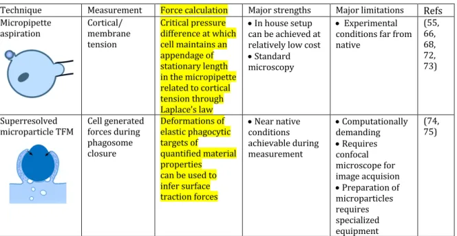
capacitance measurements to monitor particle uptake of human monocyte-derived 338 macrophages. Their results revealed that the surface of plasma membrane, rather than 339 decreasing after engulfment of a large particle, increased. Bajno et al. (53) first 340 demonstrated that during phagocytosis, recycling vesicles accumulated at the 341 phagocytic cup, suggesting that membrane extension results from internal membranes. 342 Various internal compartments were actually demonstrated to contribute to efficient 343 phagosome formation (54). Cortical tension measurements performed by Herant et al. 344 (55) on neutrophils provided another solution. In these experiments, neutrophils were 345 partially aspirated using a micropipette and the critical pressure difference at which the 346 cell maintains an appendage of stationary lengths in the micropipette is related to the 347 cortical tension through Laplace’s law. The experimental configuration, strengths and 348 limitations of such experiments are shown in Table 3. Herant et al. determined that in 349 addition to the trafficking of internal membranes to the phagocytic cup, the extent to 350 which expansion of the plasma membrane surface during phagocytosis takes place can 351 be explained by the release of ‘spare’ plasma membrane normally sequestered in a 352 reservoir of membrane folds, also used by the cell to probe its environment. Later, 353 internal compartments were revealed to play a role also as signalling platforms 354 important to regulate the local actin dynamics and depolymerisation that is necessary 355 for membrane extension (56). 356 357 Shown schematically in Figure 1, there remains some ambiguity about how phagosome 358 closure and internalisation actually occur. Certainly, for fusion and internalisation to 359 take place, force generated by the actin driven protrusions must have directionality 360 (57). A contractile activity was identified by Evans et al. following pseudopod extension 361 during the phagocytosis of yeast (58). More recently, a TFM study performed by Kovari 362 et al. (33) measured the tractions of the J774 macrophage cell line undergoing frustrated 363 phagocytosis. The authors were able to identify and quantify a contractile phase in cells 364 undergoing frustrated phagocytosis, corresponding to maximal traction forces exerted 365 on the substrate, though it should be noted that frustrated phagocytosis is planar, and 366 therefore quite dissimilar from the 3D process. The activity of myosins (59-65) and a 367 build-up of cortical tension (55, 60, 66, 67) have been suggested as the origin of a 368 contractile force required for internalisation. Several myosins are recruited at the site of 369 phagosome formation. MyosinK plays a critical role in efficient phagocytosis of large 370 particles in Dictyostelium discoideum (62). Myosin-II was described to be important for 371 CR3- more than FcR-mediated phagocytosis (65). In a later study by Herant et al. (66), 372 the authors couple their cortical tension measurements with mathematical modelling to 373 propose a model of FcR phagosome closure. They propose several possible models to 374 explain their experimental observations and find that the model that best fits their data 375 is based on a contractile flattening force with molecular motors (possibly, but not 376 necessarily myosins) that are anchored to the cell-particle interface, pulling down on the 377 actin cytoskeleton and pulling the target particle into the cell body. 378 379 In further work, Herant et al. (68) demonstrate that Zymosan triggered phagocytosis 380 occurs via a ‘protrusive push’ mechanism, with particles only internalised after an initial 381 outward push. They contrast this with the ‘enveloping embrace’ of FcR phagocytosis 382 (66), revealing that these two pathways display different force profiles. While the 383 precise origin of differing force profiles remains unclear, it has long been established 384 that the two most widely characterised phagocytic pathways, triggered by FcR and CR3 385 receptors, vary considerably in this respect (69). In both of these studies by Herant et al., 386
the cortical tension is considered the primary driver of inward motion of the target 387 particle, contrasting with the idea of a molecular motor driven inward pulling force. The 388 authors propose that it is the cortical tension driven tendency of a cell to round up, in 389 addition to the strong adhesion between the target particle and the plasma membrane of 390 the phagocyte, that is the source of target motion into the cell. Later work is in good 391 agreement with this idea, but the biochemical basis of the contractile force remains 392 unclear (33, 70). Recently though, using a Total Internal Reflection Fluorescence (TIRF) 393 Microscopy approach in living macrophages (26, 71), Marie-Anaïs et al. demonstrated 394 that the last scission step relies on the activity of the GTPase dynamin that acts in 395 concert with F-actin to mediate membrane scission and FcR-phagosome sealing (71). 396 397
Technique Measurement Force calculation Major strengths Major limitations Refs
Micropipette aspiration Cortical/ membrane tension Critical pressure difference at which cell maintains an appendage of stationary length in the micropipette related to cortical tension through Laplace’s law • In house setup can be achieved at relatively low cost • Standard microscopy • Experimental conditions far from native (55, 66, 68, 72, 73) Superresolved microparticle TFM Cell generated forces during phagosome closure Deformations of elastic phagocytic targets of quantified material properties can be used to infer surface traction forces • Near native conditions achievable during measurement • Computationally demanding • Requires confocal microscope for image acquision • Preparation of microparticles requires specialized equipment (74, 75) Table 3: Methods for investigating phagosome closure. 398 399 More recently, Vorselen et al.(75) have made observations similar to Herant et al., while 400 presenting a new, and complementary, approach to study this critical phase of 401 internalisation of phagocytic targets. The authors describe a novel strategy, that they 402 name superresolved microparticle TFM, designed for studying ligand dependent cellular 403 interactions at high spatial resolution. Like PFM, this is a method that has its roots in 404 TFM and was developed to address certain limitations in the standard 2D configuration. 405 The method relies on the reliable fabrication of functionalised polyacrylamide spheres 406 of tunable stiffness. As a proof of concept, J774 murine macrophages were exposed to 407 the spheres functionalised with IgG to trigger phagocytosis. Cell induced deformations 408 were observed and used to determine normal and shear stresses associated with 409 phagocytosis. The experimental configuration, strengths and limitations of the method 410 are shown in Table 3. The potential of this technique to be used to determine the role of 411 various actin binding proteins involved in phagocytosis, particularly in the 412 internalisation/contractile phase, in near native conditions, is significant. 413 414 415 Conclusions 416 417 The professional phagocytes of the immune system are responsible for many crucial 418 immune functions which rely on the ability of phagocytes to migrate and adhere to sites 419
of infection, dynamically probe their environments to make contact with phagocytic 420 targets, and to perform phagocytosis, a mechanism of internalization of large particles, 421 microorganisms and cellular debris for intracellular degradation. These significant 422 mechanical feats are dependent on actin polymerization to provide the force that drives 423 membrane deformation. The role of the actin cytoskeleton at key stages of these 424 processes has been extensively studied, particularly with regard to migration, 425 membrane protrusions and pseudopod extension. In recent years, researchers of 426 phagocytosis have coupled traditional methods in medicine and cell biology with 427 methods of force measurement, such as atomic and traction force microscopies, optical 428 and magnetic tweezers and micropipette aspiration to perform mechanistic studies of 429 phagocytosis, providing great insight into this essential immune process. There is a 430 great deal that remains unknown about phagocytosis though, particularly around 431 phagosome sealing and particle internalisation. Both the origin of the contractile force 432 and its trigger are undetermined, though myosins, membrane and cortical tension 433 feature in models that have been proposed. Further studies of the forces involved in this 434 dramatic process present an exciting path forward, particularly as new variants of more 435 established techniques are developed, such as in the work of Vorselen et al. (75) and 436 Labernadie et al. (44). It is the design of such live cell experimental methodologies that 437 along with classical biological methods, may in time, be able to reveal the true variety 438 and complexity of this and other processes in the future. 439 440 Acknowledgements 441 442 The authors would like to thank Sophie Echène for the lovely illustrations in this review. 443 This work was supported by grants from Centre National de la Recherche Scientifique 444 (CNRS), Institut National de la Santé et de la Recherche Médicale (Inserm), Université 445 Paris Descartes and Agence Nationale de la Recherche (ANR 16-CE13-0007-01) that 446 supported AM for a post-doctoral position. 447 448 449 Declaration 450 451 The authors declare no conflict of interest. 452 453 454 References 455 456
1. Ronald S. Flannagan VJ, and Sergio Grinstein. The Cell Biology of Phagocytosis. 457
Annual Review of Pathology: Mechanisms of Disease. 2012;7(1):61-98. 458
2. Niedergang F. Phagocytosis. Encyclopedia of Cell Biology. Waltham: Academic Press; 459
2016. p. 751-7. 460
3. Poon IKH, Lucas CD, Rossi AG, Ravichandran KS. Apoptotic cell clearance: basic 461
biology and therapeutic potential. Nat Rev Immunol. 2014;14(3):166-80. 462
4. Bloes DA, Kretschmer D, Peschel A. Enemy attraction: bacterial agonists for leukocyte 463
chemotaxis receptors. Nat Rev Microbiol. 2015;13(2):95-104. 464
5. Heit B, Liu L, Colarusso P, Puri KD, Kubes P. PI3K accelerates, but is not required for, 465
neutrophil chemotaxis to fMLP. Journal of Cell Science. 2008;121(2):205. 466
6. Devosse T, Guillabert A, Haene N, Berton A, De Nadai P, Noel S, et al. Formyl Peptide 467
Receptor-Like 2 Is Expressed and Functional in Plasmacytoid Dendritic Cells, Tissue-Specific 468
Macrophage Subpopulations, and Eosinophils. The Journal of Immunology. 469
2009;182(8):4974. 470
7. Ridley AJ. Rho proteins, PI 3-kinases, and monocyte/macrophage motility. FEBS 471
Letters. 2001;498(2):168-71. 472
8. Ridley AJ, Schwartz MA, Burridge K, Firtel RA, Ginsberg MH, Borisy G, et al. Cell 473
Migration: Integrating Signals from Front to Back. Science. 2003;302(5651):1704. 474
9. Pelham RJ, Wang Y-l. Cell locomotion and focal adhesions are regulated by 475
substrate flexibility. Proc Natl Acad Sci U S A. 1997;94(25):13661-5. 476
10. Reinhart-King CA. Chapter 3 Endothelial Cell Adhesion and Migration. Methods in 477
Enzymology. 443: Academic Press; 2008. p. 45-64. 478
11. Reinhart-King CA, Dembo M, Hammer DA. The Dynamics and Mechanics of 479
Endothelial Cell Spreading. Biophys J. 2005;89(1):676-89. 480
12. Lo C-M, Wang H-B, Dembo M, Wang Y-l. Cell Movement Is Guided by the Rigidity of 481
the Substrate. Biophys J. 2000;79(1):144-52. 482
13. Lauffenburger DA, Linderman JJ. Receptors. [electronic resource] : models for 483
binding, trafficking, and signaling: Oxford University Press; 1993. 484
14. Bidan CM, Fratzl M, Coullomb A, Moreau P, Lombard AH, Wang I, et al. Magneto-485
active substrates for local mechanical stimulation of living cells. Sci Rep. 2018;8(1):1464. 486
15. Kohyama M, Ise W, Edelson BT, Wilker PR, Hildner K, Mejia C, et al. Role for Spi-C in 487
the development of red pulp macrophages and splenic iron homeostasis. Nature. 488
2009;457(7227):318-21. 489
16. Bilzer M, Roggel F, Gerbes AL. Role of Kupffer cells in host defense and liver disease. 490
Liver International. 2006;26(10):1175-86. 491
17. Gerner Michael Y, Torabi-Parizi P, Germain Ronald N. Strategically Localized Dendritic 492
Cells Promote Rapid T Cell Responses to Lymph-Borne Particulate Antigens. Immunity. 493
2015;42(1):172-85. 494
18. Flannagan RS, Harrison RE, Yip CM, Jaqaman K, Grinstein S. Dynamic macrophage 495
“probing” is required for the efficient capture of phagocytic targets. The Journal of Cell 496
Biology. 2010;191(6):1205. 497
19. Miller YI, Chang M-K, Funk CD, Feramisco JR, Witztum JL. 12/15-Lipoxygenase 498
Translocation Enhances Site-specific Actin Polymerization in Macrophages Phagocytosing 499
Apoptotic Cells. Journal of Biological Chemistry. 2001;276(22):19431-9. 500
20. Nimmerjahn A, Kirchhoff F, Helmchen F. Resting Microglial Cells Are Highly Dynamic 501
Surveillants of Brain Parenchyma in Vivo. Science. 2005;308(5726):1314. 502
21. Jubrail J, Montauban K, Regis-Burgel P, Kurian N, Niedergang F. Characterisation of 503
defective phagocytosis by chronic obstructive pulmonary disease alveolar macrophages. 504
European Respiratory Journal. 2016;48(suppl 60). 505
22. Tabas I, Glass CK. Anti-Inflammatory Therapy in Chronic Disease: Challenges and 506
Opportunities. Science. 2013;339(6116):166. 507
23. Jubrail JK, Nisha; Niedergang, Florence. Macrophage phagocytosis cracking the defect 508
code in COPD. Biomedical Journal. 2017;In press. 509
24. Flannagan RS, Cosio G, Grinstein S. Antimicrobial mechanisms of phagocytes and 510
bacterial evasion strategies. Nat Rev Microbiol. 2009;7(5):355-66. 511
25. Pollard JW. Trophic macrophages in development and disease. Nature Reviews 512
Immunology. 2009;9:259. 513
26. Mularski A M-AF, Mazzolini J and Niedergang, F. Observing frustrated phagocytosis 514
and phagosome formation and closure using total internal reflection fluorescence 515
microscopy (TIRFM). In: Rousselet G, editor. Methods in Molecular Biology. In press: 516
Springer; 2018. 517
27. Polacheck WJ, Chen CS. Measuring cell-generated forces: a guide to the available 518
tools. Nat Meth. 2016;13(5):415-23. 519
28. Legant WR, Choi CK, Miller JS, Shao L, Gao L, Betzig E, et al. Multidimensional traction 520
force microscopy reveals out-of-plane rotational moments about focal adhesions. Proc Natl 521
Acad Sci U S A. 2013;110(3):881-6. 522
29. Toyjanova J, Bar-Kochba E, López-Fagundo C, Reichner J, Hoffman-Kim D, Franck C. 523
High Resolution, Large Deformation 3D Traction Force Microscopy. PLOS ONE. 524
2014;9(4):e90976. 525
30. Maskarinec SA, Franck C, Tirrell DA, Ravichandran G. Quantifying cellular traction 526
forces in three dimensions. Proc Natl Acad Sci U S A. 2009;106(52):22108-13. 527
31. Legant WR, Miller JS, Blakely BL, Cohen DM, Genin GM, Chen CS. Measurement of 528
mechanical tractions exerted by cells in three-dimensional matrices. Nat Meth. 529
2010;7(12):969-71. 530
32. Martiel J-L, Leal A, Kurzawa L, Balland M, Wang I, Vignaud T, et al. Measurement of 531
cell traction forces with ImageJ. Methods in Cell Biology. 2015;125:269-87. 532
33. Kovari DT, Wei W, Chang P, Toro J-S, Beach RF, Chambers D, et al. Frustrated 533
Phagocytic Spreading of J774A-1 Macrophages Ends in Myosin II-Dependent Contraction. 534
Biophys J. 2016;111(12):2698-710. 535
34. Hind LE, Dembo M, Hammer DA. Macrophage motility is driven by frontal-towing 536
with a force magnitude dependent on substrate stiffness. Integrative Biology. 2015;7(4):447-537
53. 538
35. Hind LE, Lurier EB, Dembo M, Spiller KL, Hammer DA. Effect of M1–M2 Polarization 539
on the Motility and Traction Stresses of Primary Human Macrophages. Cellular and 540
Molecular Bioengineering. 2016;9(3):455-65. 541
36. Smith LA, Aranda-Espinoza H, Haun JB, Dembo M, Hammer DA. Neutrophil Traction 542
Stresses are Concentrated in the Uropod during Migration. Biophys J. 2007;92(7):L58-L60. 543
37. Jannat Risat A, Dembo M, Hammer Daniel A. Traction Forces of Neutrophils Migrating 544
on Compliant Substrates. Biophys J. 2011;101(3):575-84. 545
38. Gupta M, Kocgozlu L, Sarangi BR, Margadant F, Ashraf M, Ladoux B. Chapter 16 - 546
Micropillar substrates: A tool for studying cell mechanobiology. In: Paluch EK, editor. 547
Methods in Cell Biology. 125: Academic Press; 2015. p. 289-308. 548
39. Ricart Brendon G, Yang Michael T, Hunter Christopher A, Chen Christopher S, 549
Hammer Daniel A. Measuring Traction Forces of Motile Dendritic Cells on Micropost Arrays. 550
Biophys J. 2011;101(11):2620-8. 551
40. Cougoule C, Van Goethem E, Le Cabec V, Lafouresse F, Dupré L, Mehraj V, et al. Blood 552
leukocytes and macrophages of various phenotypes have distinct abilities to form 553
podosomes and to migrate in 3D environments. European Journal of Cell Biology. 554
2012;91(11):938-49. 555
41. Buccione R, Orth JD, McNiven MA. Foot and mouth: podosomes, invadopodia and 556
circular dorsal ruffles. Nat Rev Mol Cell Biol. 2004;5(8):647-57. 557
42. Dufrene YF, Ando T, Garcia R, Alsteens D, Martinez-Martin D, Engel A, et al. Imaging 558
modes of atomic force microscopy for application in molecular and cell biology. Nat Nano. 559
2017;12(4):295-307. 560
43. Labernadie A, Thibault C, Vieu C, Maridonneau-Parini I, Charrière GM. Dynamics of 561
podosome stiffness revealed by atomic force microscopy. Proc Natl Acad Sci U S A. 562
2010;107(49):21016. 563
44. Labernadie A, Bouissou A, Delobelle P, Balor S, Voituriez R, Proag A, et al. Protrusion 564
force microscopy reveals oscillatory force generation and mechanosensing activity of human 565
macrophage podosomes. 2014;5:5343. 566
45. Kress H, Stelzer EHK, Griffiths G, Rohrbach A. Control of relative radiation pressure in 567
optical traps: Application to phagocytic membrane binding studies. Phys Rev E: Stat, 568
Nonlinear, Soft Matter Phys. 2005;71(6):061927. 569
46. Svitkina TM, Bulanova EA, Chaga OY, Vignjevic DM, Kojima S-i, Vasiliev JM, et al. 570
Mechanism of filopodia initiation by reorganization of a dendritic network. The Journal of 571
Cell Biology. 2003;160(3):409. 572
47. Cojoc D, Difato F, Ferrari E, Shahapure RB, Laishram J, Righi M, et al. Properties of the 573
Force Exerted by Filopodia and Lamellipodia and the Involvement of Cytoskeletal 574
Components. PLoS One. 2007;2(10). 575
48. Vonna L, Wiedemann A, Aepfelbacher M, Sackmann E. Micromechanics of filopodia 576
mediated capture of pathogens by macrophages. Eur Biophys J. 2007;36(2):145-51. 577
49. Kress H, Stelzer EHK, Holzer D, Buss F, Griffiths G, Rohrbach A. Filopodia act as 578
phagocytic tentacles and pull with discrete steps and a load-dependent velocity. Proc Natl 579
Acad Sci U S A. 2007;104(28):11633-8. 580
50. Ostrowski Philip P, Grinstein S, Freeman Spencer A. Diffusion Barriers, Mechanical 581
Forces, and the Biophysics of Phagocytosis. Developmental Cell. 2016;38(2):135-46. 582
51. Freeman SA, Grinstein S. Phagocytosis: receptors, signal integration, and the 583
cytoskeleton. Immunological Reviews. 2014;262(1):193-215. 584
52. Holevinski KO, Nelson DJ. Membrane capacitance changes associated with particle 585
uptake during phagocytosis in macrophages. Biophys J. 1998;75. 586
53. Bajno L, Peng X-R, Schreiber AD, Moore H-P, Trimble WS, Grinstein S. Focal Exocytosis 587
of Vamp3-Containing Vesicles at Sites of Phagosome Formation. The Journal of Cell Biology. 588
2000;149(3):697. 589
54. Braun V, Fraisier V, Raposo G, Hurbain I, Sibarita J-B, Chavrier P, et al. TI-590
VAMP/VAMP7 is required for optimal phagocytosis of opsonised particles in macrophages. 591
The EMBO Journal. 2004;23(21):4166-76. 592
55. Herant M, Heinrich V, Dembo M. Mechanics of neutrophil phagocytosis: behavior of 593
the cortical tension. Journal of Cell Science. 2005;118(9):1789. 594
56. Marion S, Mazzolini J, Herit F, Bourdoncle P, Kambou-Pene N, Hailfinger S, et al. The 595
NF-κB Signaling Protein Bcl10 Regulates Actin Dynamics by Controlling AP1 and OCRL-596
Bearing Vesicles. Developmental Cell. 2012;23(5):954-67. 597
57. Levin R, Grinstein S, Canton J. The life cycle of phagosomes: formation, maturation, 598
and resolution. Immunological Reviews. 2016;273(1):156-79. 599
58. Evans E, Leung A, Zhelev D. Synchrony of cell spreading and contraction force as 600
phagocytes engulf large pathogens. The Journal of Cell Biology. 1993;122(6):1295. 601
59. Araki N, Hatae T, Furukawa A, Swanson JA. Phosphoinositide-3-kinase-independent 602
contractile activities associated with Fcγ-receptor-mediated phagocytosis and 603
macropinocytosis in macrophages. Journal of Cell Science. 2002;116(2):247. 604
60. Swanson JA, Johnson MT, Beningo K, Post P, Mooseker M, Araki N. A contractile 605
activity that closes phagosomes in macrophages. Journal of Cell Science. 1999;112(3):307. 606
61. Diakonova M, Bokoch G, Swanson JA. Dynamics of Cytoskeletal Proteins during Fcγ 607
Receptor-mediated Phagocytosis in Macrophages. Molecular Biology of the Cell. 608
2002;13(2):402-11. 609
62. Boulais J, Trost M, Landry C, Dieckmann R, Levy E, Soldati T, et al. Evolutionary 610
adaptation of phagocytosis modeled the adaptive immune system. Immunity2010. 611
63. Gopaldass N, Patel D, Kratzke R, Dieckmann R, Hausherr S, Hagedorn M, et al. 612
Dynamin A, Myosin IB and Abp1 couple phagosome maturation to F-actin binding. Traffic. 613
2012;13(1):120-30. 614
64. Barger SR, Reilly NS, Shutova MS, Li Q, Maiuri P, Heddleston JM, et al. Membrane-615
cytoskeletal crosstalk mediated by myosin-I regulates adhesion turnover during 616
phagocytosis. Nature Communications. 2019;10(1):1249. 617
65. Olazabal IM, Caron E, May RC, Schilling K, Knecht DA, Machesky LM. Rho-Kinase and 618
Myosin-II Control Phagocytic Cup Formation during CR, but Not FcγR, Phagocytosis. Current 619
Biology. 2002;12(16):1413-8. 620
66. Herant M, Heinrich V, Dembo M. Mechanics of neutrophil phagocytosis: experiments 621
and quantitative models. Journal of Cell Science. 2006;119(9):1903. 622
67. Zhelev DV, Needham D, Hochmuth RM. Role of the membrane cortex in neutrophil 623
deformation in small pipets. Biophys J. 1994;67. 624
68. Herant M, Lee C-Y, Dembo M, Heinrich V. Protrusive Push versus Enveloping 625
Embrace: Computational Model of Phagocytosis Predicts Key Regulatory Role of Cytoskeletal 626
Membrane Anchors. PLoS Computational Biology. 2011;7(1):e1001068. 627
69. Allen LA, Aderem A. Molecular definition of distinct cytoskeletal structures involved 628
in complement- and Fc receptor-mediated phagocytosis in macrophages. J Exp Med. 629
1996;184. 630
70. Masters TA, Pontes B, Viasnoff V, Li Y, Gauthier NC. Plasma membrane tension 631
orchestrates membrane trafficking, cytoskeletal remodeling, and biochemical signaling 632
during phagocytosis. Proc Natl Acad Sci U S A. 2013;110(29):11875-80. 633
71. Marie-Anaïs F, Mazzolini J, Herit F, Niedergang F. Dynamin-Actin Cross Talk 634
Contributes to Phagosome Formation and Closure. Traffic. 2016;17(5):487-99. 635
72. Guevorkian K, Maître JL. Chapter 10 - Micropipette aspiration: A unique tool for 636
exploring cell and tissue mechanics in vivo. In: Lecuit T, editor. Methods in Cell Biology. 139: 637
Academic Press; 2017. p. 187-201. 638
73. Herant M, Marganski WA, Dembo M. The Mechanics of Neutrophils: Synthetic 639
Modeling of Three Experiments. Biophys J. 2003;84(5):3389-413. 640
74. Vorselen D, Wang Y, de Jesus MM, Shah PK, Footer MJ, Huse M, et al. Superresolved 641
microparticle traction force microscopy reveals subcellular force patterns in immune cell-642
target interactions. bioRxiv. 2019:431221. 643
75. Vorselen D, Wang Y, Footer MJ, Cai W, Theriot JA. Superresolved and reference-free 644
microparticle traction force microscopy (MP-TFM) reveals the complexity of the mechanical 645
interaction in phagocytosis. bioRxiv. 2018:431221. 646
647